Heavy metals, Co, Ni, Cu, Zn and Cd,
produce oxidative damage and evoke differential
antioxidant responses in spinach.
nalini pandey
1,*, girish c. pathak
1, Dharmendra K. pandey
2, ritu pandey
11 Department of Botany, University of Lucknow, Lucknow- 226 007, India
2 Department of Life Sciences, Amity School of Engineering and Technology, Amity University, Lucknow 226010, India.
* Corresponding author. Tel.:+ 919451088125; E-mail address: [email protected] Received: 18 July 2009; Returned for revision: 24 September 2009; Accepted: 30 September 2009
abstract
Exposure of 10-d-old spinach (Spinacea oleracea L.) plants to excess (500 µM) concentrations of Co, Ni, Cu, Zn and Cd in sand culture inhibited growth, induced toxicity symptoms, oxidative damage and changes in the antioxidant defense system. The severity of the metal-induced effects varied with the metals and the duration of exposure to excess supply of the metals. Each metal induced chlorosis. In addition, excess Co, Ni and Cd also produced metal specific toxic effects. Excess supply of each metal caused lipid peroxidation (TBARS). Their effectiveness in producing oxidative damage was in the order: Ni > Co > Cd > Cu >Zn. Of all the metals, Ni was also most effective in lowering the concentration of the chloroplast pigments (Chl, Car). While each metal increased the concentration of ascorbate and activated the key enzymes of the ascorbate–glutathione cycle, excess Cd and Zn were more effective in this regard. Each metal increased the activity of SOD and POD and decreased the activity of CAT. Enhancement in SOD activity and inhibition of CAT activity suggested high build-up of H2O2, possibly the main cause of oxidative stress, induced in response to excess supply of the heavy metals.
Key words: Ascorbate-glutathione cycle, heavy metal exposure, Spinacia oleracea L
introDUction
Increasing environmental pollution caused by heavy metals, released by industrial and agricultural activities, is a major problem in the world (Prasad, 2004). Plants grown on soils with parent material rich in heavy metals or polluted by industrial effluents are known to absorb heavy metals in quantities that may be toxic to plant growth and metabolism (Alloway, 1990; Prasad, 2004). Excess of heavy metals cause phytotoxic effects in several ways, one of these being the excessive production of reactive oxygen species (ROS) which disturb the cellular redox environment causing oxidative stress (Erdei et al., 2002; Shaw et al., 2004; Nada et al., 2007). While it is well established that the ROS are a major factor contributing to heavy metal
Materials anD MethoDs
Plant material: Spinach (Spinacia oleracea L.) was grown in sand culture under greenhouse conditions (Sharma, 1996). During the period of the study maximum light intensity PFFD at 1200 h ranged between 900 to 1050 µmol m-2 s-1. The temperature during the 24 h period ranged between 18 to 22oC (maximum) and 8.5 to 12oC (minimum), and the RH (9:30 A.M.) between 60% to 78%. Average day length was around 10.30±0:20 h. Plants were raised in 5 L polyethylene pots. Each pot was provided with a central drainage hole, covered with an inverted watch glass with glass wool under the rim to allow free drainage of nutrient solution. All pots were supplied 1 L of full nutrient solution (control), containing 4 mM Ca(NO3)2, 4 mM KNO3, 2 mM MgSO4, 1.33 mM NaH2PO4, 0.33 mM H3BO3, 0.1 mM Fe-EDTA, 10 µM MnSO4, 1 µM ZnSO4, 1 µM CuSO4, 0.1 µM Na2MoO4, 0.1 mM NaCl, 0.1 µM CoSO4 and 0.1 µM NiSO4 daily at 1800 h except for weekends when pots were flushed with glass-distilled water (GDW) to avoid accumulation of nutrients. After 20 d of complete nutrient supply, pots were separated into 6 lots of 3 pots each and the number of plants in each pot was reduced to 6. The 1st lot of pots was maintained as such (control). The 2nd, 3rd, 4th, 5th and 6th lots of pots were supplied 500 µM CoSO
4, NiSO4, CuSO4, ZnSO4, and CdSO4 respectively, superimposed over the control nutrient solution. Plants were treated daily up to 8 days. After 5 and 8 d exposure to the heavy metals, plants were sampled in triplicate and quantified for tissue concentration of Co, Ni, Cu, Zn and Cd; activities of SOD (total and Cu/Zn SOD), APX, GR, CAT, non-specific POD and concentration of thiobarbituric acid reactive substances (TBARS), chlorophyll (Chl), carotenoids (Car) and ascorbate (Asc).
Biomass, metal concentration: Plants were uprooted from the sand, causing minimum damage to the roots, washed thoroughly with deionised water, blotted dry, separated into leaves, stems and roots, chopped into small pieces and oven dried in an electric oven at 80oC for 48 h. After dry weight determination, the oven-dried samples were ground and 1.0 g samples were digested with a mixture of HNO3 and HClO4 (10:1 v/v). The digests were used for determining the concentration of Co, Ni, Cu, Zn and Cd by atomic absorption spectrophotometry (Perkin Elmer A Analyst 300).
Plant pigments: Measurements for concentration of chlorophyll and carotenoids were made in the first fully expanded leaf. Fresh leaf tissue was extracted in 80% acetone
and the extracts were measured for chlorophylls (a, b) at 645 and 663 nm and for carotenoids at 480 and 510 nm by a Perkin Elmer Lambda Bio 20 UV/VIS Spectrophotometer (Lichtenthaler, 1987).
Ascorbate: Ascorbate (Asc) was extracted by grinding fresh leaf tissue in 10% trichloroacetic acid (Law et al., 1983). The assay is based on the reduction of Fe3+ to Fe2+ by ascorbic acid and formation of a pink color complex between Fe2+ and
αα -bipyridyl, with absorption max at 525 nm.
Lipid peroxidation: To determine the lipid peroxide concentration, fresh leaf material was extracted in 1% trichloroacetic acid. The supernatant (after centrifugation at 10,000g for 10 min.) was treated with 0.5% thiobarbituric acid (TBA) in 20% TCA and the mixture was incubated in a boiling water-bath for 30 min. The thiobarbituric acid reactive substances (TBARS) thus formed were measured spectrophotometrically at 532 nm after adjusting for non-specific absorbance at 600 nm (Heath and Packer, 1968).
Enzyme activities: For assay of SOD and GR, fresh leaf tissue was ground in potassium phosphate buffer (50 mM, pH 7.0), containing EDTA (1 mM) and PVP (2%) at 40C. The extracts were centrifuged at 15,000 g for 10 min and the supernatant was assayed for the enzyme activities. SOD was assayed by monitoring the inhibition of nitroblue tetrazolium (Beauchamp and Fridovich, 1971). The difference after inhibition of the enzyme activity with KCN (3 mM) was taken as the measure of Cu/Zn SOD activity. For determining APX activity, 1 mM ascorbate was added to the above grinding mixture. The enzyme was assayed by monitoring the oxidation of ascorbate by following the decrease in absorbance at 290 nm per min. The reaction mixture contained 50 mM potassium phosphate buffer pH 7.0, 0.5 mM ascorbate, 1 mM EDTA, and 0.1 mM hydrogen peroxide and a suitable volume of enzyme extract (Nakano and Asada, 1981). Assay for GR (Jablonski and Anderson, 1978) was carried out in a reaction mixture containing 100 mM phosphate buffer pH 7.5, 1 mM oxidized glutathione, 1 mM EDTA, 0.1 mM NADPH and 50 µl of enzyme extract. The oxidation of NADPH was followed by monitoring the decrease in absorbance per min at 340 nm.
modification of the method described by Pandey and Sharma (2002). The reaction mixture for CAT contained 0.5 mmol hydrogen peroxide, 0.01 mmol potassium phosphate buffer (pH 7.0) and 1 ml of suitably diluted enzyme extract in a final volume of 10 ml. After incubation at 25oC for 5 min, the reaction was stopped with 5 ml 2 N H2SO4. Corresponding blanks were run simultaneously, in which H2SO4 was added prior to the enzyme extract. The mixture was titrated against 0.1 N KMnO4 and the amount of H2O2 decomposed was calculated. The reaction mixture for POD contained 0.5 mmol phosphate buffer pH 6.0, 0.01% (v/v) H2O2, 5 mg p-phenylenediamine-HCl and the enzyme extract in a final volume of 8 ml. The reaction was maintained at 25oC for 5 min and was stopped with 2 ml of 2 N H2SO4. Blanks were run simultaneously. Enzyme activity was
measured spectrophotometrically as ΔOD at 485 nm. Soluble protein in the enzyme extracts was measured by the method of Bradford (1976) using bovine serum albumin (Sigma) as standard. All enzyme activities are expressed on mg-1 protein basis.
Statistical analysis: All measurements were made on samples drawn in triplicate and the data were statistically analyzed (ANOVA) for significance (LSD at P=0.05).
resUlts
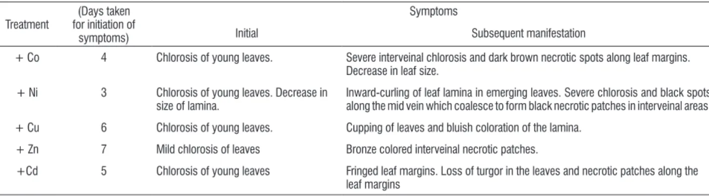
Growth, visible symptoms: Excess (500 µM) supply of each of the metals Co, Ni, Cu, Zn and Cd for 8 days inhibited growth and induced visible symptoms. However, the time taken for the initial appearance of the symptoms and the rapidity with which they intensified varied from metal to metal. Excess supply of each metal produced chlorosis of young leaves but chlorosis appeared first in plants exposed to excess Ni (3rd d) and last (7th d) in plants exposed to excess Zn. Severity of chlorosis increased with time of exposure to the metal treatment in the case of Co, Ni, Cu and Cd but not for Zn. Excess Co, Ni and Cd also produced other symptoms specific to these metals (Table 1).
table 1. Visible symptoms induced in spinach (Spinacia oleracea L.) on exposure to 500 µM supply of Co, Ni, Cu, Zn and Cd.
Treatment
(Days taken for initiation of
symptoms)
Symptoms
Initial Subsequent manifestation
+ Co 4 Chlorosis of young leaves. Severe interveinal chlorosis and dark brown necrotic spots along leaf margins.
Decrease in leaf size.
+ Ni 3 Chlorosis of young leaves. Decrease in
size of lamina.
Inward-curling of leaf lamina in emerging leaves. Severe chlorosis and black spots along the mid vein which coalesce to form black necrotic patches in interveinal areas.
+ Cu 6 Chlorosis of young leaves. Cupping of leaves and bluish coloration of the lamina.
+ Zn 7 Mild chlorosis of leaves Bronze colored interveinal necrotic patches.
+Cd 5 Chlorosis of young leaves Fringed leaf margins. Loss of turgor in the leaves and necrotic patches along the
leaf margins
The effect of excess supply of heavy metals on growth was reflected in shoot and root dry matter yield (Table 2). On d 5 and d 8, growth inhibition of the top parts of plants as well as total
dry matter production was in the order Ni>Co>Cd>Cu>Zn (Table 2). Both at d 5 and d 8, root dry matter production was inhibited in the order Co>Ni>Cd>Cu>Zn.
table 2. Dry weight yield of spinach (Spinacia oleracea L.) plants following exposure to 500 µM supply of Co, Ni, Cu, Zn and Cd in sand culture.
Days of treatment Plant part Treatment LSD
(P=0.05)
Control +Co +Ni +Cu +Zn +Cd
g dry wt plant-1
Tops 2.85 1.23 1.01 1.62 2.01 1.62 0.25
5 Roots 0.88 0.32 0.34 0.51 0.53 0.39 0.04
Total 3.83 1.55 1.35 2.12 2.54 2.01 0.32
Tops 3.32 0.92 0.86 1.37 1.54 1.20 0.80
8 Roots 0.92 0.29 0.33 0.53 0.57 0.44 0.03
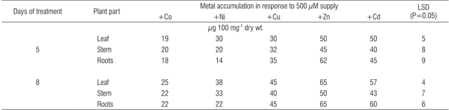
Metal uptake: Both at d 5 and d 8, leaf tissue concentration of Co and Cd in control plants was less than 1 µg g-1 dry wt., that of Ni was less than 5 µg g-1 dry wt and that of Cu and Zn around 8 µg g-1 and 28 µg g-1 dry wt. respectively. Supply of
each of the metals at a concentration of 500 µM led to their increased accumulation (Table 3). Cadmium showed much higher concentration in roots than leaves. The leaf and root concentration of the other metals did not differ markedly.
table 3. Accumulation of Co, Ni, Cu, Zn and Cd in leaves of spinach (Spinacia oleracea L.) plants exposed to 500 µM supply of the metals.
Days of treatment Plant part Metal accumulation in response to 500 µM supply (P=0.05)LSD
+Co +Ni +Cu +Zn +Cd
µg 100 mg-1 dry wt.
Leaf 19 30 30 50 50 5
5 Stem 20 20 32 45 40 8
Roots 18 14 35 62 45 9
8 Leaf 25 38 45 65 57 4
Stem 22 33 40 50 43 7
Roots 22 22 45 65 60 6
TBARS: Excess (500 µM) supply of each of the five metals led to increased accumulation of TBARS but the magnitudes of their accumulation differed from metal to metal. On d 5, excess supply of Co, Ni and Cd caused significant increase in TBARS. By d 8, marked increase in TBARS was observed in response to excess supply of each of the metals, but the magnitude of TBARS accumulation in response to Co, Ni and Cd was more than in response to Cu or Zn (Figure 1A). Both at d 5 and d 8, maximum content of TBARS was found in plants exposed to excess Ni.
Chlorophyll, carotenoids: Excess supply of each of the metals caused a decrease in the concentration of Chl and Car (Figure 1B, C). The decrease in both Chl and Car became greater with time of plant exposure to excess supply of the metals. In general, Co and Ni were more effective in decreasing Chl and Car concentrations than Cd, Cu or Zn.
Ascorbate: Excess metal effects on ascorbate (Asc) differed with the metals and the duration of exposure to the excess metal supply. Exposure to excess Cu and Zn for 5 d caused a severe decrease in Asc content, but continued (8 d) exposure to these metals led to total reversal of the effect (Figure 1D). On d 8, Asc concentration in the leaves of Cu- and Zn-treated plants became equal or higher than the control. Exposure to Ni and Cd for 5 d produced little change in Asc
content but on 8 d exposure to these metals, Asc concentration showed a significant increase. The effect of excess Cd was particularly marked. Cobalt caused a significant increase in Asc concentration on d 5 but subsequently (d 8) this came down to near control values.
Antioxidative enzyme activities: Exposure of plants to excess supply of each of the metals for 5 d led to an increase in total SOD activity (Figure 2A). The increase in SOD activity in response to excess Co, Ni and Cd was particularly marked. On d 8, total SOD activity leveled down to that of control values in the case of Co and Ni, but for Cd it still remained significantly higher than the control. At this stage, the increase in total SOD activity due to excess supply of Cu and Zn also became marked. Activity of Cu-Zn SOD followed a similar trend (Figure 2B).
Excess supply of Co, Ni, Cu and Cd caused a significant decrease in catalase activity, with little difference in the severity of the effect on d 5 and d 8 (Figure 2C). Excess Zn did not lead to any significant change in CAT activity on d 5, but inhibited it on d 8.
(5 or 8d) did not make much difference to the severity of the excess metal effects on POD activity.
The effect of plant exposure to excess supply of the metals on APX activity was similar to that on POD, except that the increase in APX activity on exposure to excess Cd was more than two-and-a-half times that of the control at d 5 (Figure 2E). Plant exposure to 500 µM Zn increased APX activity more than two times the control. The APX activity at 500 µM Ni increased marginally at d 5 but failed to show any increase at d 8.
Excess supply of each of the metals Co, Ni, Cu, Zn and Cd caused a marked and significant increase in GR activity (Figure 2F). Five days exposure to excess Co, Ni, Zn and Cd increased it to more than double that in control plants. Increase in GR activity in response to excess Co (248%) and Cd (277%) was particularly marked. Except in plants supplied excess Cu, longer (8 d) duration of exposure to each of the metals Co, Ni, Zn and Cd caused partial reversal of the effect but activity still remained significantly higher than in control plants with the lowest activity being observed in plants receiving excess Ni (Figure 2 F).
B. Chlorophyll
0 40 80 120
*
*
*
*
*
*
*
*
*
C. Carotenoids
0 40 80 120
C Co Ni Cu Zn Cd
C Co Ni Cu Zn Cd
C Co Ni Cu Zn Cd
C Co Ni Cu Zn Cd
*
*
*
*
*
*
*
*
*
*
A. TBARS
0 80 160 240
R.C R.C
R.C R.C
*
*
*
*
*
*
*
*
D. Ascorbate
0 50 100 150 200
*
*
*
*
*
*
*
*
figure 1. Relative concentration (R.C.) of TBARS (A), Chl (B), Car (C) and Asc (D) in the leaves of spinach (Spinacia oleracea L.) plants following exposure to 500
µM supply of Co, Ni, Cu, Zn and Cd in sand culture for 5 d (□) and 8 d (■). The control values taken as 100% are represented by a line and significant (P=0.05)
E. Ascorbate peroxidase
0 100 200 300
*
*
*
*
*
*
*
F. Glutathione reductase
0 100 200 300
*
*
*
*
*
*
*
A. Total SOD
0 80 160 240
*
*
*
*
*
*
*
*
B. Cu /Zn SOD
0 40 80 120 160
*
*
*
*
*
*
*
*
C. Catalase
0 40 80 120
R
. A
R
. A
R
. A R. A
R
. A R. A
*
*
*
*
*
*
*
*
D. Peroxidase
0 80 160 240
*
*
*
* *
*
*
*
C Co Ni Cu Zn Cd C Co Ni Cu Zn Cd
C Co Ni Cu Zn Cd C Co Ni Cu Zn Cd
C Co Ni Cu Zn Cd C Co Ni Cu Zn Cd
figure 2. Relative activities (R.A.) of total SOD (A), Cu/Zn SOD (B), CAT (C), POD (D), APX (E) and GR (F) in the leaves of spinach (Spinacia oleracea L.) plants
following exposure to 500 µM supply of Co, Ni, Cu, Zn and Cd in sand culture for 5 d (□) and 8 d (■). The control values taken as 100%are represented by a line
and significant (P=0.05) differences are marked with an asterisk (*).
DiscUssion
Exposure of plants to excess (500 µM) supply of the heavy metals – Co, Ni, Cu, Zn and Cd inhibited growth, induced visible symptoms and caused enhanced accumulation of TBARS. The exposure to heavy metal stress caused significant reduction in biomass accumulation both at d 5 and d 8 to
a varying extent and reflected the plant’s sensitivity and the cumulative effects of damage due to inhibited physiological functions. The decrease in biomass under heavy metal stress
has been reported earlier (Krovaćević et al., 1999; Pandey
al. (2002). The higher accumulation of metals in roots than leaves and stem as recorded in the case of Cd stress was probably due to its rapid absorption by the roots and its slow translocation to shoot (Nada et al., 2007).
Heavy metal exposure caused a decrease in Chl and Car contents of the test plant both at d 5 and d 8. The decreased concentration of the chloroplastic pigments may be an outcome of reduced synthesis and/or enhanced oxidative degradation of these pigments by the imposed oxidative stress. Chlorosis is one of the most common symptoms of heavy metal toxicity (Myśliwa-Kurdziel et al., 2002). Interference of heavy metals with normal iron metabolism is known to induce physiological iron deficiency which is expressed in the form of chlorosis due to decreased concentration of chloroplastic pigments. Excess of divalent cationic heavy metals compete with iron for uptake (Pandey and Sharma, 2002) by binding with biomolecules of which iron is a constituent. Noriega et al. (2007) recently reported that Cd caused inhibition of ALA dehydratase, an enzyme catalyzing the rate-limiting step of the pathway of haem and chlorophyll synthesis from its precursor ALA. Reduced Chl concentration due to toxicity of heavy metals in different plant species has been well documented for Co (Pandey and Sharma, 2002), Ni (Pandey and Pathak, 2006), Cu (Lombardi and Sebastiani, 2005), Zn (Pathak et al., 2005), and Cd (Nada et al., 2007). Th higher decrease in Chl concentration in plants treated with excess Ni followed by excess Co and Cd is in agreement with our earlier reports for cabbage (Pandey and Sharma, 2002). The lower decrease in Chl by excess Cd relative to excess Ni and Co has been attributed to a comparatively low inhibition of Fe uptake by Cd relative to Ni and Co (Pandey and Sharma, 2002). The Car are known to be potent quenchers of ROS, particularly singlet oxygen species. As the Car protect chlorophyll from photo-oxidative destruction (Middleton and Teramura, 1993), a differential reduction in Car under excess of different heavy metals might be a reason for the differential decrease in chlorophyll being greater in Ni-starved plants.
The measurement of MDA, a product of lipid peroxidation, is routinely used as an index of lipid peroxidation under stress conditions. That toxicity of heavy metals contributes to enhanced generation of ROS, causing peroxidative damage to membrane lipids, has been reported earlier in the case of Co (Li et al., 2005), Ni (Rao and Sresty, 2000; Pandey and Pathak, 2006), Cu (Mazhoudi et al., 1997; Lombardi
and Sebastiani et al., 2005), Zn (Chaoui et al., 1997; Rao and Sresty, 2000) and Cd (Chaoui et al., 1997; Dey et al., 2007). Most of these studies involved excess supply of only one or two metals. In the present study, we compared the effectiveness of an equimolar (500 µM) supply of Co, Ni, Cu, Zn and Cd in causing lipid peroxidation and inducing changes in antioxidants and antioxidative enzymes. As with the intensity of visible toxicity effects and decrease in dry matter yield, the effectiveness in causing lipid peroxidation was in the order: Ni>Co>Cd>Cu>Zn.
Of the antioxidants found in plants, Asc is the most abundant and has diverse physiological roles. In addition to being a substrate for APX it directly scavenges singlet oxygen, superoxide and hydroxyl radicals (Noctor, 2006). Enhanced concentration of Asc under excess supply of Co, Ni, Cu, Zn and Cd indicated the involvement of Asc in the antioxidant response of this plant and is in consonance with earlier reports (Pathak et al., 2005; Gonçalves, 2007). Ascorbate peroxidase is a key enzyme of the glutathione-ascorbate pathway and eliminates peroxides by converting ascorbic acid to dehydroascorbate (Asada, 1992; Foyer and Noctor, 2005). The increase in the Asc pool along with the increase in activity of APX, indicates that de novo synthesis of Asc was enhanced under heavy metal stress.
this could impair efficacious detoxification of H2O2 (Kono and Fridovich, 1982). Cho and Seo (2005) reported that oxidative stress in response to Cd toxicity is due to H2O2 accumulation. While Co, Ni, Cu, Zn and Cd each induced oxidative stress and consequential changes in the antioxidant enzyme activities, Co was next only to Ni in causing oxidative damage and being more effective in activating the antioxidative defense than Ni. Copper was inhibitory to CAT but relatively less effective in producing oxidative damage probably due to the fairly enhanced levels of carotenoids, Cu/Zn SOD, POD and GR.
In conclusion, the present findings affirm that, in common with other abiotic stresses excess intake of the heavy metals produce oxidative stress, and trigger antioxidative responses, but differ in their effectiveness to do so. At equimolar concentrations (500 µM), Ni induced the most severe visual toxicity effects and exhibited maximum oxidative damage as observed by accumulation of TBARS and lower antioxidant capacity than the plants exposed to Co, Cd, Cu and Zn. This was especially so in SOD and H2O2-eliminating enzymes (POD, CAT and APX) which showed lower activity in Ni excess plants. Further, the study suggests that the degree of oxidative damage may also be assessed by the manifestation of external visual toxicity effects both of which were found to be in the order Ni>Co>Cd>Cu>Zn.
Acknowledgements: The authors are grateful to Dr Vivek Pandey, Scientist, Stress Physiology, National Botanical Research Institute, Lucknow, for his help during the course of this investigation. DKP is grateful to CSIR for financial support in the form of a fellowship.
references
Alloway BJ (1990) Heavy Metals in Soils. John Wiley, New york.
Asada K (1992) Ascorbate peroxidase – a hydrogen peroxide scavenging enzyme in plants. Physiol. Plant. 85:235-241.
Beauchamp C, Fridovich I (1971) Superoxide dismutase: Improved assays and an assay applicable to acrylamide gel. Anal. Biochem. 44:276-287. Bradford MM (1976) A rapid and sensitive method for the quantitation of microgram quantities of protein utilizing the principle of protein-dye binding. Anal. Biochem. 72:248-254.
Chaoui A, Mazhoudi S, Ghorbal MH, El Ferjani E (1997) Cadmium and zinc induction of lipid peroxidation and effect on antioxidant enzyme activities in
bean (Phaseolus vulgaris L.). Plant Sci. 127:139-147.
Cho U-H, Seo N-H (2005) Oxidative stress in Arabidopsis thaliana exposed
to cadmium is due to hydrogen peroxide accumulation. Plant Sci. 168:113-120.
Dey SK, Dey J, Patra S, Pothal D (2007) Changes in the antioxidative enzyme activities and lipid peroxidation in wheat seedlings exposed to cadmium and lead stress. Braz. J. Plant Physiol. 19:53-60.
Erdei S, Hegedus A, Hauptmann G, Szali J, Horvath G (2002) Heavy metal induced physiological changes in the antioxidative response system. Acta Biologica Szegediensis. 46:89-90.
Foyer CH, Noctor G (2005) Redox homeostasis and antioxidant signaling: metabolic interface between stress perception and physiological responses. Plant Cell 17:1866-1875.
Gonçalves JF, Becker AG, Cargnelutti D, Tabaldi LA, Pereira LB, Battisti V, Spanevello RM, Morsch MV, Nicoloso FT, Schetinger MRC (2007) Cadmium toxicity causes oxidative stress and induces response of the antioxidant system in cucumber seedlings. Braz. J. Plant Physiol. 19: 223-232.
Gratão PL, Polle A, Lea PJ, Azevedo RA (2005) Making the life of heavy metal-stressed plants a little easier. Funct. Plant Biol. 32: 481-494.
Heath RL, Packer L (1968) Photoperoxidation in isolated chloroplasts. I. Kinetics and stiochiometry of fatty acid peroxidation. Arch. Biochem. Biophys. 125:189-198.
Jablonski PB, AndersonJW (1978) Light-dependent reduction of oxidized
glutathione by ruptured chloroplasts. Plant Physiol. 61:221-225.
Kono y, Fridovich I (1982) Superoxide radical inhibits catalase. J. Biol. Chem. 257:5751-5754.
Krovaćević G, Kastori R, Merkulov LJ (1999) Dry matter and leaf structure
in young wheat plants as affected by cadmium, lead and nickel. Biol. Plant. 42:119-123.
Law My, Charles SA, Halliwell B (1983) Glutathione and ascorbic acid in
spinach (Spinacia oleracea) chloroplasts. The effect of hydrogen peroxide
and of paraquat. Biochem. J. 210:899-903.
Li C-Z, Wang D, Wang G-X (2005) The protective effects of cobalt on potato seedling leaves during osmotic stress. Bot. Bul. Acad. Sin. 46: 119-125. Lichtenthaler HK (1987) Chlorophylls and carotenoids: Pigments of photosynthetic biomembranes. In: Packer L, Douce R (eds), Methods in Enzymology, pp. 350-382. 148, Academic Press, New york.
Lombardi L, Sebastiani L (2005) Copper toxicity in Prunus cerasifera:
growth and antioxidant enzyme responses of in vitro grown plants. Plant Sci.
168:797-802.
Mazhoudi S, Chaoui A, Ghorbal,S, El Ferjani E (1997) Response of antioxidant
enzymes to excess copper in tomato (Lycopersicon esculentum Mill.). Plant
Sci. 127:129-137.
Middleton EM, Teramura AH (1993) The role of flavonol glycosides and carotenoids in protecting soybean from UV-B damage. Plant Physiol. 103:741-752.
Myśliwa-Kurdziel B, Strzatka K (2002) Influences of heavy metals on
biosynthesis of photosynthetic pigments. In: Prasad MNV, Strzatka K (eds) Physiology and biochemistry of metal toxicity and tolerance in plants, pp. 201-227. Kluwer Academic Publishers, Dordrecht, The Netherlands. Nada E, Ferjani BA, Ali R, Bechir BR, Imed M, Makki B (2007) Cadmium induced growth inhibition and alteration of biochemical parameters in almond seedlings grown in solution culture. Act. Physiol. Plant. 29:57-62.
Nakano y, Asada K (1981) Hydrogen peroxide is scavenged by ascorbate specific peroxidase in spinach chloroplasts. Plant Cell Physiol. 21:1295-1307.
Noriega GO, Balestrasse KB, Battle A, Tomaro ML (2007) Cadmium induced oxidative stress in soybean plants also by the accumulation of δ-aminolevulinic acid. Biometals 20:841-851.
Pandey N, Pathak GC (2006) Nickel alters antioxidative defense and water status in green gram. Indian J. Plant Physiol. 11:113-118.
Pandey N, Sharma CP (2002) Effect of heavy metals Co2+, Ni2+ and Cd2+ on
Pathak GC, Pandey N, Sharma CP (2005) Zinc regulation of antioxidant
defence in green gram (Vigna radiata L.). J. Plant Biol. 32:211-216.
Prasad MNV (2004) Heavy metal stress in plants: from biomolecules to
ecosystems. 2nd ed. Narosa Publishing House, 22 Dyryaganj, New Delhi.
Rao KVM, Sresty, TVS (2000) Antioxidative parameters in the seedlings
of pigeon pea (Cajanus cajan) in response to Zn and Ni stress. Plant Sci.
157:113-128.
Sharma CP (1996) Deficiency Symptoms and Critical Concentration of Micronutrients in Crop Plants. Micronutrients Project (ICAR) Bulletin, Lucknow University Center, Lucknow, pp.1-12.
Shaw BP, Sahu SK, Mishra RK (2004) Heavy Metal Induced Oxidative Damage in Terrestrial Plants. In: Prasad MNV (ed), Heavy Metal Stress in Plants: From
Biomolecules to Ecosystems (2nd edition), pp. 84-126. Narosa Publishing