v. 48 – no.3 – jul./set. 2011 Arq Gastroenterol 171
AR
TIGO ORIGINAL / ORIGINAL AR
TICLE
ARQGA/1557
HIGH PREVALENCE OF DUODENAL ULCER
IN A TERTIARY CARE HOSPITAL
IN THE CITY OF SÃO PAULO, SP, BRAZIL
Sergio B.
MARQUES
, Rejane
MATTAR
, Everson L. A.
ARTIFON
, Paulo
SAKAI
and
Flair José
CARRILHO
ABSTRACT – Context - In spite of Helicobacter pylori infection being the etiological cause of peptic ulcer and its high prevalence in Brazil, the prevalence of peptic ulcer disease has been poorly studied. Objectives - To verify the peptic ulcer disease prevalence in
patients of a tertiary care hospital. Methods - Diagnostic indings from 1,478 consecutive endoscopies were correlated with the urease
test results for H. pylori infection diagnosis and demographic data in a total of 3,779 endoscopies performed in 2005. The mean age of the patients was 51.14 ± 16.46, being 613 (41.5%) men. Results - Peptic ulcer was diagnosed in 494 (33.4%) patients with a mean age of 54.86 ± 14.53, 205 (52%) were men, being 391 (26.5%) duodenal ulcer and 103 (7%) gastric ulcer. Normal endoscopy was found in 272 (18.4%) patients with a mean age of 38.4 ± 15.22, being 49 (18%) men. The comparison of peptic ulcer group with the patients that had normal endoscopy revealed that H.pylori infection [P = 0.005; OR = 1.70; 95% CI = 1.17-2.47], male gender [P<0.0001; OR = 5.53; 95%CI = 3.67-8.34] and older age [P<0.0001; OR = 1.08; 95%CI = 1.06-1.09] increased the risk of peptic ulcers. The overall H. pylori prevalence was 53% (786). Conclusions - Prevalence of duodenal ulcer is high in a Brazilian population that had H. pylori infection associated with older age and male gender as important determinants to gastrointestinal diseases outcome. Future prospective studies should conirm these indings.
HEADINGS - Peptic ulcer, epidemiology. Helicobacter infections.
INTRODUCTION
The evidence that H. pylori infection was the etiological cause of peptic ulcer disease and that the triple therapy with antibiotics would cure most of the H. pylori infections changed peptic ulcer disease conceptually and in practice, as an infectious disease(6).
Since then, the epidemiology of H. pylori infection has been studied among Brazilians, being dependent on age, socioeconomic status, and ethnicity, as well as sanitation conditions. High prevalence has been reported in the low income-community in Fortaleza (Northeastern Brazil), with 73.3% positive cases in individuals 11-20 years old, and up to 87% in those over 60 years old(20). In the city of São Paulo (Southern
Brazil) that has high standard of living the prevalence of H. pylori was lower (65.6%)(30).
It was estimated that the lifetime risk for ulcer was higher in H. pylori-positive subjects, 10%-15% developed ulcer disease during long term follow-up in studies in developed areas of the world. It is unknown whether H. pylori-positive subjects in developing countries have similar disease risks(11). Prevalence of peptic ulcer
in Brazil has been poorly studied in spite of the high prevalence of H. pylori infection. A report from the South part of Brazil found low prevalence of duodenal
Departamento de Gastroenterologia, Faculdade de Medicina da Universidade de São Paulo, SP, Brasil
Correspondence: Dr. Sergio B. Marques - Department of Gastroenterology - University of São Paulo School of Medicine - Av. Dr. Enéas de Carvalho Aguiar, 255 - 9º andar - sala 9159 - 05403-000 - São Paulo, SP, Brasil. E-mail:[email protected]
ulcer(24), being more frequent in the male gender(15, 24),
despite the preponderance of women in the peptic ulcer group in our studied population(14).
As the peptic ulcer prevalence may be decreasing; the purpose of this study was to determine peptic ulcer prevalence in patients that had undergone upper gastrointestinal endoscopy in a tertiary care medical academic center.
METHODS
Study population
This study was approved by the local Ethics Committee. Diagnostic indings and demographic data were searched in the records of 1,478 consecutive gastrointestinal endoscopies in a total of 3,779 that were performed in 2005 for diagnostic purpose of dyspeptic patients at the Endoscopy Unit of the Hospital das Clínicas, a tertiary care academic medical center. The mean age of the patients was 51.14 ± 16.46, being 613 (41.5%) men. Patients with peptic ulcer and those with normal endoscopy were grouped and their demographic data and H. pylori status were analyzed, and compared. Ulcer stage was classiied according to Sakita(21). Urease tests of H. pylori infection status
Marques SB, Mattar R, Artifon ELA, Sakai P, Carrilho FJ. High prevalence of duodenal ulcer in a tertiary care hospital in the city of São Paulo, SP, Brazil
Arq Gastroenterol
172 v. 48 – no.3 – jul./set. 2011
biopsies placed into the homemade urease test according to a previously described technique(14).
Statistical analysis
The correlation of age, gender, H. pylori status, and endoscopic indings were determined by the Student t-test, chi-square, and Fischer exact tests using SPSS software version 15.0 for Windows (Chicago, Illinois, USA). A P value of less than 0.05 was considered statistically signiicant. Signiicant factors were subjected to multivariate stepwise logistic regression analysis, comparing peptic ulcer group with the one that had normal endoscopy.
RESULTS
In 1,478 endoscopies, peptic ulcer was found in 494 (33.4%) patients and normal endoscopy in 272 (18.4%) patients. Among the peptic ulcer group 391 (26.5%) patients with a mean age of 54.86 ± 14.53, being 205 (52%) men had duodenal ulcer. Gastric ulcer was diagnosed in 103 (7%) patients with a mean age of 59.68 ± 15.29, being 56 (54%) men. Patients with normal endoscopy had a mean age of 38.4 ± 15.22, being 49 (18%) men (Table 1).
DISCUSSION
Gastric colonization with H. pylori induces chronic gastritis that may be asymptomatic through all lifelong, being intriguing that only 10% to 20% of infected individuals have lifetime risk of developing ulcer disease(11). However, infection with more
pathogenic H. pylori isolates with positive cagPAI genes(15, 17) was previously shown as risk of ulcer disease outcome.
Other factors were being between 40 and 60 years of age, being male, cigarette smoking, alcohol consumption, use of aspirin or NSAIDs, heavy social psychological stress, frequent mobility, irregular working hours, low socioeconomic status, higher acid output, and heredity(4, 5, 29).
The worldwide ulcer prevalence differs, with duodenal ulcers dominating in Western populations and gastric ulcers being more frequent in Asia, especially in Japan(29). In the
present study 33.4% of the patients had peptic ulcer disease, mainly, duodenal ulcer (26.5%) that was high in comparison to Southern Brazil (3.3%)(24), Philippines (10.3%)(27) and
Turkey (16.8%)(23). Even though the overall prevalence of H.
pylori infection was lower than previous reported (65%) in the city of São Paulo(30), in Southern Brazil (63.4%)(22), and Endoscopic
inding
Mean age
Male gender
H. pylori
positive Total (%)
Normal 38.4±15.22 49 (18%) 143 (53%) 272 (18.4) Duodenal ulcer 54.86±14.53 205 (52%) 252 (64%) 391(26.5) Gastric ulcer 59.68±15.29 56 (54%) 59 (57%) 103 (7%)
TablE 1. Demographic data and H. pylori status in 494 patients with peptic ulcer and 272 with normal endoscopy in a total of 1,478 consecutive endoscopies performed
Sakita Gastric ulcer (%) Duodenal ulcer (%)
A 35 (23) 49 (13)
H 35 (23) 46 (12)
S 81 (54) 296 (54)
Total 151 391
TablE 2. Stage of the peptic ulcer groups according to the Sakita classiication
The stage of gastric and duodenal ulcers is included in gastric ulcer group and in the duodenal ulcer group
There was a signiicant difference between the mean age of patients with normal endoscopy and the peptic ulcer group (38.4 ± 15.22; 55.87 ± 14.80; P<0.001) and the mean age of patients with gastric ulcer and the patients with duodenal ulcer (59.68 ± 15.29; 54.83 ± 14.79; P = 0.004).
The overall prevalence of H. pylori infection was 53% (786) out of a total of 1,478 individuals. H. pylori infection was positive in 252 (64%, P<0.05) of the patients with duodenal ulcer, 59 (57%) of the patients with gastric ulcer and in 143 (53%) of those with normal endoscopy (Table 1). The comparison of peptic ulcer group with the patients that had normal endoscopy revealed that H.pylori infection [P = 0.005; OR = 1.70; 95% CI = 1.17-2.47], male gender [P<0.0001; OR = 5.53; 95% CI = 3.67-8.34] and older age [P<0.0001; OR = 1.08; 95% CI = 1.06-1.09] increased the risk of peptic ulcers.
Most of the gastric (81; 54%) and duodenal ulcers (296; 54%) were in the scar stage (S) (Table 2). Within the duodenal ulcer group, 15 patients with serious comorbidities had H. pylor i-negative active (A) ulcer. H. pylori was negative in 9 (20%) of those in healing (H) and 115 (39%) in scar stages (S).
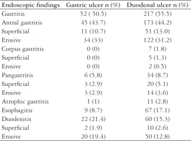
The analysis of endoscopic indings associated with gastric and duodenal ulcers (Table 3), showed that antral erosive gastritis was the most frequent endoscopic inding and the
Endoscopic indings Gastric ulcer n (%) Duodenal ulcer n (%)
Gastritis 52 ( 50.5) 217 (55.5)
Antral gastritis 45 (43.7) 173 (44.2)
Supericial 11 (10.7) 51 (13.0)
Erosive 34 (33) 122 (31.2)
Corpus gastritis 0 (0) 7 (1.8)
Supericial 0 (0) 5 (1.3)
Erosive 0 (0) 2 (0.5)
Pangastritis 6 (5.8) 34 (8.7)
Supericial 3 (2.9) 20 (5.1)
Erosive 3 (2.9) 14 (3.6)
Atrophic gastritis 1 (1) 11 (2.8)
Esophagitis 9 (8.7) 67 (17.1)
Duodenitis 22 (21.4) 60 (15.3)
Supericial 2 (1.9) 10 (2.6)
Erosive 20 (19.4) 50 (12.8)
TablE 3. Endoscopic indings associated with gastric and duodenal ulcers
Marques SB, Mattar R, Artifon ELA, Sakai P, Carrilho FJ. High prevalence of duodenal ulcer in a tertiary care hospital in the city of São Paulo, SP, Brazil
Arq Gastroenterol 173
v. 48 – no.3 – jul./set. 2011
Marques SB, Mattar R, Artifon ELA, Sakai P, Carrilho FJ. Alta prevalência de úlcera duodenal num hospital terciário na cidade de São Paulo. Arq Gastroenterol. 2011;48(3):171-4.
RESUMO – Contexto - Apesar da infecção pelo Helicobacter pylori ser causa de úlcera péptica e de sua alta prevalência no Brasil, a prevalência da úlcera péptica tem sido pouco estudada. Objetivos - Veriicar a prevalência da doença ulcerosa péptica em pacientes de um hospital terciário. Métodos - Achados diagnósticos de 1.478 endoscopias consecutivas foram correlacionados com os resultados de testes de urease para diagnóstico de infecção pelo H. pylori e dados demográicos num total de 3.779 endoscopias realizadas em 2005. A média de idade dos pacientes foi 51,14 ± 16,46, sendo 613 (41,5%) homens. Resultados - A úlcera péptica foi diagnosticada em 494 (33,4%) pacientes com média de idade de 54,86 ± 14,53, 205 (52%) eram homens, sendo 391 (26,5%) úlceras duodenais e 103 (7%) úlceras gástricas. Endoscopia normal foi achada em 272 (18,4%) pacientes com média de idade de 38,4 ± 15,22, sendo 49 (18%) homens. A comparação do grupo de úlcera péptica com os pacientes que tinham endoscopia normal revelou que a infecção pelo H.pylori [P = 0,005; OR = 1,70; 95% CI = 1,17-2,47], gênero masculino [P<0,0001; OR = 5,53; 95% CI = 3,67-8,34] e idade mais avançada [P<0,0001; OR = 1,08; 95% CI = 1,06-1,09] aumentaram o risco de úlceras pépticas. A prevalência de infecção pelo H. pylori no total foi de 53% (786). Conclusões - A prevalência da úlcera duodenal é alta numa população brasileira de um hospital terciário que teve a infecção pelo H. pylori, idade avançada e gênero masculino como determinantes importantes de evolução para doença gastrointestinal. Estudos futuros prospectivos devem conirmar esses achados.
DESCRITORES - Úlcera péptica, epidemiologia. Infecções por helicobacter. in Northeastern Brazil (92% in subjects 46-60 years)(19, 20); the
prevalence of peptic ulcers has not changed in countries with a low prevalence of H. pylori infection due to eradication regimens(13).
The risk of infected individuals to develop peptic ulcer was found to be 1.7 [95% CI = 1.17-2.47] which is similar to other reports that showed a relative risk of 1.6–5.7 (mean, 3.3) for infected people of developing peptic ulcer, compared with non-infected people(5). Male gender [OR = 5.53; 95% CI
= 3.67-8.34] and aging, as previously reported(26, 28), were also
risks of peptic ulcer development. Within the peptic ulcer group, gastric ulcer group had signiicantly higher age; the incidence of gastric ulcer has been relatively higher in the elderly people, compared to that of duodenal ulcers(26) that may be
due to lower defensive factors such as poor gastrin-elevating action capable of healing by its trophic effect(8), decreased
mucus, bicarbonate secretion and prostaglandins(12).
Endoscopic erosive antral gastritis was more frequent than corpus gastritis in both gastric and duodenal ulcer groups, in our previous report the activity of inlammation was also more intense in the antrum of peptic ulcer patients(14);
in addition, gastritis was more intense in the peptic ulcer group than in those with normal endoscopy and H. pylori positive(15). Endoscopic diagnosis has been 62% as sensitive as
histological diagnosis for erosive gastritis and 84% for atrophic gastritis in the antrum(10). In India, the antral gastritis also
predominated in 82% of the cases, and H. pylori was usually found in gastric biopsies taken from the antrum(16).
Association of duodenal ulcer with esophagitis was already described at higher rates of 33%(1) than in the present study
(17%). One possible explanation is higher acid secretion that has a role in the etiology of duodenal ulcer(9) and gastroesophageal
relux disease and its complications(7) more frequently found
in the male gender(3). The unexpected cases of duodenal ulcer
with atrophic gastritis may be the result of a long-term use of proton pump inhibitors(18).
Most of the gastric (54%) and duodenal ulcers (54%) were in the S stage which may explain the prevalence of H. pylori infection of 57% and 64%, respectively; which may indicate that were eradication-control patients. Within the duodenal ulcer group, 15 patients with serious co-morbidities had H. pylori-negative A ulcer. The clinical characteristics described of patients with H. pylori-negative duodenal ulcers were more likely to be older, have concomitant medical problem, pre-existing malignancy, recent surgery, underlying sepsis, or taken non-steroidal anti-inlammatory drugs(2).
This study has some limitations as the information concerning proton pump inhibitors, aspirin or NSAIDs intake, cigarette smoking, and alcohol consumption was not available. In addition, H. pylori infection was based in a single test (urease) and may be underestimated by the use of proton pump inhibitors, even though biopsies were taken from the antrum and the corpus, presenting 97.4% sensitivity and 100% speciicity(25). A prospective study should further
clarify these factors.
CONCLUSION
We conclude that the prevalence of duodenal ulcer is high in a Brazilian population that had H. pylori infection associated with older age and male gender as important determinants to gastrointestinal diseases outcome.
ACKNOWLEDGMENTS
Marques SB, Mattar R, Artifon ELA, Sakai P, Carrilho FJ. High prevalence of duodenal ulcer in a tertiary care hospital in the city of São Paulo, SP, Brazil
Arq Gastroenterol
174 v. 48 – no.3 – jul./set. 2011
REFERENCES
1. Boyd EJ. The prevalence of esophagitis in patients with duodenal ulcer or ulcer-like dyspepsia. Am J Gastroenterol. 1996;91:1539-43.
2. Chu KM, Kwok K-F, Law S, Wong KH. Patients with Helicobacter pylori
positive and negative duodenal ulcers have distinct clinical characteristics. World J Gastroenterol. 2005;11:3518-22.
3. Derakhshan MH, El-Omar E, Oien K, Gillen D, Fyfe V, Crabtree JE, McColl KE. Gastric histology, serological markers and age as predictors of gastric acid secretion
in patients infected with Helicobacter pylori. J Clin Pathol. 2006;59:1293-9.
4. Dong WG, Cheng CS, Liu SP, Yu JP. Epidemiology of peptic ulcer disease in Wuhan
area of China from 1997 to 2002. World J Gastroenterol. 2004,10:3377-9.
5. Duggan JN, Duggan AE. The possible causes of the pandemic of peptic ulcer
in the late 19th and early 20th century. Med J Aust. 2006;185:667-9.
6. Graham D. Foreword. In: Lee A, Mégraud F, editors. Helicobacter pylori:
techniques for clinical diagnosis & basic research. London: Saunders; 1996. 7. Hirschowitz BI, Simmons JL, Johnson LF, Mohnen J. Risk factors for esophagitis
in extreme acid hypersecretors with and without Zollinger-Ellison syndrome. Clin Gastroenterol Hepatol. 2004;2:220-9.
8. Ishihara M, Ito M. Inluence of aging on gastric ulcer healing activities of cimetidine and omeprazole. Eur J Pharmacol. 2002;444:209-15.
9. Jacobson K, Chiba N, Chen Y, Barrientos M, James C, Riddell RH, Hunt RH.
Gastric acid secretory response in Helicobacter pylori-positive patients with
duodenal ulcer disease. Can J Gastroenterol. 2001;15:29-39.
10. Kaminishi M, Yamaguchi H, Nomura S, Oohara T, Sakai S, Fukutomi H, Nakahara A, Kashimura H, Oda M, Kitahora T, Ichikawa H, Yabana T, Yagawa Y, Sugiyama T, Itabashi M, Unakami M, Oguro Y, Sakita T. Endoscopic classiication of chronic gastritis based on a pilot study by the research society for gastritis. Dig Endosc. 2002;14:138-51.
11. Kusters JG, van Vliet AHM, Kuipers EJ. Pathogenesis of Helicobacter pylori
Infection. Clin Microbiol Rev. 2006;49:449-90.
12. Laine L, Tokeuchi K, Tarnawski A. Gastric mucosal defense and cytoprotection: bench to bedside. Gastroenterology. 2008;135:41-60.
13. Manuel D, Cutler A, Goldstein J, Fennerthy MB, Brown K. Decreasing prevalence
combined with increasing eradication of Helicobacter pylori infection in the United
States has not resulted in fewer hospital admissions for peptic ulcer disease-related complications. Aliment Pharmacol Ther. 2007;25:1423-7.
14. Mattar R, dos Santos AF, Eisig JN, Rodrigues TN, Silva FM, Lupinacci RM,
Iriya K, Carrilho FJ. No correlation of babA2 with vacA and cagA genotypes
of Helicobacter pylori and grading of gastritis from peptic ulcer disease patients
in Brazil. Helicobacter. 2005;10:601-8.
15. Mattar R, Marques SB, Monteiro Mdo S, Dos Santos AF, Iriya K, Carrilho FJ.
Helicobacter pylori cag pathogenicity island genes: clinical relevance for peptic
ulcer disease development in Brazil. J Med Microbiol. 2007;56:9-14.
16. Misra V, Misra S, Dwivedi M, Singh UP, Bhargava V, Gupta SC. A topographic
study of Helicobacter pylori density, distribution and associated gastritis. J
Gastroenterol Hepatol. 2000;15:737-43.
17. Pacheco AR, Proença-Módena JL, Sales AI, Fukuhara Y, da Silveira WD,
Pimenta-Módena JL, de Oliveira RB, Brocchi M. Involvement of the Helicobacter
pylori plasticity region and cag pathogenicity island genes in the development
of gastroduodenal diseases. Eur J Clin Microbiol Infect Dis. 2008;27:
1053-9.
18. Raghunath AS, O’Morain C, McLoughlin RC. Review article: the long-term use of proton-pump inhibitors. Aliment Pharmacol Ther. 2005;22:55-63. 19. Rodrigues MN, Queiroz DM, Rodrigues RT, Rocha AM, Braga Neto MB, Braga
LL. Helicobacter pylori infection in adults from a poor urban community in
northeastern Brazil: demographic, lifestyle and environmental factors. Braz J
Infect Dis. 2005;9:405-10.
20. Rodrigues MN, Queiroz DM, Rodrigues RT, Rocha AM, Luz CR, Braga LL.
Prevalence of Helicobacter pylori infection in Fortaleza, Northeastern Brazil.
Rev Saúde Publica. 2005;39:847-9.
21. Sakita T. Endoscopy in diagnosis of early gastric cancer. Clin Gastroenterol. 1973;2:345-60
22. Santos IS, Boccio J, Santos AS, Valle NC, Halal CS, Bachilli MC, Lopes RD.
Prevalence of Helicobacter pylori infection and associated factors among adults in
Southern Brazil: a population-based cross-sectional study. BMC Public Health.
2005;5:118.
23. Sari YS, Sander E, Erkan E, Tunalı V. Endoscopic diagnoses and CLO test
results in 9239 cases, prevalence of Helicobacter pylori in Istanbul, Turkey. J
Gastroenterol Hepatol. 2007;22:1706-11.
24. Saul C, Teixeira CR, Pereira-Lima JC, Ronaldo J, Torresini RJ. Redução da prevalência de úlcera duodenal: um estudo brasileiro (análise retrospectiva na última década: 1996-2005). Arq Gastroenterol. 2007;44:320-4.
25. Vaira D, Vakil N, Gatta L, Ricci C, Perna F, Saracino I, Fiorini G, Holton J.
Accuracy of a new ultrafast rapid urease test to diagnose Helicobacter pylori
infection in 1000 consecutive dyspeptic patients. Aliment Pharmacol Ther. 2010;31:331-8.
26. Watanabe Y,Kurata JH, Kawamoto K, Kawai K. Epidemiological study of
peptic ulcer disease among Japanese and Koreans in Japan. J Clin Gastroenterol.
1992;15:68-74.
27. Wong S N, Sollano JD, Chan MM, Carpio RE, Tady CS, Ismael AE, Judan-Ruiz EA, Ang VN, Go JT, Lim VY, Perez JY, Alvarez SZ. Changing trends in peptic ulcer prevalence in a tertiary care setting in the Philippines: A seven-year study. J Gastroenterol Hepatol. 2005;20:628-32.
28. Wu HC, Tuo BG, Wu WM, Gao Y, Xu QQ, Zhao K. Prevalence of peptic ulcer in
dyspeptic patients and the inluence of age, sex, and Helicobacter pylori infection.
Dig Dis Sci. 2008;53:2650-6.
29. Yuan Y, Padol IT, Hunt RH. Peptic ulcer disease today. Nat Clin Pract Gastroenterol
Hepatol. 2006;3:80-9.
30. Zaterka S, Eisig JN, Chinzon D, Rothstein W. Factors related to Helicobacter
pylori prevalence in an adult population in Brazil. Helicobacter. 2007;12:82-8.