GASTROENTEROLOGIA EXPERIMENT
AL / EXPERIMENT
AL GASTROENTEROLOGY
ANALYSIS OF MYOSIN-V
IMMUNOREACTIVE MYENTERIC NEURONS
FROM ARTHRITIC RATS
Ivan Domicio da Silva
SOUZA
1, Janine Silva
RIBEIRO
1,
Ciomar Aparecida
BERSANI-AMADO
2and Jacqueline Nelisis
ZANONI
1ABSTRACT – Context- The inlammatory response itself and the consequent oxidative stress are able to promote neurodegeneration. So, it is possible that enteric nervous system is affected by inlammatory diseases threatening quality of life of patients. However, gastrointestinal symptoms of arthritis are usually attributed to anti-inlammatory drugs rather than neural damage. Objective -To
conirm if the general population of myenteric neurons from the ileum and jejunum of rats is affected by arthritis. Methods - Twenty Holtzmann rats, 58-day-old male, were used and divided in four groups: control group (C30), arthritic group (Art30), older control group (C60) and older arthritic group (Art60). At 58 days old, the animals in groups Art30 and Art60 received an injection of the complete Freund’s adjuvant in order to induce arthritis. The whole-mount preparations of ileum and jejunum were processed for myosin-V immunohistochemistry. Quantitative and morphometric analyses were performed. Results - Groups Art30 and Art60 presented, respectively, a reduction of 2% and 6% in intestinal area when compared to their control groups. No signiicant differences were observed in general neuronal density among the four groups (P>0.05). Group C60 presented a reduction of 14.4% and 10.9% in mean neuronal cell body area when compared to group C30 (P<0.05), for the ileum and jejunum, respectively. The other groups had
a similar mean neuronal cell body area (P>0.05). Conclusion - Arthritis does not promote quantitative or morphological damages in general myenteric population. However, studies in progress have revealed some signiicant alterations in myenteric neurons subpopulations (nitrergic and VIP-ergic neurons).
HEADINGS - Arthritis. Myenteric plexus. Myosin type V. Rats.
INTRODUCTION
The enteric nervous system (ENS) consists of two sets of ganglionated plexi, the myenteric and the submucous, connected to central nervous system through sympathetic and parasympathetic nerves. In spite of these extensive communications to central nervous system, ENS is perfectly able to work autonomously(15, 16, 17). Once it is relatively independent of central nervous
system, ENS is responsible to coordinate complex functions such as motility, secretion, mucosal growth and blood low in the gastrointestinal tract(15, 16, 38).
Myenteric ganglia range in number, size, shape, and orientation from species to species and from an intestinal segment to another(15, 16, 37). These neurons
comprehend a complex and diversiied population that should be studied in order to understand the mechanisms related to digestive physiology. In addition, digestion and absorption may be seriously affected by injuries in enteric neurons, threatening
1 Department of Morphological Sciences, Universidade Estadual de Maringá; 2 Department of Pharmacology, Universidade Estadual de Maringá, Maringá, PR, Brasil.
Correspondence: Dr. Jacqueline Nelisis Zanoni - Department of Morphological Sciences, Universidade Estadual de Maringá - Avenida Colombo, 5790- Bloco H79- 87020-900, Maringá, PR, Brazil. E-mail: [email protected]
quality of life of patients. Inlammatory response and oxidative stress have been elucidated as the main mechanisms that promote alterations in enteric neuroplasticity(8, 9, 10, 21, 39), leading to secondary
complications like: gastroparesis, dysphagia, emesis, diarrhea and constipation.
Rheumatoid arthritis (RA) is a multisystemic proliferative auto-immune chronic inlammatory disease(20, 26). Implications of RA arise from the
elevated production of inlammatory mediators, the action of activated inlammatory cells and from the consequent increase in the rate of oxidant species(2, 3, 18). Hence, primary effects of RA may possibly
damage neural tissues promoting pathological neurodegenerative disorders. However, few is known about RA effects on ENS, since its gastrointestinal symptoms are commonly attributed to medicines rather than neuronal damages(12, 25, 40).
is an experimental immunopathology similar to human RA(31, 32). We used Holtzmann rats because some studies
report that they present higher genetic susceptibility to AIA and develop all characteristic symptoms of RA(4, 5).
Our objective was to assess if implications of AIA may damage myenteric neurons in the ileum and jejunum of rats after 30 and 60 days from the induction of inlammatory reaction.
METHODS
Animals
All experimental procedures were conducted in accordance with the ethical principles of the Brazilian Academy of Animal Experimentation and approved by the Committee of Ethics in Animal Experimentation from Universidade Estadual de Maringá, PR, Brazil.
Twenty, 58-day-old male, albine Holtzmann’s strain rats obtained from the central vivarium of Universidade Estadual de Maringá were used. The animals were divided into four groups: control group (C30), old arthritic group (Art30), older control group (C60) and older arthritic group (Art60). Groups C30 and Art30 went through a 30-days experimental period, whereas groups C60 and Art60 went through a 60-days experimental period. The animals were kept in polypropylene boxes at the sectorial vivarium in a light (lights on from 6:00 am to 6:00 pm) and temperature (24ºC ± 2ºC) controlled environment. Tap water and chow (Nuvital; Nuvilab, Colombo, PR, Brazil) were available ad libitum.
Adjuvant arthritis was induced by an intradermal injection of 100 µL of a suspension of Freund’s adjuvant (heat-inactivated Mycobacterium tuberculosis) suspended in mineral oil at a concentration of 0.5% (w/v) into the left hind paw(28).
Material resection and immunohistochemistry
At the end of the experimental period, animals were weighed, anesthetized with a 40 mg/kg body weight intraperitoneal dose of thiopental (Abbott Labs, Chicago, IL, USA) and killed. Each ileum and jejunum was resected and then processed according to an immunohistochemistry technique for myosin-V(11), a general myenteric population
marker.
The animals were perfused with cold 1.1% saline solution followed by fixative solution(24) containing sodium periodate
(10 mM), lysine (75 mM) and paraformaldehyde (1%) in phosphate buffer, pH 7.4 (37 mM). Immediately after perfusion, ileums and jejunums were resected, measured (circumference and length), washed with saline solution up to complete removal of feces, carefully inflated with the fixative solution such that the segments were not distended, and tied at the extremities with cotton thread. Afterward, ileums and jejunums were maintained in the fixative solution for 1 h, dehydrated in an increasing series of alcohols (v/v; 50%, 70%, 80%, 90% and 100%), clarified in xylol and rehydrated in a decreasing series
of alcohols (v/v; 100%, 95%, 90%, 80% and 70%). Then, ileums and jejunums were subdivided into segments of approximately 1 cm in width. These segments were dissected under stereomicroscope to obtain whole-mount muscular layer preparations through the removal of mucous and submucous layer.
Subsequently, the preparations were washed four times in phosphate buffer saline (PBS), pH 7.4 (0.1 M), and blocked for 1.5 h in a solution containing bovine serum albumin (BSA, 2%; Sigma, St. Louis, MO, USA), goat serum (1:50), Triton X-100 (0.5%; Sigma), and PBS. Preparations were sequentially incubated at room temperature in solution containing the primary antibody (1:750) speciic for the medial tail of myosin-V (donated by PhD Enilza Maria Espreaico, Universidade de São Paulo, Ribeirão Preto, SP, Brazil). After 48 h, the tissues were washed twice in PBS solution containing Triton X-100 (0.1%) and twice in PBS solution containing Tween-20 (0.05%; Sigma). The tissues then were incubated for 24 h with secondary antibody conjugated with peroxidase (1:1000; Pierce, Rockford, IL, USA) at room temperature. Finally, tissues were washed 4 times with PBS solution containing Tween-20 (0.05%). The immunoreaction was revealed by diaminobenzidine (DAB; Sigma), and the preparations were mounted in glycerol gel. Negative control was performed by omitting the primary antibody.
Quantitative analysis
The quantiication of myosin-V immunoreactive myenteric neurons was performed using digital images obtained randomly from the intermediate region (60º-120º and 240º-300º) of the ileum and jejunum intestinal circumferences for each animal, with 0º as the mesenteric insertion(43).
Images were captured by a high resolution camera (Olympus QColor 3; Olympus, Melville, NY, USA) coupled to an optical microscope (Olympus BX 41, Tokyo, Japan) at 400X magniication, digitalized on a computer using the software QCapture Pro 5.1.1.14 (Media Cybernetics, Silver Spring, MD, USA) and recorded onto a compact disc. The image analysis software Image-Pro Plus 4.5.0.29 (Media Cybernetics) was used for manual neuronal quantiication of the images.
For each animal, the neurons present in 62 images were counted, except by the ones overlapping the lower and right edges of the image. The total area of the 62 images, measured with Image-Pro Plus, was approximately 5.8 mm2. Results were expressed as the mean number of
neurons per cm2. A correction factor (C
f, equation 1) was
employed onto the neuronal density of groups Art30 and Art60 to keep the same proportion of intestinal area of their respective control groups(29).
Morphometrical analysis
The areas of myosin-V immunoreactive myenteric neuronal cell bodies were measured using the same images in the quantitative analysis. The area (µm2) of 100 neuronal
TABLE 1. Mean intestinal area, length and circumference
Group Area (cm2) Length (cm) Circumference (cm)
C30 89.8 ± 7.4 88.7 ± 3.7 1.02 ± 0.09
Art30 88.0 ± 5.2 97.6 ± 3.8 0.90 ± 0.03 C60 158.4 ± 12.9* 101.5 ± 2.8 1.56 ± 0.17* Art60 148.8 ± 3.2** 99.2 ± 2.1 1.50 ± 0.00** Labels: C30) 88-day-old control group; Art30) 88-day-old arthritic group; C60) 118-day-old
control group and Art60) 118-day-old arthritic group. Mean ± SE. n = 5 rats per group
* P<0.001 when compared to group C30
** P<0.001 when compared to group Art30
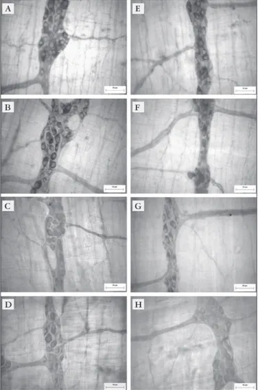
Labels: A, E) 88-day-old control group (C30); B, F) 88-day-old arthritic group (Art30); C, G) 118-day-old control group (C60) and D, H) 118-118-day-old arthritic group (Art60). Scale bar: 50 µm
FIGURE 1. Myosin-V immunoreactive myenteric neurons from the intermediate region in the ileum (A, B, C, D) and jejunum (E, F, G, H)
Plus, with a total of 500 areas per group. Results were presented as frequency distribution and mean areas were compared.
Statistical analysis
Data were statistically analyzed using Statistica 7.1 (StatSoft, Tulsa, OK, USA) and GraphPad Prism 5.01 (GraphPad Software, San Diego, CA, USA) and were expressed as mean ± standard error. Morphometric data were set in delineation blocks followed by Tukey’s test. For all the other data, we applied one-way analysis of variance (ANOVA) followed by Tukey’s test. Values of P<0.05 were considered statistically signiicant.
RESULTS
Between the 8th and the 13th day after Freund’s adjuvant
injection, arthritic animals developed the characteristic symptoms of AIA, such as an intense inlammatory reaction in the left paw and in the non-injected paws. Regarding secondary lesions, featuring nodules in the ears and tail and an increase in the volume of the front paws, they emerged from the 10th day onward in all arthritic animals
reaching maximum severity on the 30th day. We observed
that the gain of weight in arthritic animals was inferior when compared to the respective control groups (data not presented).
Mean intestinal length was similar in the four groups studied (P>0.05). However, we observed an increase in mean intestinal area of groups C60 and Art60, in relation to groups C30 and Art30, once they presented a larger intestinal circumference (P<0.001; Table 1). Intestinal circumference in groups Art30 and Art60 was 11.8% and 3.8% smaller than in their respective control groups. Animals in groups Art30 and Art60 exhibited a 2% and 6% retraction in intestinal area, respectively, when compared to their control groups (Table 1). Correction factors for intestinal retraction in groups Art30 and Art60 were 0.98 and 0.94, respectively.
In this study, the immunohistochemistry technique for myosin-V allowed us to distinguish the primary and secondary components, as well as the small caliber ibers in the tertiary component of myenteric plexus from the ileum and jejunum (Figure 1). It was observed neuronal
cell bodies mostly in the ganglia and rarely throughout the interganglionary ibers.
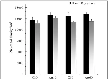
No signiicant differences were found in neuronal density when we compared the four groups, in the ileum and in the jejunum (P>0.05). We found a higher number of neurons in the ileum (15680 ± 381) than in the jejunum (14400 ± 311) when comparing the mean neuronal density of the four groups in each segment (P<0.05, Figure 2). Morphometrical analysis showed that most of the myenteric neurons studied presents a neuronal cell body area between 100 and 200 µm2 in all groups, in the ileum and in the
DISCUSSION
The alterations in physiologic routine and the inlammation signals observed in the arthritic animals (Art30 and Art60) are in accordance with the literature about rats induced to arthritis(31, 32, 36). The slight retraction in intestinal area
of the arthritic animals suggests an atrophic effect of AIA on intestinal development, such as the one observed in undernourished rats(6, 33). Alterations in intestinal area
have been reported in other clinical situations like diabetes mellitus(41) and aging(19, 23).
The immunohistochemistry technique for myosin-V has been used as a pan-neuronal marker of myenteric neurons once it does not distinguish the subpopulations (nitrergic, VIP-ergic, CGRP-ergic neurons, among others). Myosin-V is a structural motor protein related to neuronal vesicle transportation present all over the myenteric plexus(22), so that
one can consider it as a general marker of myenteric neurons. Many authors have employed this technique and obtained good results in different segments of the gastrointestinal tract (colon(7, 35), duodenum(23) and ileum(43)).
The arrangement of myenteric plexus in Holtzmann rats is similar to the one irstly described in 1864(1). The
myenteric neurons of the ileum and jejunum in all groups studied presented mainly excentric nuclei and were mostly placed inside the ganglia, arranged in longitudinal rows, similar to those observed in previous investigations(16, 37).
The ganglia were abundant and showed predominantly an elongated shape, as described for Wistar rats(13).
The density of myenteric neurons myosin-V immunoreactive in the ileum was higher than in the jejunum. The higher number of neurons found in the ileum when compared to jejunum is in accordance with the literature data. The neuronal density in the ileum and jejunum from Holtzmann rats was lower than those found to Wistar rats(34) using the same immunohistochemistry.
For instance, a previous study(30) observed a myenteric neuronal
density closer to ours (14060 ± 156 neurons per cm2)in the
ileum of 90-day-old Fischer rats, stained by cuprolinic blue. Another study(19) warned to the fact that different strains
may present different patterns of enteric neuronal density. In the same study, they found 19600 ± 2030 neurons per cm2
in the ileum of 120-day-old Sprague Dawley rats, using an immunohistochemistry for the protein gene product 9.5 (PGP 9.5). We believe that differences in neuronal density are a result of the animal species, since the age of the animals in the other studies were similar to those used by us.
Neuronal density/cm
2
18000
15000
12000
9000
6000
3000
0
C30 Art30 C60 Art60
Ileum Jejunum
Labels: C30) 88-day-old control group; Art30) 88-day-old arthritic group; C60) 118-day-old
control group and Art60) 118-day-old arthritic group. Mean ± SE. n = 5 rats per group
FIGURE 2. Neuronal density of myosin-V immunoreactive myenteric neurons observed in the intermediate region in the ileum and jejunum
0.4
0.3
0.2
0.1
0.0
Relative Frequency
Neuronal cell body area (µm2)
C30 Art30 C60 Art60
0 100 200 300 400 500 600 700
FIGURE 3. Relative frequency distribution of neuronal cell bodies area of myosin-V immunoreactive myenteric neurons from the ileum
Labels: C30) 88-day-old control group; Art30) 88-day-old arthritic group; C60) 118-day-old control group and Art60) 118-day-old arthritic group. n = 5 rats per group
0.4
0.3
0.2
0.1
0.0
Relative Frequency
Neuronal cell body area (µm2)
C30 Art30 C60 Art60
0 100 200 300 400 500 600 700
Labels: C30) 88-day-old control group; Art30) 88-day-old arthritic group; C60) 118-day-old control group and Art60) 118-day-old arthritic group. n = 5 rats per group
FIGURE 4. Relative frequency distribution of neuronal cell bodies area of myosin-V immunoreactive myenteric neurons from the jejunum
TABLE 2. Mean areas of neuronal cell bodies of myosin-V immunoreactive myenteric neurons
Group Ileum (µm2) Jejunum (µm2)
C30 205.1 ± 4.8 188.0 ± 4.4
Art30 196.4 ± 5.2 186.3 ± 4.4
C60 175.5 ± 3.9* 167.5 ± 3.7*
Art60 190.6 ± 4.3 172.1 ± 3.7
Labels: C30) 88-day-old control group; Art30) 88-day-old arthritic group; C60) 118-day-old
control group and Art60) 118-day-old arthritic group. Mean ± SE. n = 5 rats per group
According to the literature, arthritis increases the production of oxidant species and of inlammation mediators that affect the enteric neurons(2, 3, 18, 39). However, in this study
we observed that arthritic condition did not promoted any alterations in general neuronal density, in the ileum and in the jejunum, after 30 and 60 days of arthritis induction.
In the graphic representation of the frequency distribution of neuronal cell bodies areas, for the ileum or jejunum, we can see uniformity among the four groups once their curves are similar. The fact that there is a similarity in the morphometry of arthritic and control groups suggests the maintenance of cell metabolism normality. When a pathology affects this normality, it is possible to observe some alterations such as swelling of cell bodies, axons and varicosities(30, 42). The neuronal cell body area in group C60
was reduced when compared to group C30, for the ileum or jejunum. These results are not in accordance with the expected for other strains of rats(23), however, this is the
irst study describing the morphometry and density of myenteric neurons in Holtzmann rats. Additional studies are necessary to clarify this behavior.
Arthritic patients frequently present gastrointestinal symptoms that are soon associated to the use of anti-inlammatory drugs(12, 14, 25, 40). Our results refer only to the
general population of myenteric neurons, so it is important
to evaluate if any myenteric subpopulation is affected before rejecting the hypothesis that gastrointestinal symptoms in RA are related to neural damage as well. Some authors have reported alterations in myenteric subpopulations as a consequence of diabetes(27, 29, 42) and aging(30). Our group
is conducting further studies that have indicated some signiicant alterations in nitrergic and VIP-ergic myenteric neuronal subpopulations in Holtzmann arthritic rats (data not yet published).
CONCLUSIONS
Data presented allow us to conclude that arthritis does not promote quantitative or morphological damages in the general population of myenteric neurons in the ileum or jejunum of rats. Further studies are necessary to assess any possible alterations in myenteric neurons subpopulations.
ACKNOWLEDGEMENTS
We wish to thank the excellent technical support of Mr. Jailson Dantas Araujo, Mrs. Ana Paula de Santi Rampazzo, Mrs. Maria Euride do Carmo Cancino and Mrs. Maria dos Anjos Fortunato.
Souza IDS, Ribeiro JS, Bersani-Amado CA, Zanoni JN. Análise dos neurônios mioentéricos miosina-V imunoreativos de ratos artríticos. Arq Gastroenterol. 2011;48(3):205-10.
RESUMO - Contexto -A resposta inlamatória e o estresse oxidativo acentuados em decorrência da artrite reumatóide são capazes de promover neurodegeneração. Nessas condições, é possível que o sistema nervoso entérico seja afetado, diminuindo a qualidade de vida dos pacientes. No entanto, os sintomas da artrite no trato gastrointestinal são geralmente associados ao uso de medicamentos anti-inlamatórios do que a um possível dano neural. Objetivo - Veriicar se a população geral de neurônios mioentéricos do íleo e do jejuno de ratos artríticos é afetada pela artrite. Métodos
- Foram utilizados 20 ratos Holtzmann, inicialmente com 58 dias de idade, divididos em 4 grupos: controle com 88 dias (C30); artrítico com 88 dias (Art30); controle com 118 dias (C60) e artrítico com 118 dias (Art60). Os animais dos grupos Art30 e Art60 receberam aos 58 dias de idade o adjuvante completo de Freund para indução da artrite. Os preparados totais de íleo e jejuno foram submetidos a imunoistoquímica para a proteína miosina-V. Realizou-se análises quantitativas e morfométricas dos neurônios. Resultados - Os animais Art30 e Art60 apresentaram, respectivamente, redução de 2% e 6% na área intestinal em relação aos seus controles. Não foram observadas diferenças na densidade neuronal geral entre os quatro grupos (P>0,05). O grupo C60 apresentou redução de 14,4% e 10,9% na área média do corpo celular neuronal em relação ao grupo C30 (P<0,05). Os demais grupos apresentaram área média de corpo celular semelhante (P>0,05). Conclusão - A artrite não provocou alterações quantitativas ou
morfológicas na população mioentérica geral, entretanto, estudos em andamento revelam alterações signiicativas em subpopulações de neurônios mioentéricos (nitrérgicos e VIP-érgicos).
DESCRITORES - Artrite. Plexo mientérico. Miosina tipo V. Ratos.
REFERENCES
1. Auerbach L. Fernere vorlauige Mitteilung über den Nervenapparat des Darmes. Arch Pathol Anat Physiol. 1864;30:457-60.
2. Babior BM. Phagocytes and oxidative stress. Am J Med. 2000;109:33-44. 3. Badolato R, Oppenheim JJ. Role of cytokines, acute-phase proteins, and
chemokines in the progression or rheumatoid arthritis. Semin Arthritis Rheum. 1996;26:526-38.
4. Bersani-Amado CA, Barbuto JA, Jancar S. Comparative study of adjuvant induced arthritis in susceptible and resistant strains of rats. I. Effect of cyclophosphamide. J Rheumatol. 1990;17:149-52.
5. Bersani-Amado CA, Duarte AJ, Tangi MM, Cianga M, Jancar S. Comparative study of adjuvant induced arthritis in susceptible and resistant strains of rats. III. Analysis of lymphocyte subpopulations. J Rheumatol. 1990;17:153-8.
6. Brandão MCS, De Angelis RC, De-Souza RR, Fróes LB, Liberti EA. Effects of pre- and postnatal protein energy deprivation on the myenteric plexus of the small intestine: a morphometric study in weanling rats. Nutr Res. 2003;23: 215-23.
7. Buttow NC, Zucoloto S, Espreaico EM, Gama P, Álvares EP. Substance P enhances neuronal area and epithelial cell proliferation after colon denervation in rats. Dig Dis Sci. 2003;48:2069-76.
8. Cameron NE, Cotter MA. Effects of antioxidants on nerve and vascular dysfunction in experimental diabetes. Diabetes Res Clin Pract. 1999;45:137-46.
9. Cui K, Luo X, Xu K, Ven Murthy MR. Role of oxidative stress in neurodegeneration: new developments in assay methods for oxidative stress and nutraceutical antioxidants. Prog Neuropsychopharmacol Biol Psychiatry. 2004;28: 771-99.
11. Drengk AC, Kajiwara JK, Garcia SB, Carmo VS, Larson RE, Zucoloto S, Espreaico EM. Immunolocalisation of myosin-V in the enteric nervous system of the rat. J Auton Nerv Syst. 2000;78:109-12.
12. Ehab S El-Desoky. Pharmacotherapy of rheumatoid arthritis: an overview. Curr Ther Res. 2001;62:92-112.
13. Fiorini A, Molinari SL, Natali MRM, Miranda-Neto MH. Quantitative morphological analysis of the myenteric neurons of the ileum in rats under experimental desnutrition. Acta Sci. 1999;21:409-13.
14. Fung HB, Kirschenbaum HL. Selective cyclooxygenase-2 inhibitors for the treatment of arthritis. Clin Ther. 1999;21:1131-57.
15. Furness JB, Costa M. Types of nerves in the enteric nervous system. Neuroscience. 1980;5:1-20.
16. Furness JB, Costa M. The enteric nervous system. Edinburgh: Churchill Livingstone; 1987.
17. Gershon MD. The enteric nervous system. Annu Rev Neurosci. 1981;4:227-72.
18. Hitchon CA, El-Gabalawy HS. Oxidation in rheumatoid arthritis. Arthritis Res Ther. 2004;6:265-78.
19. Johnson RJR, Schemann M, Santer RM, Cowen T. The effects of age on the overall population and on sub-populations of myenteric neurons in the rat small intestine. J Anat. 1998;192:479-88.
20. Kumar V, Abbas AK, Fausto N. Robbins e Cotran. Patologia – bases patológicas da doença. 7a ed. Rio de Janeiro: Elsevier; 2005.
21. Kuyvenhoven JP, Meinders AE. Oxidative stress and diabetes mellitus. Pathogenesis of long-term complications. Eur J Intern Med. 1999;10:9-19.
22. Langford GM. Myosin-V, a versatile motor for short-range vesicle transport. Trafic. 2002;3:859-65.
23. Marese AC, de Freitas P, Natali MR. Alterations of the number and the proile of myenteric neurons of Wistar rats promoted by age. Auton Neurosci. 2007;137:10-8.
24. McLean IW, Nakane PK. Periodate-lysine-paraformaldehyde ixative. A new ixation for immunoelectron microscopy. J Histochem Cytochem. 1974;22:1077-83.
25. Moore RA, Derry S, Phillips CJ, McQuay HJ. Nonsteroidal anti-inlammatory drugs (NSAIDs), cyxlooxygenase-2 selective inhibitors (coxibs) and gastrointestinal harm: review of clinical trials and clinical practice. BMC Musculoskelet Disord. 2006;7:79.
26. Odeh M. New insights into the pathogenesis and treatment of rheumatoid arthritis. Clin Immunol Immunopathol. 1997;83:103-16.
27. Pasricha PJ, Pehlivanov ND, Gomez G, Vittal H, Lurken MS, Farrugia G. Changes in the gastric enteric nervous system and muscle: a case report on two patients with diabetic gastroparesis. BMC Gastroenterol. 2008;8:21.
28. Pearson CM, Wood FD. Studies of arthritis and other lesions induced in rats by the injection of mycobacterial adjuvant: VII. Pathologic details of the arthritis and spondylitis. Am J Pathol. 1963;42:73-95.
29. Pereira RVF, de Miranda-Neto MH, da Silva Souza ID, Zanoni JN. Vitamin E supplementation in rats with experimental diabetes mellitus: analysis of myosin-V and nNOS immunoreactive myenteric neurons from terminal ileum. J Mol Histol. 2008;39:595-603.
30. Phillips RJ, Kieffer EJ, Powley TL. Aging of the myenteric plexus: neuronal loss is speciic to cholinergic neurons. Auton Neurosci. 2003;106:69-83.
31. Rainsford KD. Adjuvant polyarthritis in rats: is this a satisfactory model for screening anti-arthritic drugs? Agents Actions. 1982;12:452-8.
32. Rosenthale ME, Capetola RJ. Adjuvant arthritis: immunopathological and hyperalgesic features. Fed Proc 1982;41:2577-82.
33. Sant’ana DM, Miranda-Neto MH, de Souza RR, Molinari SL. Morphological and quantitative study of the myenteric plexus of the ascending colon of rats subjected to proteic desnutrition. Arq Neuropsiquiatr. 1997;55:687-95. 34. Schneider LC, Perez GG, Banzi SR, Zanoni JN, Natali MR, Buttow NC. Evaluation
of the effect of Ginkgo biloba extract (EGb 761) on the myenteric plexus of the small intestine of Wistar rats. J Gastroenterol. 2007;42:624-30.
35. Schoffen JP, Soares A, de Freitas P, Buttow NC, Natali MR. Effects of a hypoproteic diet on myosin-V immunostaneid myenteric neurons and the proximal colon wall of aging rats. Auton Neurosci. 2005;122:77-83.
36. Silva MA, Ishii-Iwamoto EL, Bracht A, Caparroz-Assef SM, Kimura E, Cuman RK, Bersani-Amado CA. Eficiency of combined methotrexate/chloroquine therapy in adjuvant-induced arthritis. Fundam Clin Pharmacol. 2005;19:479-89. 37. Sternini C. Structural and chemical organization of the myenteric plexus. Annu
Rev Physiol. 1988;50:81-93.
38. Surprenant A. Control of the gastrointestinal tract by enteric neurons. Annu Rev Physiol. 1994;56:117-40.
39. Vasina V, Barbara G, Talamonti L, Stanghellini V, Corinaldesi R, Tonini M, De Ponti F, De Giorgio R. Enteric neuroplasticity evoked by inlammation. Auton Neurosci. 2006;126-127:264-72.
40. Wolfe F, Hawley DJ. The comparative risk and predictors of adverse gastrointestinal events in rheumatoid arthritis and osteoarthritis: a prospective 13 year study of 2131 patients. J Rheumatol. 2000;27:1668-73.
41. Zanoni JN, de Miranda-Neto MH, Bazotte RB, de Souza RR. Morphological and quantitative analysis of the neurons of the myenteric plexus of the cecum of streptozotocin-induced diabetic rats. Arq Neuropsiquiatr. 1997;55:696-702. 42. Zanoni JN, Buttow NC, Bazotte RB, Miranda-Neto MH. Evaluation of the
population of NADPH-diaphorase-stained and myosin-V myenteric neurons in the ileum of chronically streptozotocin-diabetic rats treated with ascorbic acid. Auton Neurosci. 2003;104:32-8.
43. Zanoni JN, De Freitas P, Pereira RV, Dos Santos Pereira MA, De Miranda-Neto MH. Effects of supplementation with ascorbic acid for a period of 120 days on the myosin-V and NADPHd positive myenteric neurons of the ileum of rats. Anat Histol Embryol. 2005;34:149-53.