330
LETTERS
DOI: 10.1590/0004-282X20130030
Expanded endonasal approach to skull
base meningiomas
Acesso endonasal expandido para meningiomas da base do crânio
Bernardo Assumpção de Monaco1, Henrique Faria Ramos2, Marcos Queiroz Telles Gomes1, Marcelo
Prudente do Espirito Santo1, Luciano Foroni1, Luiz Ubirajara Sennes2, Manoel Jacobsen Teixeira1
1Neurosurgery Division, Hospital das Clínicas, Universidade de São Paulo (USP), São Paulo SP, Brazil;
2Otorhinolaryngology Department, Hospital das Clínicas, USP, São Paulo SP, Brazil.
Correspondence: Bernardo Assumpção de Monaco; Instituto Central HCFMUSP Neurocirurgia; Avenida Doutor Enéas de Carvalho Aguiar 255 / sala 5084; 05403-900 São Paulo SP - Brasil; E-mail: [email protected]
Confl ict of interest: There is no conl ict of interest to declare.
Received 25 February 2012; Received in i nal form 18 September 2012; Accepted 25 September 2012.
Expanded endonasal endoscopic approach is an alterna-tive technique for management of meningiomas of the ante-rior skull base, allowing complete resection with an accept-able morbidity rate1-3.
We report the i rst three cases of anterior skull base me-ningiomas managed by this technique.
CASE REPORT
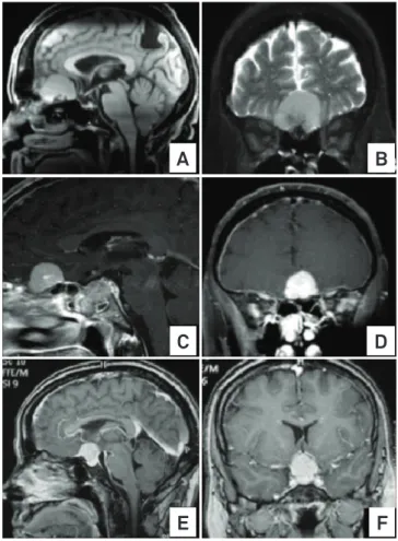
Case 1 – 79 year-old female with a small olfactory groove meningioma, during follow-up, presented visual impairment and frontalization signs, along with an increase in the volume of the tumor on MRI (Figs 1A and B)
Case 2 – 58 years-old male presented with right oculo-motor nerve partial palsy and right facial hypoesthesia. MRI showed a planum sphenoidale and olfactory groove menin-gioma, associated with a small sellar lesion extending to the sphenoidal sinus (Figs 1C and D).
Case 3 – 39 years-old female, complaining of decreased vi-sual acuity bilaterally, worse on the right side for the past eight months. MRI revealed a tuberculum sellae lesion (Figs 1E and F).
OPERATIVE TECHNIQUE
Endonasal approach was performed with a 0o 4 mm
endo-scope. Angled endoscopes were used at the end of the proce-dure to inspect the operative i eld as a whole. h e procedure was carried out by a neurosurgeon and an otorrhinolaryngol-ogist working simultaneously.
h e procedure was initiated with anterior and posterior ethmoidectomies and wide sphenoidotomies, exposing the anterior skull base, lamina papyracea and the frontal recess. Bilateral middle turbinectomy were carried out, since the
olfactory groove is located above its insertion on the skull base. A nasoseptal pedicled l ap was harvested and placed
Fig 1. (A and B) Case 1 Magnetic Resonance Imaging (MRI) T1 without contrast showing lesion on olfactory groove and encephalomalacia area in parietal region secondary to a previous ischemic insult. (C and D) Case 2 MRI T1 with gadolinium. Planum sphenoidale and olfactory groove meningioma, associated to a small lesion on the sellar region and sphenoidal sinus. (E and F) Case 3 MRI T2/T1 gadolinium with a tuberculum sellae meningioma.
A
B
C
E
D
331
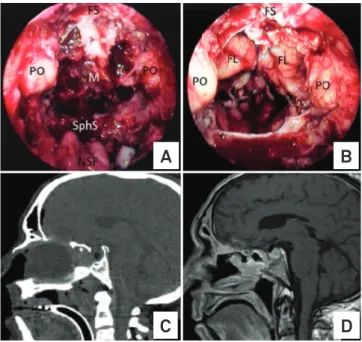
Arq Neuropsiquiatr 2013;71(5):330-335 into the nasopharynx for posterior reconstruction of the skull
base defect4 (Fig 2A).
At this point, the superior half of the posterior nasal septum was removed, communicating both nasal cavities and exposing the entire anterior skull base. Anterior and posterior ethmoidal arteries were identii ed and coagulated to devascularize the tu-mor3. h e boundaries of the approach were as follows: anteriorly,
posterior wall of the frontal sinuses; laterally, the medial wall of both orbits; and posteriorly, planum sphenoidale and sellar area5.
h e cribriform plate was drilled and removed bilaterally. Tumor was debulked and surrounding arachnoid was dis-sected away from the tumor capsule. Once tumor has been freed from any adherence, the tumor capsule could be gently peeled away from surrounding structures and removed piece-meal or en bloc, achieving gross total resection (Fig 2B).
Reconstruction of the skull base was accomplished with one un-derlay layer of fascia lata, followed by the nasoseptal l ap covering the entire defect and i brin glue. A Foley ballon catheter was inl ated with saline solution under endoscopic vision (Figs 2C and D).
OUTCOMES
Case 1 improved her visual symptoms. Case 2 showed slight im-provement of diplopia without new deficits. Case 3 visual complaints improved one day after surgery, but experienced cerebrospinal fluid (CSF) leakage ten days after surgery and, forty days after surgery, deep venous thrombosis, massive lung embolism and death.
CONCLUSION
Expanded endonasal endoscopic approach is a feasi-ble option to remove selected skull base meningiomas with
minor injury to other structures, allowing complete removal and consistent reconstruction of skull base l oor.
CSF leak is the major complication related with this approach2.
1. Al-Mefty O, Holoubi A, Rifai A, Fox JL. Microsurgical removal of suprasellar meningiomas. Neurosurgery 1985;16:364-372.
2. Gardner PA, Kassam AB, Thomas A, et al. Endoscopic endonasal resection of anterior cranial base meningiomas. Neurosurgery 2008;63:36-52.
3. de Divitiis E, Esposito F, Cappabianca P, Cavallo LM, de Divitiis O, Esposito I. Endoscopic transnasal resection of anterior cranial fossa meningiomas. Neurosurg Focus 2008;25:E8.
4. Hadad G, Bassagasteguy L, Carrau RL, et al. A novel reconstructive technique after endoscopic expanded endonasal approaches: vascular pedicle nasoseptal flap. Laryngoscope 2006;116:1882-1886.
5. Kassam A, Snyderman CH, Mintz A, Gardner P, Carrau RL. Expanded endonasal approach: the rostrocaudal axis. Part I. Crista galli to the sella turcica. Neurosurg Focus 2005;19:E3.
References
Fig 2. (A) Exposure of the anterior skull base meningioma (M), limited anteriorly by the frontal sinus (FS), laterally by the periorbits (PO) and posteriorly by the sphenoidal sinus (SphS). The nasoseptal flap is disp laced into the nasopharynx. (B) After removal of the meningioma, the frontal lobes (FL) can be observed. (C) Immediate postoperative CT scan with an insuflated 24F Foley catheter within the nasal cavity retaining the nasoseptal pediculated flap against the dural defect. Small pneumoencephalus in the frontal and sellar region may be noted. (D) MRI T1 showing no residual lesions, after Foley catheter removal, seven days after surgery.