313
ARTICLE
Brain tissue oxidative damage as a possible
mechanism for the deleterious effect of a
chronic high dose of estradiol on learning and
memory in ovariectomized rats
Dano oxidativo ao tecido cerebral como possível mecanismo de efeito deletério da alta
dose crônica de estradiol no aprendizado e memória de ratas ooforectomizadas
Fatimeh Khodabandehloo1, Mahmoud Hosseini2, Ziba Rajaei3, Mohammad Soukhtanloo4, Esmaeil Farrokhi2,
Mohsen Rezaeipour2
Ovarian hormones afect a wide variety of nervous sys-tem functions especially the cognitive abilities, learning and memory1. It has been suggested that estrogen may prevent
cognitive impairments due to aging2. Increasing in the risk
of Alzheimer’s disease due to estrogen deiciency has been reported3. It was also found that estrogens have enhancing
action on memory tasks4. Controversially, some researchers
observed no efects or even a negative efect of estrogens on the learning and memory functions5,6. It seems that these
dif-ferential efects are related to the used dose and serum es-tradiol level7. Low levels of exogenous estradiol improves
learning and memory tasks7, whereas administration of high
1Department of Physiology, School of Medicine, AJA University of Medical Sciences, Tehran, Iran;
2Neurocognitive Research Center and Department of Physiology, School of Medicine, Mashhad University of Medical Sciences, Iran; 3Department of Physiology, School of Medicine, Isfahan University of Medical Sciences, Isfahan, Iran;
4Department of Biochemistry, School of Medicine, Mashhad University of Medical Sciences, Iran.
Correspondence: Mahmoud Hosseini; Department of Physiology, School of Medicine, Mashhad University of Medical Sciences; Mashhad-Iran; E-mail: [email protected]
Conflict of interest: There is no conlict of interest to declare.
Received 18 September 2012; Received in inal form 05 December 2012; Accepted 12 December 2012.
ABSTRACT
In addition to antioxidative effects, estrogens also exert pro-oxidative actions. The effect of chronic administration of a high dose of estradiol valerate on Morris water maze tasks and brain tissues oxidative damage was investigated. The Sham-Est and OVX-Est groups were treated with estradiol valerate (4 mg/kg) for 12 weeks. Escape latency and traveled path in the Sham-Est and OVX-Est groups were signiicantly higher than in the Sham and OVX groups (p<0.01 and p<0.001). In the probe trial, the animals of the Sham-Est and OVX-Est groups spent lower time in Q1 compared to Shamand OVX groups (p<0.05 and p<0.001). In Sham-Est and OVX-Est groups, the brain tissue total thiol con-centration was signiicantly lower, and malondialdehyde (MDA) concon-centrations were higher than in the Sham and OVX groups (p<0.05 and p<0.001). It is concluded that administration of high exogenous levels of estradiol impairs performance and enhances oxidative stress.
Key words: learning, memory, ovariectomy, estradiol, morris water maze, oxidative stress.
RESUMO
Além dos efeitos antioxidantes, os estrógenos também têm ação pró-oxidativa. Foi investigado o efeito da administração crônica de alta dose de valereato de estradiol no desempenho do labirinto aquático de Morris e o dano oxidativo ao tecido cerebral. Os grupos Sham-Est e OVX-Est foram tratados com valereato de estradiol (4 mg/kg) por 12 semanas. O tempo de latência para escapada e o caminho percorrido foram signiicativa-mente maiores nos grupos Sham-Est e OVX-Est em relação aos grupos Sham e OVX (p<0,01 e p<0,001). No estudo probe, os animais dos grupos Sham-Est e OVX-Est levaram menos tempo no Q1 emcomparação aos grupos Shame OVX (p<0,05 e p<0,001). Nos grupos Sham-Est e OVX-Est, a concentração total de tiol foi signiicativamente menor, enquanto a concentração de malondialdehydo (MDA) for maior do que aquela dos grupos Sham e OVX (p<0,05 e p<0,001). Concluiu-se que a administração de altas doses de estradiol exógeno compromete o desempenho e aumenta o estresse oxidativo naqueles animais.
Palavras-Chave: aprendizado, memória, ovariectomia, estradiol, Labirinto Aquático de Morris, estresse oxidativo.
314 Arq Neuropsiquiatr 2013;71(5):313-319
exogenous pharmacological or physiological levels of estra-diol impair learning and memory performances7,8. he results
of our previous studies showed that treatment of ovariecto-mized rats by 2 mg/kg/week of estradiol valerate improved spatial learning and memory while in sham operated rats the efect of estradiol was negative4.
he central nervous system (CNS) tissues contain a high level of membranes and fatty acids. It has also been shown that the susceptibility of membrane lipid constituents in the CNS to oxidative injury is very high9. It has also been well
documented that oxidative damage plays an important role in the pathogenesis of various CNS disorders and neurobe-havioral impairments9. here is good evidence that oxidative
stress and reactive oxygen species contribute in the learning and memory impairments. Stress-induced lipid peroxidation afects learning and memory performances in the rat con-versely, and antioxidants have been shown to prevent memo-ry impairments in various experimental models9.
Interestingly, antioxidant actions of estrogens have been long recognized in a variety of in vitro and in vivo models10.
Estrogen has been found to protect against a wide range of toxic insults including free radical generators and excitotox-icity11. he beneicial efects of estrogen in the CNS,
especial-ly, on learning and memory have been frequently attributed to its protective properties against oxidative damage10.
he aim of present study was to evaluate the efect chron-ic administration of a high dose of estradiol valerate on spa-tial learning and memory tasks and brain tissues oxidative damage in ovariectomized rats.
METHODS
Animals and drugs
hirty two female Wistar rats with 16 weeks old and weighing 240±10 g were used. he animals were housed in 4–5 per standard cages, at room temperature (22±2ºC), on a 12 h light/dark cycle. Food and water were available
ad libitumproperly. Animal handling and all related
proce-dures were approved by the Mashhad Medical University Committee on Animal Research. he animals were divid-ed into four groups: (1) Sham, (2) ovariectomizdivid-ed (OVX), (3) Sham-estradiol (Sham-Est) and (4) ovariectomized-es-tradiol (Est). he animals in the Sham-Est and OVX-Est groups were treated by weekly injections of estradiol valerate (4 mg/kg; S.c.) for 12 weeks. he animals of Sham and OVX groups received 1 mL/kg of saline instead of estra-diol. All animals were tested in Morris water maze. Ketamin was purchased from Alfasan Company (Holand). β- estra-diol was kindly provided by Iran - Hormone Pharm (Tehran, Iran). Other chemicals which were used for malondialde-hyde (MDA) and total thiol (SH) concentrations were pur-chased from Merck Company.
Surgery
Before surgery, the rats were permitted 15 days for ac-climatization to the animal house. he animals were ovari-ectomized under ketamine anesthesia (150 mg/kg, i.p.). Anesthesia was conirmed by reduced respiratory rate and no response to gentle pinching of foot pad. Abdominal inci-sion was made through the skin of the lank of the rats and ovaries and ovarian fats were removed. Ovaries were iso-lated by ligation of the most proximal portion of the oviduct before removal. he same procedure was performed on the Sham rats, except that the wound was closed without re-moving the ovaries.
Apparatus
he Morris water maze was a black circular pool with a diameter of 136 cm and a height of 60 cm, illed with 24±1ºC water to a depth of 30 cm. he maze was divided geographi-cally into four equal quadrants and release points were de-signed at each quadrant as North (N), East (E), South (S) and West (W). A hidden circular platform (10 cm in diameter), made of plexiglass, was located in the center of the south-east quadrant, submerged 1.5 cm beneath the surface of the water. Fixed, outside of the maze, visual cues were present at various locations around the maze (i.e. computer, hardwares and posters). A tracking system was used to measure the es-cape latency and traveled path.
Behavioral assessment
he Morris water maze task for testing spatial memory was assessed in a water tank as described previously. he animals received a block of four trials during ive daily sessions. During ive days, the platform, situated in the center of the southeast quadrant, was submerged 1.5 cm below the surface of water and therefore invisible. he platform position remained stable during ive days. A trial was started by placing a rat into the pool, facing the wall of the tank. Each of four starting positions (N, E, S, W) was used once in a series of four trials and their order was randomized. Each trial was terminated as soon as the rat had climbed onto the platform or when 60 seconds had elapsed. he animal was allowed to stay on the platform for 15 seconds. hen, it was taken from the platform and the next trial was started after 20 seconds. he rats which did not ind the platform within 60 s were put on the platform by the experimenter and were allowed to stay there for 15 seconds. After completion of the 4th trial, the animals were kept warm for an hour, and then
315
Fatimeh Khodabandehloo et al. Estradiol: brain damage rats
traveled path, were compared between groups. All tests were conducted between 16h and 18h o’clock.
Biochemical assessment
Finally, the animals were sacriiced and the brain tis-sues (cortical tistis-sues) were removed, weighed and submit-ted to determination of SH groups and MDA concentrations. Total SH groups were measured using DTNB (2, 2’-dinitro- 5, 5’-dithiodibenzoic acid) as the reagent. his reagent re-acts with the SH groups to produce a yellow colored com-plex which has a peak absorbance at 412 nm. Briely, 1 mL Tris-EDTA bufer (pH=8.6) was added to 50 μL brain homog-enate in 1 mL cuvettes, and sample absorbance was read at 412 nm against Tris-EDTA bufer alone (A1). hen, 20 μL DTNB reagents (10 mM in methanol) were added to the mix-ture and, after 15 min (stored in laboratory temperamix-ture), the sample absorbance was read again (A2). he absorbance of DTNB reagent was also read as a blank (B). Total thiol con-centration (mM) was calculated from the following equation.
Total thiol concentration (mM) = (A2-A1-B) × 1.07/0.05 × 13.6
MDA levels, as an index of lipid peroxidation, were mea-sured. MDA reacts with thiobarbituric acid (TBA) as a thiobar-bituric acid reactive substance (TBARS) to produce a red col-ored complex which has peak absorbance at 535 nm. Two mL from reagent of TBA/trichlorooacetic acid (TCA)/hydrochloric acid (HCL) was added to 1 mL of homogenate, and the solution was heated in a water bath for 40 minutes. After cooling, the whole solutions were centrifuged within 1000 g for 10 minutes.
he absorbance was measured at 535 nm. he MDA concentra-tion was calculated as follows: C(m) = Absorbance/(1.56×105). Statistical analysis
All data were expressed as means±standard error medi-an (SEM). he data of diferent groups during ive days were compared using repeated measures ANOVA test with Tukeys’ post hoc between groups. he data obtained from probe tri-al was compared using one-way ANOVA and post hoc test. Data for MDA and total SH groups were evaluated by one-way ANOVA and post hoc test. Diferences were considered statistically signiicant when p<0.05.
RESULTS
Escape latency and traveled path in the OVX group were signiicantly higher than in the Sham group (p<0.05 and p<0.001, respectively) (Figs 1 and 2). he animals of the Sham-Est group had signiicantly higher time and path length to reach the platform compared to Sham group (bothp<0.001) (Figs 1 and 2). In the OVX-Est group, escape latency and trav-eled path were also signiicantly higher than in the OVX group (p<0.01 and p<0.001, respectively) (Figs 1 and 2). here were no diferences between the Sham-Est and OVX-Est groups in escape latency and path length (Figs 1 and 2).
In the probe trial, there were no significant differences in the time spent in target quadrant (Q1) between Sham and OVX groups (Fig 3). However, the animals of OVX group spent more time in non-target quadrant (Q4) in
Fig 1. Comparison of time latency (sec) to reach the platform between Sham, OVX, Sham-Est and OVX-Est groups.
OVX: ovariectomized; Sham-Est: Sham-estradiol; OVX-Est: ovariectomized-estradiol; SEM: standard error median. Data are presented as mean±SEM (n=8 in each group). The animals of Sham-Est and OVX-Est groups were treated by 4 mg/kg estradiol valerate for 12 weeks. The Sham and OVX groups were injected by saline instead of estradiol. The latency time was signiicantly higher in OVX group compared to Sham group (p<0.05). In Sham-Est and OVX-Sham-Est groups, the time latency was signiicantly higher than Sham and OVX groups respectively (p<0.001 and p<0.01).
60
40
20
0
0 2 4 6
Day
Sham
Ovx
Sham-Est
Ovx-Est
Time (sec)
Fig 2. Comparison of the length of swimming path (cm) between Sham, OVX, Sham-Est and OVX-Est groups.
OVX: ovariectomized; Sham-Est: Sham-estradiol; OVX-Est: ovariectomized-estradiol; SEM: standard error median. Data are presented as mean±SEM (n=8 in each group). The animals of Sham-Est and OVX-Est groups were treated by 4 mg/kg estradiol valerate for 12 weeks. The Sham and OVX groups were injected by saline instead of estradiol. The length of swimming path was signiicantly higher in OVX group compared to group 1 (p<0.001). In Sham-Est and OVX-Est groups, the length of swimming path was signiicantly higher than Sham and OVX groups respectively (p<0.001).
1200 1600
800
400
0
0 2 4 6
Day
Sham
Ovx
Sham-Est
Ovx-Est
Dis
316 Arq o q tr 2013;71(5):313-319
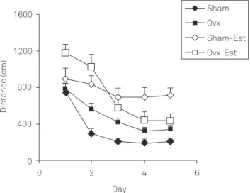
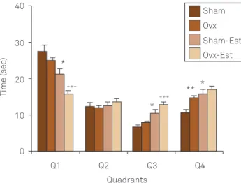
comparison with Sham — operated rats (p<0.01) (Fig 3). The animals of the Sham-Est group spent lower time in Q1compared to Shamgroup (p<0.05) (Fig 3). The time spent in Q1 by the animals of OVX-Est group was signifi-cantly lower than of OVX group (p<0.01) (Fig 3). The an-imals of Sham-Est group spent more time in non-target quadrants (Q3 and Q4) when compared with Sham group (bothp<0.05) (Fig 3). The animals of OVX-Est group also spent more time in non-target quadrants (Q3) in compari-son with OVX group (p<0.001) (Fig 3). Fig 4 showed the animals of OVX group swam more distance in non target quadrant (Q4) in comparison with Sham group (p<0.01). The traveled path length by the animals of OVX-Est group in target quadrant was lower than OVX group (p<0.001) (Fig 4). However, the animals of OVX-Est group traveled longer distance in non target quadrant(Q3) compared to OVX group (p<0.001). The animals of Sham-Est group traveled longer distances in non-target quadrants (Q3and Q4) in comparison Sham group (p<0.01) (Fig 4).
he total thiol concentration in cortical tissues of ovari-ectomized rats were signiicantly lower than Sham animals (p<0.01). In both OVX-Est and Sham-Est groups, the total thiol concentration were signiicantly lower than OVX and Sham groups respectively (p<0.001 and p<0.05 respectively). here was no signiicant diference between OVX-Est and Sham-Est groups in thiol group (Fig 5). MDA concentration in cortical tissues of ovariectomized animals was higher than sham operated ones (p<0.05). Chronic treatment of both the ovariectomized and sham-operated rats by 4 mg/kg estradi-ol increased MDA concentration in cortical tissues in com-parison with saline treated Sham and OVX rats (p<0.05 and
p<0.01 respectively) (Fig 6). he results also showed that the serum 17 β-estradiol concentration in OVX group was sig-niicantly higher than that of sham operated rats (p<0.001) (Fig 6). Fig 7 shows treatment by 4 mg/kg estradiol increased 17 β-estradiol serum concentration in ovariectomized and sham operated animals (p<0.001).
DISCUSSION
he results of the present study indicated that OVX rats had more time and path length to reach the platform in comparison with Sham group. he results also showed that
Fig 3. The results of the time (sec) spent in each quadrant during the probe trial on day 6 (24 h after the last secession of learning).
OVX: ovariectomized; Sham-Est: Sham-estradiol; OVX-Est: ovariectomized-estradiol; SEM: standard error median. Data are shown as mean±SEM of seven animals per group. The platform was removed, and the time spent in the target quadrant (Q1) and non target quadrants (Q2–Q4) was compared between the groups. *p<0.05, **p<0.01 compared to Sham group, +++p<0.001
compared to OVX group.
30 40
20
10
0
Q1 Q2 Q3 Q4
Quadrants
Sham
Ovx
Sham-Est Ovx-Est
Time (sec)
+++ *
+++ *
* **
600 800
400
200
0
Q1 Q2 Q3 Q4
Quadrants
Sham
Ovx
Sham-Est
Ovx-Est
Dis
tance (cm)
+++
+++ ** **
**
Fig 4. The results of the length of the swimming path (cm) in each quadrant during the probe trial on day 6 (24 h after the last secession of learning).
OVX: ovariectomized; Sham-Est: Sham-estradiol; OVX-Est: ovariectomized-estradiol; SEM: standard error median. Data are shown as mean±SEM of eight animals per group. The platform was removed, and the length of the swim path in the target quadrant (Q1) and non target quadrants (Q2–Q4) was compared between the groups. **p<0.01 compared to Sham group,
+++p<0.001 compared to OVX group.
Fig 5. The total thiol concentrations in cortical tissues of four groups.
OVX: ovariectomized; Sham-Est: Sham-estradiol; OVX-Est: ovariectomized-estradiol; SEM: standard error median. Data are shown as mean±SEM of eight animals per group. **p<0.05, ***p<0.001 compared to Sham group,
+p<0.05 compared to OVX group.
30
20
10
0
Groups
Sham
Ovx
Sham-Est
Ovx-Est
To
tal thiol Conc
.(mM)
+
**
317
Fat aa ta. t a a a a at deprivation of rats from ovarian hormones afects retention
phase of Morris water maze in rats. However, there were no signiicant diferences between OVX and Sham-operated rats in the time spent and traveled distance in target quadrant in probe trial when the platform was removed from the tank; the animals of OVX rats spent more time and traveled lon-ger distance in non target quadrant (Q4) in comparison with sham animals. he results of the present study conirmed the results of previous studies showing that ovariectomy impairs spatial memory4. However, it was found no efect of surgical
menopause on cognitive functioning3.
Estrogen replacement therapy has been reported to pre-vent or delay cognitive decline in postmenopausal women and in estrogen-depleted animals3,4. It has been suggested
that the efect of exogenous administration of estradiol on learning and memory of ovariectomized female rats depends on the dose and the estradiol plasma concentration. Low, but physiological, levels of exogenous estradiol enhances perfor-mance whereas administration of high exogenous pharma-cological or physiological levels of estradiol impairs perfor-mance7. he results of our previous study also showed that
treatment by 2 mg/kg estradiol valerate for eight weeks im-proves Morris water maze tasks of ovariectomized rats, how-ever deleterious efects of estradiol was seen in the same dose and time when administered to Sham-operated rats4. It has
also been suggested that, during the breeding season (high estradiol), female voles show poor performance in compar-ison with males in the spatial version of the water escape task12. Besides of the dose of estradiol, the discrepancies of
the data may be due to the diferences in type of memory which is studied or the age of OVX animals.
Estrogen exerts its memory-related effect via mul-tiple pathways including antioxidant properties, which may also have a role in its neuroprotective effects10. The
antioxidant property of the estradiol has been attributed to its free phenolic hydroxyl group on the A-ring of the ste-roid. Removal or blocking of the phenolic hydroxyl group eliminates the antioxidant effect, as well the neuroprotec-tion properties10. The results of the present experiment
demonstrated that SH contents in cortical tissues were decreased and MDA concentration increased after ovari-ectomy. Ovariectomy model has been widely used as an
in vivo model to mimic post-menopausal
pathophysiologi-cal changes in women related to learning and memory and brain oxidative stress13. Therefore, it is suggested that
learning and memory impairments which were seen in ovariectomized rats in comparison to the sham animals at least in part may be due to brain tissues oxidative damage. The cognitive impairments due to human menopausal conditions may also be related to brain oxidative damage. The results of previous studies have also confirmed that ovariectomy of animals or postmenopausal conditions in women increase lipid peroxidation in brain, erythrocytes and plasma13.
Neuroprotective effect of estrogen has been attribut-ed to its antioxidant properties of estrogen. It has been shown that the low nanomolar doses of estrogen has neu-roprotection effect in rodent models and reduces brain lip-id peroxlip-idation14. It was also shown that estrogens prevent
intracellular peroxide accumulation in an endoplasmic re-ticulum (ER)-independent manner15, decrease reactive
ox-ygen species (ROS) production16, limit lipid peroxidation
and decrease hydrogen peroxide concentrations17. Indirect
anti-oxidative effects of estrogens have been reported, in-cluding attenuation of microglial superoxide release18,
Fig 6. f!A" "# $ $ " $ "%$ & #
b#$ '# #f& (& .
MDA: a a yd)VX:variectomized; Sham-Est: Sham-estradiol;
OVX-Est: ovariectomized-estradiol; SEM: standard error median. Data are shown as mean±SEM of eight animals per group. *p<0.05, **p<0.01 compared to Sham group, ++p<0.01 compared to OVX group.
40
30
20
10
0
Groups
Sham Ovx
Sham-Est Ovx-Est
MD
A Conc
. (nmol/
g
Tissue)
++
* *
Fig 7. Comparison of serum estradiol level between four groups.
OVX: ovariectomized; Sham-Est: Sham-estradiol; OVX-Est: ovariectomized-estradiol; SEM: standard error median. Data are shown as mean±SEM of eight animals per group. ***p<0.001 compared to Sham group,
+++p<0.01 compared to OVX group. 8000
5000 7000
6000
4000
3000
2000
1000
0
Groups
Sham Ovx Sham-Est Ovx-Est
Ser
um 17
β
-es
tr
adiol concentr
a
tion (pg
/mL)
+++ ***
318 *+,- . /+012 3, /345+67 8 9 :; 8 <= >? 98 9@ 98 B
increase of glutathione reductase, gamma-glutamylcys-tein synthetase, glutaredoxin and glutathione10, increased
manganese superoxide dismutase (MnSOD) activity19 and
reduction of free radical production via an increase mito-chondrial efficiency20. The ability of estrogens for
regen-eration of vitamin E has also been suggested21. It has also
been shown that estrogens potentiated lipid peroxidation inhibitors22. Estrogens are highly lipid soluble and
large-ly reside in the membrane component of cells23. In fact,
there is evidence that estrogen prevents lipid peroxidation by sacrificing itself to oxidation24. This estrogen redox
cy-cle is operative in the brain and serves, together with the classic antioxidant mechanism, as a defense mechanism against ROS.
he results of the present study indicated that administra-tion of a high dose (4 mg/kg) of exogenous estradiol valerate impaired learning and memory in sham and ovariectomized which was accompanied by low level of total SH groups in cortical tissues.
his result was in contrast with the indings mentioned and showed beneicial efects of estradiol on learning and memory and its antioxidant properties. In consistent with the results of present study, it has been shown that high dos-es of dos-estradiol have deleterious efects on memory and learn-ing. Varga et al. showed that Sprague-Dawley ovariectomized rats treated by the high dose of estradiol displayed a week performance in the water maze task in comparison with non treated ones25. It has also been reported that chronic
admin-istration of high doses of estradiol impairs performance on a working/reference memory in the eight-arm radial arm maze7. Fugger et al. also showed that treatment of the female,
but not male, mice with estradiol impairs acquisition of wa-ter maze tasks26.
We hypothesized that the negative efects of a high dose of estradiol seen in the present study may be related to its pro-oxidative efects. he results showed that in both Sham-Est and OVX-Est rats the MDA concentrations was higher and total thiol concentration was lower than that of
sham and ovariectomized rats. It should be noted that in at least one study, estradiol administrationwasresulted in a time-dependent increase in lipid peroxidation27. It seems
that the differential actions of estrogens as a pro-oxidant or an antioxidant depends in part on their metabolism and the subsequent actions of the metabolites28. It has been
shown that estrogen administration to rodents results in the initiation of tumors in several organs29. The induction
of renal tumors in Syrian hamsters due to chronic expo-sure to estrogens is an extensively studied model of carci-nogenesis. It has been shown that estrogen-induced free radicals play a tumor-initiating role in this process27,30. It
was shown that renal lipid peroxide concentrations were raised by chronic treatment with estradiol27. It was also
shown that certain metabolites of estradiol or estrone can directly produce DNA damage in target tissues, indepen-dent of their interaction with the estrogen receptor29. The
genotoxic effect of hydroxyestrogens has also been relat-ed to the generation of free radicals29. Indeed,
hydroxyes-trogen-semiquinone intermediates can react in vitro with
molecular oxygen yielding the superoxide anion (O2•−).
Finally it seems that estrogens have dual actions in refer-ence to their oxidative effects31.
In conclusion, the results of present study show that a high dose of estradiol impairs spatial learning and mem-ory. Our results also indicate that the oxidative stress in brain cortical tissues might take part in spatial learning and memory impairments due to high levels of estradiol, but further investigations using more methods need to be done.
ACKNOWLEDGMENTS
The results described in this paper were from a M.Sc. student thesis proposal. The authors would like to thank the Vice Chancellor of Research Affairs of Mashhad University of Medical Science for financial assistance.
1. *[email protected] HI H02 2 .3J3 DI K0/eL54J E00 DI K4G.ML34J NI
F.+. 3G0J3DIO L0G4F4J G.L E0 0P.Q3R R. +.J5.ff.S52 0R2S010E4T3J.
0J E.4 +J3J MI T.T0+U, 4JG J35+ 3S 0xide metabolite levels in
hippocampal tissues of ovariectomized and Sham-operated rats. Arq Neuropsiquiatr 2012;70:447-452.
2. Frick KM. Estrogens and age-related memory decline in rodents: What have we learned and where do we go from here? Horm Behav 2009;55:2-23.
3. Vearncombe KJ, Pachana NA. Is cognitive functioning detrimentally affected after early, induced menopause? Menopause 2009;16:188-198.
4. Hosseini M, Headari R, Oryan S, Hadjzadeh MA, Saffarzadeh F, Khazaei M. The effect of chronic administration of L-arginine on the learning and memory of estradiol-treated ovariectomized rats tested in the morris water maze. Clinics 2010;65:803-807.
5. Chesler EJ, Juraska JM. Acute administration of estrogen and progesterone impairs the acquisition of the spatial morris water maze in ovariectomized rats. Horm Behav 2000;38:234-242.
6. Galea LA, Kavaliers M, Ossenkopp KP, Hampson E. Gonadal hormone levels and spatial learning performance in the Morris water maze in male and female meadow voles, Microtus pennsylvanicus. Horm Behav 1995;29:106-125.
7. Holmes MM, Wide JK, Galea LAM. Low levels of estradiol facilitate, whereas high levels of estradiol impair, working memory performance on the radial arm maze. Behav Neurosc 2002;116:928-934.
8. Galea LAM, Wide JK, Paine TA, Holmes MM, Ormerod BK, Floresco SB. High levels of estradiol disrupt conditioned place preference learning, stimulus response learning and reference memory but have limited effects on working memory. Behav Brain Res 2001;126:115-126.
319
FVWXYZ [\ [] ^V_V` ^Z[c]]ZWVcghi Wj V^X]c k_j VX `^V YVlZj VWi
9g FjِcXm[n,pXZ^Z jZ jrgsjZ Zj V ^XmVcYZm[ V` X iY iX`^Z YZ ` WXV]uvcw[ZXYZj
Wx yZ V ` ^ W[Z y] WZ` WXVc u]j V ` WX]xidative treatment.
Arzneimittel-Forschung 1995;45:443-446.
10. Niki E, Nakano M. Estrogens as antioxidants. Meth Enzymol 1990;186:330-333.
11. Vedder H, Anthes N, Stumm G, Würz C, Behl C, Krieg JC. Estrogen hormones reduce lipid peroxidation in cells and tissues of the central nervous system. J Neurochemistry 1999;72:2531-2538.
12. Galea LAM, Kavaliers M, Ossenkopp KP, Hampson E. Gonadal hormone levels and spatial learning performance in the Morris water maze in male and female meadow voles, Microtus pennsylvanicus. Horm Behav 1995;29:106-125.
13. Monteiro SC, Matté C, Bavaresco CS, Netto CA, Wyse ATS. Vitamins E and C pretreatment prevents ovariectomy-induced memory deicits in water maze. Neurobiol Learn Mem 2005;84:192-199.
14. Green PS, Gridley KE, Simpkins JW. Nuclear estrogen receptor-independent neuroprotection by estratrienes: a novel interaction with glutathione. Neuroscience 1998;84: 7-10.
15. Behl C, Skutella T, Lezoualc’h F, et al. Neuroprotection against oxidative stress by estrogens: structure-activity relationship. Mol Pharmacol 1997;51:535-541.
16. Culmsee C, Vedder H, Ravati A, et al. Neuroprotection by estrogens in a mouse model of focal cerebral ischemia and in cultured neurons: evidence for a receptor-independent antioxidative mechanism. J Cerebral Blood Flow Metabolism 1999;19:1263-1269.
17. Kii N, Adachi N, Liu K, Arai T. Acute effects of 17beta-estradiol on oxidative stress in ischemic rat striatum. J Neurosurg Anesthesiol 2005;17:27-32.
18. Bruce-Keller AJ, Keeling JL, Keller JN, Huang FF, Camondola S, Mattson MP. Antiinlammatory effects of estrogen on microglial activation. Endocrinology 2000;141: 3646-3656.
19. Gottipati S, Cammarata PR. Mitochondrial superoxide dismutase activation with 17beta-estradiol-treated human lens epithelial cells. Mol Vis 2008;14:898-905.
20. Nilsen J, Irwin RW, Gallaher TK, Brinton RD. Estradiol in vivo regulation of brain mitochondrial proteome. J Neurosc 2007;27:14069-14077.
21. Mukai K, Daifuku K, Yokoyama S, Nakano M. Stopped-low
investigation of antioxidant activity of estrogens in solution. Biochim Biophys Acta 1990;1035:348-352.
22. Wang X, Simpkins JW, Dykens JA, Cammarata PR. Oxidative damage to human lens epithelial cells in culture: estrogen protection of mitochondrial potential, ATP, and cell viability. Invest Ophthalmol Vis Sci 2003;44:2067-2075.
23. Liang Y, Belford S, Tang F, Prokai L, Simpkins JW, Hughes JA. Membrane luidity effects of estratrienes. Brain Res Bull 2001;54:661-668.
24. Prokai L, Prokai-Tatrai K, Perjesi P, et al. Quinol-based cyclic antioxidant mechanism in estrogen neuroprotection. Proc Nat Acad Sci USA 2003;100:11741-11746.
25. Varga H, Németh H, Tَth T, Kis Z, Farkas T, Toldi J. Weak if any effect of estrogen on spatial memory in rats. Acta Biol Szegediensis 2002;46:13-16.
26. Fugger HN, Cunningham SG, Rissman EF, Foster TC. Sex differences in the activational effect of ERalpha on spatial learning. Horm Behav 1998;34:163-170.
27. Wang MY, Liehr JG. Induction by estrogens of lipid peroxidation and lipid peroxide-derived malonaldehyde-DNA adducts in male Syrian hamsters: role of lipid peroxidation in estrogen-induced kidney carcinogenesis. Carcinogenesis 1995;16:1941-1945.
28. Nathan L, Chaudhuri G. Antioxidant and prooxidant actions of estrogens: potential physiological and clinical implications. Semin Reprod Med 1998;16:309-314.
29. Liehr JG. Dual role of oestrogens as hormones and pro-carcinogens: tumour initiation by metabolic activation of oestrogens. Eur J Cancer Prev 1997;6:3-10.
30. Kirkman H. Estrogen-induced tumors of the kidney. III. Growth characteristics in the Syrian hamster. Nat Cancer Inst Monograph 1959;1:1-57.