High-Intensity Inspiratory Protocol Increases
Heart Rate Variability in Myocardial
Revascularization Patients
Flavia Cristina Rossi Caruso
1, PhD; Rodrigo Polaquini Simões
1, PhD; Michel Silva Reis
2, PT, MSc, PhD; Solange Guizilini
3,
PT, MSc, PhD; Vera Lucia dos Santos Alves
4, PT, MSc, PhD; Valeria Papa
5, PT, MSc; Ross Arena
6, PhD; Audrey Borghi-Silva
1,
PT, MSc, PhD
Abstract
Objective: To evaluate heart rate variability during an inspiratory muscle endurance protocol at three different load levels [30%, 60% and 80% of maximal inspiratory pressure], in patients who had previously undergone coronary artery bypass grafting.
Methods: Nineteen late postoperative myocardial revascularization patients participating in a cardiovascular rehabilitation program were studied. Maximal inspiratory pressure maneuvers were performed. An inspiratory muscle endurance protocol at 30%, 60% and 80% of maximal inspiratory pressure was applied for four minutes each, in random order. Heart rate and RR intervals were recorded and heart rate variability was analyzed by time (RMSSD-the mean of the standard deviations for all R-R intervals, and RMSM-root-mean square differences of successive R-R intervals) and frequency domains indices (high and low frequency) in normalized units. ANOVA for repeated measurements was used to compare heart rate variability indices
and Student t-test was used to compare the maximal inspiratory pressure and maximal expiratory pressure values.
Results: Heart rate increased during performance of maximal respiratory pressures maneuvers, and the maximal inspiratory pressure and maximal expiratory pressure mean values were significantly lower than predicted values (P<0.05). RMSSD increased significantly at 80% in relation to rest and 30% of maximal inspiratory pressure and RMSM decreased at 30% and 60% of maximal inspiratory pressure in relation to rest (P<0.05). Additionally, there was significant and progressive decrease in low frequency and increase in high frequency at 30%, 60% and 80% of maximal inspiratory pressure in relation to the resting condition.
Conclusion: These results suggest that respiratory muscle training at high intensities can promote greater parasympathetic activity and it may confer important benefits during a rehabilitation program in post-coronary artery bypass grafting.
Keywords: Autonomic Nervous System. Respiratory Muscles. Heart Rate. Physical Therapy Modalities. Coronary Artery Bypass.
DOI: 10.5935/1678-9741.20160007
1 Laboratory of Cardiopulmonary Physiotherapy at Federal University of São Carlos
(UFSCar), São Carlos, SP, Brazil.
2Department of Physiotherapy at Faculty of Medicine at Federal University of Rio
de Janeiro (FMUFRJ), Rio de Janeiro, RJ, Brazil.
3Department of Sciences of Human Movement at Federal University of São Paulo
(UNIFESP), Santos, SP, Brazil.
4Faculty of Medical Sciences at Santa Casa de São Paulo (FCMSCSP), São Paulo,
SP, Brazil.
5Hospital São Francisco of Ribeirão Preto (SF), Ribeirão Preto, SP, Brazil.
6Department of Physical Therapy and Integrative Physiology Laboratory, College
of Applied Health Sciences, University of Illinois Chicago, Chicago, IL, USA.
This study was carried out at the Laboratory of Cardiopulmonary Physiotherapy at Federal University of São Carlos (UFSCar), São Carlos, SP, Brazil.
Financial support: FAPESP - 2009-01842-0
Correspondence Address: Audrey Borghi Silva
Laboratório de Fisioterapia Cardiopulmonar/Departamento de Fisioterapia Universidade Federal de São Carlos (UFSCar)
Rodovia Washington Luis, Km: 235 – Monjolinho – São Carlos, SP, Brazil Zip code: 13565-905
E-mail: [email protected]
Article received on September 8th, 2015
Article accepted on February 5th, 2016
Abbreviations, acronyms & symbols
ANOVA BMI CABG CAD COPD ECG HFun HR HRV LFnu
= Analysis of variance = Body mass index
= Coronary artery bypass grafting = Coronary artery disease
= Chronic obstructive pulmonary disease = Electrocardiogram
= High frequency in normalized units = Heart rate
= Heart rate variability
= Low frequency in normalized units
MEP MIP MRP RMS RMSM RMSSD RMT RSA RV TPC
= Maximal expiratory pressure = Maximal inspiratory pressure = Maximal respiratory pressure = Respiratory muscle strength
= Root-mean square differences of successive R-R intervals = Mean of the standard deviations for all R-R intervals = Respiratory muscle training
= Respiratory sinus arrhythmia = Residual volume
INTRODUCTION
The evaluation of autonomic cardiac function by heart rate variability (HRV) in patients with coronary artery disease (CAD)
has been widely used for risk stratification[1-3]. Studies have been
developed to evaluate HRV in patients with CAD who have
undergone coronary artery bypass grafting (CABG) surgery[4,5];
the benefits being HRV assessment is non-invasive, low cost and predicts cardiovascular morbidity and mortality in early and late
phases of surgery procedure[2,6,7].
Respiratory muscle strength (RMS) evaluation by maximal respiratory pressure (MRP) is used as an important diagnostic
and prognostic measure in patients with neuromuscular[8],
pulmonary[9] and cardiovascular disease[10]. Moreover, respiratory
muscle training (RMT) have proven to be a valuable treatment
approach in preventing pulmonary complications after CABG[11].
Collectively, the simultaneous evaluation of autonomic cardiac function and RMS may play an important role in the evaluation
of cardiorespiratory integrity in patients following CABG[12].
CABG produces an important negative impact on autonomic
cardiac function[13,14] and RMS[15]. Previous studies have reported
that return of RMS takes several months following CABG[16].
Participation in cardiac rehabilitation induces a host of benefits,
including improved cardiac autonomic function[17] and RMS[18].
To our knowledge, no previous study has assessed the cardiac autonomic system by HRV during different loads resistance loads imposed upon the respiratory musculature in patients post-CABG; this has important implications for establishing RMT
intensities that optimally improve HRV[19-21]. Therefore, the aim
of this study was to assess HRV during an inspiratory muscle endurance protocol at three different levels of effort [30%, 60% and 80% of maximal inspiratory pressure (MIP)] in patients post-CABG. We hypothesized that the application of a high-intensity inspiratory muscle endurance protocol may induce greater changes in HRV when contrasted to moderate and low loads.
METHODS
Subjects
Twenty eight patients who had previously undergone CABG were recruited to participate in the present study and 19 male patients satisfied all inclusion criteria. All subjects participated in a cardiovascular physical therapy program [60 minute sessions three times a week for at least 6 months at 70-85% of maximum heart rate (HR)]. Exclusion criteria were emergent or concomitant surgery, implanted pacemaker, unstable angina, recent myocardial infarction (less than 6 months), chronic disturbances in heart rhythm that could compromise HRV analysis, chronic obstructive pulmonary disease (COPD), valvular heart disease, severe non-cardiac diseases, and the inability to perform the study protocol. Patients who were obese [body mass index (BMI)
> 30 kg/m2], active smokers, had evidence of left ventricular
dysfunction, neurological and respiratory disturbances, visible alterations in thoracic and/or abdominal mobility, or accentuated structural deviations in the spine that might alter the respiratory dynamic were excluded.
Time from completion of surgery to the entry in the study protocol was 180±12 days. All subjects were oriented to the
experimental procedures to be performed and they signed a written informed consent agreement in accordance with resolution 196/96 of the National Health Council. This study was reviewed and approved by the Ethics Committee for Human Research (number 109/2006).
Experimental Design
The study procedures were performed in the cardiopulmonary laboratory of our institution, in the morning to avoid any circadian variations, with a room temperature controlled at 22 to 24°C and at a relative air humidity of 50% to 60%.
The patients were instructed to not ingest alcohol or other stimulants the night before and day of the experimental procedures, to not do any heavy physical exercise, to avoid heavy meals for two hours before the experimental procedures and to get a good night’s sleep the night before.
Clinical Evaluation
All patients underwent clinical evaluation which consisted of: 1) anamneses; 2) past medical and surgical history; 3) family medical history; 4) risk factor profile; 5) lifestyle habits; 6) visual inspection to identify possible alterations in the thoracic and abdominal regions such as cutaneous folds and accentuated structural deviations in the spine that might alter respiratory dynamics; 7) anthropometric evaluation measuring height and body mass by stadiometer and scale (Welmy, São Paulo, SP, Brazil); 8) 12-lead standard electrocardiogram (ECG) measurement of HR (cardiac monitor - Ecafix TC 500, São Paulo, SP, Brazil); 9) arterial blood pressure (sphygmomanometer BD, São Paulo, SP, Brazil); 10) maximum dynamic physical effort test; and 11) laboratory exams (fasting glycemia, total and fractions cholesterol, triglycerides, uric acid, creatine and type 1 urine).
Respiratory Muscle Strength
To obtain values for MIP and maximal expiratory pressure (MEP), an aneroid type manovacuometer (GER-AR, São Paulo,
SP, Brazil) with an operational interval of ± 300 cmH2O was used.
A plastic mouthpiece was coupled with a tube attached to the
manovacuometer. This mouthpiece had a leak[22] with a diameter
of approximately 2 mm that permitted a small amount of air to escape to avoid any elevation of pressure within the oral cavity
by contraction of the facial muscles[23]. Each individual used a
rubber mouthpiece with a diameter of 32 mm over the plastic mouthpiece.
Before the measurements were taken, patients remained seated while being familiarized with the equipment and instructed on how to perform the maneuvers. Immediately following, the HR and R-R intervals were registered for three periods: 1) during the first minute at rest; 2) for approximately three seconds of the maneuver; and 3) during the final minute of the procedure.
before the maneuver was performed, a nasal clip was placed on the patient (to not allow air to escape from the nostrils), and the patient was instructed to keep their lips tightly closed over the mouth while performing the forced inspiration maneuver from residual volume (RV) and the forced expiration maneuver from total pulmonary capacity (TPC) to maintain maximum respiratory
effort for approximately one second[23].
The greatest values obtained from three correctly performed repetitions (with a 10% or less difference between values) for each maneuver were registered. It is important to emphasize that a single evaluator performed the manovacuometry for all individuals and verbal encouragement was given to all patients during the maneuvers to reach the maximal effort of the patient. The MIP and MEP measurement were compared to the predicted
values for Brazilian population according to Simões et al.[23].
Inspiratory Muscle Endurance Protocol
The inspiratory muscle endurance protocol consisted of maneuvers at three pressure levels: 30%, 60% and 80% of MIP. The loads were applied in a random order by drawing of shuffled, opaque, coded envelopes that were opened by one investigator. First, the subjects were maintained at rest for 10 minutes and the HR data was obtained while subjects rested quietly, breathing spontaneously in the seated position.
During the protocol, the patient remained seated in a chair, using a nose clip and performed inspiratory efforts using the manovacuometer which had previously shown to the value that corresponded to the individual’s pressure percentage of MIP (30%, 60% or 80%). Each effort level was performed for four minutes and the patient was oriented to make an inspiratory effort and maintain the equipment indicator on the demarcated line, which corresponded to the percentage being tested, for two seconds followed by expiration through the mouth for three seconds; this corresponded to a total of 12 respiratory cycles per
minute[24]. To ensure that the maneuver was performed correctly
and at the correct times for inspiration and expiration as previously instructed, one of the evaluators used a chronometer
to give verbal commands to the patient[24].
The HR was continually registered one minute before the beginning of effort, during four minutes at each effort of intensity, and during the first minute after the effort. During this period, the ECG signal and HR were observed in real time on the computer monitor to verify signal quality.
Heart Rate and R-R Intervals Data Collection
The ECG signal, R-R intervals and the HR were obtained on a beat to beat basis in real time through a cardiac monitor registering the derivation of the CM5 lead, using disposable self adhesive activated carbon electrodes (red electrode positioned
on the esternal manubrium, yellow electrode in the 5th left
intercostals space on the anterior axillary line, and black electrode
in the 5th right intercostals space).
The ECG signal was transferred to a microcomputer (PC-AT 486 DX-4, 100MHz) through an analog-digital converter (Lab. PC + National Instruments, Co.), that constituted an interface between the cardiac monitor and the microcomputer. The
analog signals taken from the ECG were converted into binary values and processed by software specifically designed to
capture ECG and calculate the R-R intervals[25].
Data Analysis
The HRV was analyzed by the time and frequency domain methods. In the time domain, the R-R intervals were analyzed by the root-mean square differences of successive R-R intervals (RMSM) and mean of the standard deviations for all R-R intervals (RMSSD) indices. The RMSM index corresponded to the square root of the sum of the square of the differences of the individual values in relation to the mean value, divided by
the number of R-R intervals in a specified time period[26]. The
RMSSD corresponded to the square root of the sum of the square of the differences between the R-R intervals registered, divided by the number of R-R intervals in a specified time period
less one[26]. To the frequency domain analysis, 256 points of the
four minutes (endurance protocol) was selected as the criterion required for application of the spectral methods (i.e., Fast Fourier Transforming). Then, the power spectral components were obtained at low and high frequencies in normalized units [low frequency in normalized units (LFnu) and high frequency in normalized units (HFnu), respectively].
Statistical Analysis
On the basis of the results of the pilot study (n=8), we estimated that a sample size of 15 individuals would have a power >80% to detect a 5 ms difference in the RMSSD index (main outcome) amongst the imposed inspiratory loads. The level of significance was set at 5%. The data distribution was verified by the Shapiro-Wilk test, and when normality was confirmed the data were expressed in mean and standard deviation. Analysis of variance (ANOVA) for repeated measurements was used to compare the indexes of HRV (RMSM, RMSSD, LFnu and HFnu) obtained during the endurance protocol at four different situations: rest (pre-effort), 30%, 60% and 80% of MIP. The Student
t-test for dependent samples was used to compare the MIP and
MEP values obtained by the manovacuometer to the predicted
values[23] and to compare the HR and R-R intervals values
obtained during rest (pre-effort) with peak values achieved during MIP. The probability of a type I error was established at 5% for all tests (α=0.05). The data was analyzed using the STATISTICA for Windows software program (Stat Soft Inc, 2000).
RESULTS
internal mammary artery grafting. None of the subjects presented with pulmonary or hemodynamic complications in the perioperative period. Mean of cardiopulmonary bypass time was 78±22 min, time of surgery procedure was 178±60 min, time to mechanical ventilation was 10±5.8 h, intensive care unit stay was 2.3±1.2 days and total time in the hospital was 7±2 days. All patients underwent grafting with mammary artery.
Table 1 lists the anthropometric characteristics of the patients studied in relation to age, weight, height and BMI, as well as risk factors and number and type of grafts used in CABG. The medications taken by the patients during the evaluation period and the respective number of patients were: 1) acetyl salicylic acid (12 patients); 2) anti-arrhythmic (2 patients); 3) angiotensin converting enzyme (15 patients); 4) β–blockers (12 patients); 5) calcium-channel blockers (5 patients); 6) nitrates (7 patients); 7) angiotensin II receptor antagonists (2 patients); 8) antihypercolesterolemics (9 patients); and 9) diuretics (5 patients).
Subjects presented with significantly lower MIP and MEP
values (P<0.05) when compared to predicted values (Table 2). In
relation to HR during the MIP and MEP maneuvers, a significant increase was found at peak when compared to rest. This fact can be confirmed by the reduction in R-R intervals during the MIP and MEP, in comparison to the pre-effort rest period (Table 2).
The HRV evaluated during the endurance protocol at the different MIP percentages showed a significant increase in
Table 1. Age, anthropometric characteristics, risk factors and number and type of grafts used in coronary artery bypass surgery.
Age and Anthropometric
Characteristics n=19
Age (years) 68±5
Weight (kg) 78±25
Height (m) 1.69±0.08
BMI (kg/m2) 27±4.2
Risk Factors n
Hypertension 12
History of CAD 16
Dyslipidemia 11
Ex-smoker 13
Grafts n (quantity and type)
4 grafts 4 (3v and 1a)
3 grafts 8 (2v and 1a)
2 grafts 5 (1v and 1a)
1 graft 2 (a)
Table 2. Comparison of maximal respiratory pressures, heart rate and R-R intervals values under pre effort rest conditions and during maximal inspiratory and expiratory pressures maneuvers.
Variables MIP
(n=19)
MEP (n=19)
MRP obtained (cmH2O) 68±15 78±20
MRP predicted (cmH2O) 78±10* 75±12*
HR rest (bpm) 65±10 66±9
HR peak (bpm) 98±12† 102±10†
R-Ri rest (ms) 1012±220 1112±176
R-Ri peak (ms) 613±101† 615±180†
Data expressed in mean and standard deviation.
MIP=maximal inspiratory pressure; MEP=maximal expiratory pressure; MRP=maximal respiratory pressure; HR=heart rate; R-Ri=R-R intervals
*P<0.05 between obtained and predicted values; †P<0.05
between rest and peak conditions.
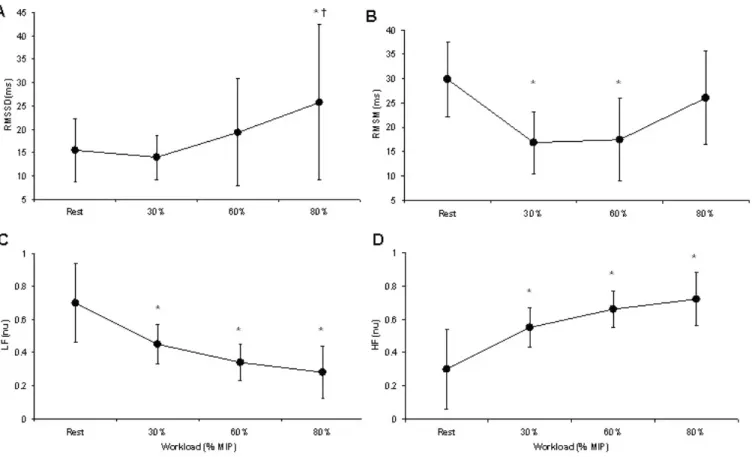
the RMSSD at 80% (Figure 1A) and significant decrease in the RMSM index at 30% and 60% of MIP in relation to rest (Figure 1B), and significant decrease and increase in the LFnu and HFnu, respectively, at 30%, 60% and 80% of MIP in relation to the resting condition (Figures 1C and 1D). However, only the 80% MIP maneuver demonstrated a significant increase in RMSSD and high frequency as well as reduced low frequency when contrasted with the 30% MIP maneuver.
DISCUSSION
The main findings of the present study showed that after 6 months of CABG, reductions in MIP and MEP compared to predicted values were observed. In addition, we observed that the increase in inspiratory muscle workload (% of MIP) produced higher parasympathetic and lower sympathetic cardiac modulation when contrasted with moderate and low loads. These findings may have important implications for establishing RMT strategies following CABG.
Our results showed a significant increase in the RMSSD and HFnu index was observed (Figures 1A and 1D, respectively), indicating an increase in parasympathetic activity when the protocol was performed at a higher load. There was also a decrease in sympathetic activity with the increase of workload, as indicated by LFnu behavior (Figure 1C). In relation to the RMSSD, this index reflects both sympathetic and parasympathetic activity, therefore reduced values observed at 30% and 60% of MIP could be explained by a decrease in sympathetic modulation. Additionally, it was possible to observe an increase in the values of this index at 80% of MIP, this fact revealed a predominance of parasympathetic modulation at this load in the present study cohort.
A possible explanation for the increase in parasympathetic and decrease sympathetic modulation during a higher inspiratory workload is the increase in tidal volume, as well as the Data expressed in mean and standard deviation.
Fig. 1 – Comparison of heart rate variability index at rest, 30%, 60% and 80% of maximal inspiratory pressure during endurance protocol. Data expressed in mean and standard deviation. MIP=maximal inspiratory pressure; LFnu=low frequency in normalized units; HFun=high frequency in normalized units.
*P<0.05 in relation to rest condition; †P<0.05 in relation to 30% of MIP.
increase in inspiratory effort. In this context, some studies have demonstrated that the magnitude of autonomic responses of HR during respiratory sinus arrhythmia (RSA) maneuver is directly
proportional to the tidal volume[19].
The endurance protocol applied in the current study has a certain similarity to the RSA maneuver in which it was performed with controlled inspiratory and expiratory times. On the other hand, the protocol offers resistance to the inspiratory muscles. Also, a greater accentuation of the RSA is achieved because the maneuver performed at a higher frequency (i.e., 12 cycles per minute). However, we believe that the mechanisms involved in cardiac autonomic control with the respiratory maneuver are the same, and we base this on the probability of tidal volume having increased with the increase in workload, which in turn influenced
the autonomic cardiac responses. Kautzner[27] verified that there
is evidence that the variations in tidal volume can potentially interfere with the reproducibility of RSA and in the capacity of the test to measure differences in vagal control.
Several experimental studies[28,29] have shown that an
increase in ventilation through an increase in tidal volume may cause fluctuations in autonomic cardiac modulation. For this reason, one speculates whether a RMT program in humans can
influence total HRV. Laoutaris et al.[30] verified in their study with
chronic heart failure patients undergoing RMT at 60% of MIP that HR at rest was significantly reduced after 10 weeks of respiratory
training (three times a week). According to these authors[30],
these findings may reflect a relation between the improvement in respiratory function and the changes in autonomic balance, favoring an increase in vagal activity. Another hypothesis is the relative attenuation of HR due to RMT would be related to the reflex mediated by the diaphragm muscle, that may influence
the sympathetic tone; recent research[31,32] has demonstrated
that when muscle fatigue is induced in healthy individuals, a decrease in blood flow and increase in vascular resistance to the lower limbs is observed, resulting from sympathetic responses to diaphragmatic stress.
As to the MRP, the lower values for MIP as well as MEP in comparison to the predicted values indicate that the RMS in these patients is still reduced, suggesting that despite myocardial revascularization and participation in a cardiovascular rehabilitation program for 6 months, a decrease
in respiratory muscle performance persists. Several studies[33-37]
have demonstrated that the reduction in RMS after myocardial
to the damage caused by mechanical respiration with thoracic incision, reducing the capacity of the respiratory muscles to generate sufficient tension to perform respiratory work imposed
due to a mechanical disadvantage. Borghi-Silva et al.[34] found
that RMS suffers a significant reduction after CABG and that the pressure values verified on the day of hospital discharge remain
lower than preoperative values. KristjAnsdottir et al.[37] found
that thoracic mobility is reduced even one year after surgical intervention. For this reason, we believe that these alterations in respiratory mechanics caused by thoracic incision could reduce the respiratory muscle efficiency of the patients evaluated.
However, some authors[36] have demonstrated that pulmonary
function returns to normal values after six months of surgery. In this context, an important aspect to note is that RMT has not been commonly employed in cardiovascular rehabilitation programs. However, based on the results of the present study, we believe that RMT should be applied not only in late phase
post-CABG, but also in the immediate postoperative phase[33]
aiming at re-establishing RMS. In this context, RMT applied at higher intensities could be an important strategy to enhance vagal tone in parallel to aerobic exercise training programs in these patients. In particular, the positive effects of RMT on vagal tone could produce a cardioprotective effect, reducing risks to arrhythmias and fatal events.
Some limitations in this study should be taken into consideration. Although the patients were in a stable postoperative phase after surgical intervention, the sample was relatively small. However, the sample size to answer the main outcome was powered >80%. In addition, it was not possible to perform tidal volume measurement at the different percentages of MIP during the endurance protocol and it also was not possible to compare thoracic mobility during the protocol, aspects that would establish a more solid foundation for the findings observed in this study. Finally, the results of the present study are restricted to patients in the late phase of cardiac surgery. In this context, the impact of high intensity MIP maneuvers to produce a marked parasympathetic modulation during the early postoperative phase, when RMS is more profoundly impacted, requires further investigation.
CONCLUSION
In conclusion, we found that after 6 months post-CABG reductions in MIP and MEP were persisted. In addition a high-intensity inspiratory protocol promoted a greater parasympathetic modulation in comparison to maneuvers at lower loads. These results provide important implications for rehabilitation procedures following CABG, in particular including a RMT component as a standard of care.
Authors’ roles & responsibilities
FCRC
RPS
MSR
SG
VLSA
VP
RA
ABS
Analysis and/or data interpretation; statistical analysis; final manuscript approval
Conception and design study; realization of operations and/or trials; statistical analysis; final manuscript approval
Conception and design study; analysis and/or data interpretation; manuscript redaction or critical review of its content; final manuscript approval
Analysis and/or data interpretation; manuscript redaction or critical review of its content; final manuscript approval Manuscript redaction or critical review of its content; final manuscript approval
Analysis and/or data interpretation; manuscript redaction or critical review of its content; final manuscript approval Manuscript redaction or critical review of its content; final manuscript approval
Conception and design study; realization of operations and/or trials; statistical analysis; analysis and/or data interpretation; manuscript redaction or critical review of its content; final manuscript approval
REFERENCES
1. Jørgensen RM, Abildstrøm SZ, Levitan J, Kobo R, Puzanov N, Lewkowicz M, et al. Heart rate variability density analysis (Dyx) and prediction of long-term mortality after acute myocardial infarction. Ann Noninvasive Electrocardiol. 2016;21(1):60-8.
2. Wulsin LR, Horn PS, Perry JL, Massaro JM, D’Agostino RB. Autonomic imbalance as a predictor of metabolic risks, cardiovascular disease, diabetes, and mortality. J Clin Endocrinol Metab. 2015;100(6):2443-8. 3. Vanderlei LC, Pastre CM, Hoshi RA, Carvalho TD, Godoy MF. Basic
notions of heart rate variability and its clinical applicability. Rev Bras Cir Cardiovasc. 2009;24(2):205-17.
4. Soares PP, Moreno AM, Cravo SL, Nobrega AC. Coronary artery bypass surgery and longitudinal evaluation of the autonomic cardiovascular function. Crit Care. 2005;9(2):R124-31.
5. Demirel S, Akkaya V, Oflaz H, Tükek T, Erk O. Heart rate variability after coronary artery bypass graft surgery: a prospective 3-year follow-up study. Ann Noninvasive Electrocardiol. 2002;7(3):247-50.
6. Lahiri MK, Kannankeril PJ, Goldberger JJ. Assessment of autonomic function in cardiovascular disease: physiological basis and prognostic implications. J Am Coll Cardiol. 2008;51(18):1725-33. 7. Hadase M, Azuma A, Zen K, Asada S, Kawasaki T, Kamitani T, et al. Very
low frequency power of heart rate variability is a powerful predictor of clinical prognosis in patients with congestive heart failure. Circ J. 2004;68(4):343-7.
8. Foglio K, Clini E, Facchetti D, Vitacca M, Marangoni S, Bonomelli M, et al. Respiratory muscle function and exercise capacity in multiple sclerosis. Eur Respir J. 1994;7(1):23-8.
9. Alvisi R, Volta CA, Righini ER, Capuzzo M, Ragazzi R, Verri M, et al. Predictors of weaning outcome in chronic obstructive pulmonary disease patients. Eur Respir J. 2000;15(4):656-62.
11. Hulzebos EH, Helders PJ, Favié NJ, De Bie RA, Brutel de la Riviere A, Van Meeteren NL. Preoperative intensive inspiratory muscle training to prevent postoperative pulmonary complications in high-risk patients undergoing CABG surgery: a randomized clinical trial. JAMA. 2006;296(15):1851-7.
12. Meyer FJ, Borst MM, Zugck C, Kirschke A, Schellberg D, Kübler W, et al. Respiratory muscle dysfunction in congestive heart failure: clinical correlation and prognostic significance. Circulation. 2001;103(17):2153-8.
13. Pantoni CB, Mendes RG, Di Thommazo-Luporini L, Simões RP, Amaral-Neto O, Arena R, et al. Recovery of linear and nonlinear heart rate dynamics after coronary artery bypass grafting surgery. Clin Physiol Funct Imaging. 2014;34(6):449-56.
14. Yavuz B, Duman U, Abali G, Dogan OF, Yazicioglu A, Sahiner L, et al. Coronary artery bypass grafting is associated with a significant worsening of QT dynamicity and heart rate variability. Cardiology. 2006;106(1):51-5.
15. Mendes RG, Simões RP, Costa FSM, Pantoni CBF, Luzzi S, Catai AM, et al. Heart rate variability and pulmonary function behavior in patients undergoing coronary artery bypass grafting and physiotherapy intervention. Crit Care. 2007;11(Suppl 3): P55.
16. Richter Larsen K, Ingwersen U, Thode S, Jakobsen S. Mask physiotherapy in patients after heart surgery: a controlled study. Intensive Care Med. 1995;21(6):469-74.
17. Mendes RG, Simões RP, Costa FSM, Pantoni CB, Di Thommazo L, Luzzi S, et al. Short-term supervised inpatient physiotherapy exercise protocol improves cardiac autonomic function after coronary artery bypass graft surgery: a randomised controlled trial. Disabil Rehabil. 2010;32(16):1320-7.
18. Renault JA, Costa-Val R, Rosseti MB, Houri Neto M. Comparison between deep breathing exercises and incentive spirometry after CABG surgery. Rev Bras Cir Cardiovasc. 2009;24(2):165-72.
19. Hirsch JA, Bishop B. Respiratory sinus arrhythmia in humans: how breathing pattern modulates heart rate. Am J Physiol. 1981;241(4):H620-9.
20. Reis MS, Arena R, Deus AP, Simões RP, Catai AM, Borghi-Silva A. Deep breathing heart rate variability is associated with respiratory muscle weakness in patients with chronic obstructive pulmonary disease. Clinics. 2010;65(4):369-75.
21. Reis MS, Arena R, Archiza B, Toledo CF, Catai AM, Borghi-Silva A. Deep breathing heart rate variability is associated with inspiratory muscle weakness in chronic heart failure. Physiother Res Int. 2014;19(1):16-24. 22. Ragnarsdottir M, KristjAnsdottir A, Ingvarsdottir I, Hannesson P,
Torfason B, Cahalin L. Short-term changes in pulmonary function and respiratory movements after cardiac surgery via median sternotomy. Scand Cardiovasc J. 2004;38(1):46-52.
23. Simões RP, Deus APL, Auad MA, Dionísio J, Mazzonetto M, Borghi-Silva A. Pressões respiratórias máximas em indivíduos saudáveis
sedentários de 20 a 89 anos da região central do Estado de São Paulo. Rev Bras Fisioter. 2010;14(1):60-7.
24. Archiza B, Simões RP, Mendes RG, Fregonezi GA, Catai AM, Borghi-Silva A. Acute effects of different inspiratory resistive loading on heart rate variability in healthy elderly patients. Braz J Phys Ther. 2013;17(4):401-8.
25. Silva E, Catai AM, Trevelin LC, Guimarães JO, Silva Jr LP, Silva LMP, et al. Design of a computerized system to evaluate the cardiac function during dynamic exercise. Anais do World Congress of Medical Physics and Biomedical Engineering; 1994;409.
26. Rossi Caruso FC, Arena R, Mendes RG, Reis MS, Papa V, Borghi-Silva A. Heart rate autonomic responses during deep breathing and walking in hospitalised patients with chronic heart failure. Disabil Rehabil. 2011;33(9):751-7.
27. Kautzner J. Reproducibility of heart rate variability measurement: heart rate variability. Armonk: Futura; 1995. p.167-71.
28. Daly MD. Some reflex cardioinhibitory responses in the cat and their modulation by central inspiratory neuronal activity. J Physiol 1991;439:559-77.
29. Gilbey MP, Jordan D, Richter DW, Spyer KM. Synaptic mechanisms involved in the inspiratory of vagal cardio-inhibitory neurones in the cat. J Physiol. 1984;356:65-78.
30. Laoutaris I, Dritsas A, Brown MD, Manginas A, Alivizatos PA, Cokkinos DV. Inspiratory muscle training using an incremental endurance test alleviates dyspnea and improves functional status in patients with chronic heart failure. Eur J Cardiovasc Prev Rehabil. 2004;11(6):489-96. 31. Vogiatzis I, Georgiadou O, Koskolou M, Athanasopoulos D, Kostikas
K, Golemati S, et al. Effects of hypoxia on diaphragmatic fatigue in highly trained athletes. J Physiol. 2007;581(1):299-308.
32. Sheel AW, Derchak PA, Pegelow DF, Dempsey JA. Threshold effects of respiratory muscle work on limb vascular resistance. Am J Physiol Heart Circ Physiol. 2002;282(5):H1732-8.
33. Borghi-Silva A, Mendes RG, Costa FS, Di Lorenzo VA, Oliveira CR, Luzzi S. The influences of positive end expiratory pressure (PEEP) associated with physiotherapy intervention in phase I cardiac rehabilitation. Clinics. 2005;60(6):465-72.
34. Borghi-Silva A, Di Lorenzo VA, Oliveira CR, Luzzi S. Comportamento da função pulmonar e da força muscular respiratória em pacientes submetidos à revascularização do miocárdio e a intervenção fisioterapêutica. Rev Bras Ter Intens. 2004;16(3):155-9.
35. Schuller D, Morrow LE. Pulmonary complications after coronary revascularization. Curr Opin Cardiol. 2000;15(5):309-15.
36. Elias DG, Costa D, Oishi J. Efeitos do treinamento muscular respiratório no pré e pós-operatório de cirurgia cardíaca. Rev Bras Ter Intens. 2000;12(1):9-18.