1Laboratory of Cardiopulmonary Physical Therapy, Physical Therapy Department, Universidade Federal de São Carlos (UFSCar), São Carlos, SP, Brazil 2PneumoCardioVascular and Respiratory Muscle Performance Laboratory, Physical Therapy Department, Universidade Federal do Rio Grande
do Norte (UFRN), Natal, RN, Brazil
Received: 11/06/2012 Revised: 12/14/2012 Accepted: 12/21/2012
a r t i c l e
Acute effects of different inspiratory resistive loading
on heart rate variability in healthy elderly patients
Bruno Archiza1, Rodrigo P. Simões1, Renata G. Mendes1,
Guilherme A. F. Fregonezi2, Aparecida M. Catai1, Audrey Borghi-Silva1
ABSTRACT | Background: The cardiovascular system is noticeably affected by respiration. However, whether different
inspiratory resistive loading intensities can inluence autonomic heart rate (HR) modulation remains unclear. Objective:
The objective was to investigate HR modulation at three different inspiratory resistive loading intensities in healthy
elderly men. Method: This was a prospective, randomized, double-blind study that evaluated 25 healthy elderly men.
Cardiac autonomic modulation was assessed using heart rate variability (HRV) indices. All of the volunteers underwent maximal inspiratory pressure (MIP) measurements according to standardized pulmonary function measurements. Three randomly-applied inspiratory resistive loading (30, 60 and 80% of MIP) intensities were then applied using an inspiratory resistance device (POWERbreathe, Southam, UK), during which the volunteers were asked to inhale for 2 seconds and exhale for 3 seconds and complete 12 breaths per minute. Each effort level was performed for 4 minutes, and HR and the distance between 2 subsequent R waves of electrocardiogram (R-R intervals) were collected at rest and at each intensity for further HRV analysis. Results: The parasympathetic HRV (rMSSD, SD1 and HF) indices demonstrated lower values at 80% (rMSSD: 19±2 ms, SD1: 13±2 ms and HF: 228±61 ms2) than at 30% MIP (rMSSD: 25±3 ms, SD1: 18±2 ms and HF: 447±95 ms2; p<0.05). Conclusions: Lower inspiratory resistive loading intensities promoted a marked and positive
improvement of parasympathetic sinus node modulation.
Keywords: physical therapy; respiratory muscle strength; maximal respiratory pressure; cardiac autonomic function; elderly.
HOW TO CITE THIS ARTICLE
Archiza B, Simões RP, Mendes RG,Fregonezi GAF, Catai AM, Borghi-Silva A. Acute effects of different inspiratory resistive loading on heart rate variability in healthy elderly patients. Braz J Phys Ther. 2013 July-Aug; 17(4):401-408. http://dx.doi.org/10.1590/ S1413-35552012005000100
Introduction
Cardiopulmonary physical therapy intervention involves techniques to help restore pulmonary volume,
respiratory muscle strength (RMS), functional
capacity and ameliorate post-surgical complications1,2.
In this context, respiratory muscle training (RMT) is
a commonly used resource that fosters improvement in both respiratory and peripheral muscle strength and
provides functional capacity beneits in the elderly as
well as in chronic cardiopulmonary disease patients3,4.
The aging process and some chronic diseases can
reduce RMS5. Previous studies have described the
importance of this strength measurement for detecting pulmonary complications, morbidity and mortality in chronic cardiopulmonary disease patients; thus, it is
an important tool in risk stratiication6,7.
During RMT, intrathoracic pressure increases
concomitant with linear load pressure4. A previous
study demonstrated that overloading respiratory
pressure produced beneficial hemodynamic and autonomic effects such as decreased sympathetic tone and improved arterial baroreceptor sensitivity8.
Additionally, diaphragm length-tension curve alterations can modify vagal and sympathetic feedback
on the sinus node via cardiovascular adjustment9. In
this context, heart rate variability (HRV) has become
an important method for assessing cardiovascular autonomic regulation10.
Moreover, Bainbridge, previously observed11 that
dog heart rates (HRs) increased during ventricular illing, which corresponded to the inspiration phase.
Thus, it is evident that respiration is a powerful
modulator of HRV as well as baroreflex and chemorelex sensitivity12.
It follows that breathing amplitude and muscle
recruitment alterations may produce autonomic
observed a signiicant association between respiratory muscle weakness and lower HRV indices in chronic obstructive pulmonary disease patients. RMT, can therefore improve autonomic HR modulation
and is an important tool for reducing sympathetic activation, which is associated with higher rates of cardiovascular events and morbidity.
Because cardiovascular system modulation is
noticeably affected by respiration12 in addition to the
fact that RMT is an important therapeutic strategy and the aging process produces remarkable respiratory
system alterations14-16, the present study investigated
HR modulation during different inspiratory resistive loading intensities (mild, moderate and high) in
healthy elderly men. The hypothesis of the present study was that lower inspiratory resistive loading
intensities would promote marked and positive autonomic inluences on the HR response, which would make it a powerful diagnostic and prognostic
tool.
Method
Study design and patient population
This prospective, randomized, double-blind study
was conducted with 33 healthy male subjects aged 60 to 75 years old. The study was approved by the Human Ethics Committee of Universidade Federal de São Carlos (UFSCar), São Carlos, SP, Brazil (109/2006) in compliance with the Declaration of Helsinki. Each volunteer was informed about the
study and signed an informed consent form before participating.
The exclusion criteria were the following: body
mass index (BMI) >35 kg/m2; systolic blood pressure (SBP)>140 mmHg or diastolic blood pressure (DBP) >90 mmHg (at rest); cardiac arrhythmias (atrial lutter or ibrillation, multiple ventricular or
atrial ectopy, second or third degree atrioventricular
block), smoking, medication use, left ventricular
dysfunction, neurological or respiratory disorders and serious postural deviation in the chest such as
severe scoliosis, kyphosis or hyperlordosis that could inluence the respiratory pattern.
Experimental procedures
The study was performed at Cardiopulmonary
Physical Therapy Laboratory and the Center for Physical Exercise Research at the UFSCar, Brazil. All of the procedures were performed between 8 a.m. and 12 p.m. with controlled temperature and relative humidity (22 to 24°C and 50 to 60%, respectively).
The subjects were familiarized with the experimental environment and research personnel before the trials.
Each volunteer was instructed to avoid caffeinated drinks on the day before and on the day of the test, to avoid physical exercise 24 hours prior to data
collection, to eat a light meal on the morning of data
collection and to sleep adequately (at least 8 hours)
the night before the test.
Clinical evaluation
All of the participants underwent an evaluation
consisting of an anamnesis involving clinical and family history and lifestyle habits, a physical evaluation to evaluate posture, vital sign
measurements (respiratory rate, heart rate and systolic and diastolic blood pressure), anthropometric measurements (weight and height), a conventional resting electrocardiogram (FUNBEC, São Paulo, SP, Brazil), laboratory exams (blood glucose,
cholesterol, triglyceride, uric acid, and creatine levels
as well as urinalysis) and a cardiologist-administered
maximal or symptom-limited exercise test to evaluate cardiovascular response integrity.
Respiratory muscle strength and inspiratory resistive loading protocol
Maximal inspiratory pressure (MIP) was obtained
by measuring the difference in the residual volume from the total lung capacity; maximal expiratory
pressure (MEP) was obtained by measuring the
difference in the total lung capacity from the
residual volume. During evaluation, the subjects
sat and wore nose clips as well as a mouthpiece that was connected to a manual shutter apparatus with maximal pressure as measured by an aneroid-gauge
manometer (±300 cmH2O) (GER-AR, São Paulo, SP, Brazil). The volunteers were asked to perform MIP and MEP efforts against an obstructed mouthpiece with a small leak to prevent them from closing their glottis during the maneuver. Patients sustained their
maximal effort for one second, and the best of three
consecutive attempts was used to determine MIP.
The percent-predicted values were derived from this measurement17.
After determining the maximal respiratory
pressures, the inspiratory effort protocol was applied, which featured three inspiratory resistive loading
intensities: 30, 60 and 80% MIP. During the protocol,
the volunteer sat in a chair, wore a nose clip and made inspiratory efforts using a previously adjusted
by lots. The volunteers and examiner were blinded to the applied loads.
The maneuver was performed during the
appropriate breathing time (inspiratory and expiratory times). To accomplish this, the volunteers were
previously instructed to begin on a verbal command, which corresponded to the appropriate respiratory cycle phase. The volunteer was verbally encouraged to
inhale for 2 seconds and exhale for 3 seconds, similar
to a physiological breathing pattern, completing
12 breaths per minute. A wall clock with a second
hand was used to maintain the cycle synchrony and
respiratory rate. Moreover, the researcher provided verbal feedback based on the ECG signal and HR plot on the computer monitor, which conirmed whether
the respiratory cycle had been performed correctly.
Each effort level was performed for 4 minutes and
was separated by 5 minutes of rest and 5 minutes of recovery.
Heart rate and R-R interval data acquisition
HR and R-R interval (R-Ri) datawere recorded
using a Polar S810i heart rate monitor (Polar Electro TM, Kempele, Finland) with a 1000 Hz sampling
frequency18 at rest and during the exercise protocol.
Subjects were asked to not participate in physical exercise or ingest caffeinated products 24 h prior to the experiment. Initially, the subjects rested in a sitting position for 10 min. Measurements were then obtained as follows: 1) Rest: Seated resting
spontaneous breathing (SB) measurements were
obtained for 5 minutes before any exercise was
performed; and 2) Exercise: Exercise measurements
were obtained for 4 minutes at each intensity (30, 60 and 80% of MIP).
Signal processing and HRV analysis
After acquisition, the signals were transferred to the Polar Precision Performance Software, visually inspected and corrected for ectopic beats (i.e., premature, supraventricular and ventricular). Periods with more than 10% correction were excluded. Time series data were processed using Kubios HRV Analysissoftware (MATLAB, version 2 beta, Kuopio, Finland). For analysis, a HR and R-Ri section at each exercise intensity that included 6
respiratory cycles was selected by visual inspection
according to the European Society of Cardiology and the North American Society of Pacing and Electrophysiology Task Force of criteria10. A 5 min
section SB in pre-exercise rest was also analyzed. The HRV was analyzed using mathematical and
statistical models within the time and frequency
domains and with nonlinear models. Time domain
analysis included: mean R-Ri, mean R-Ri for normal beats; the NN interval standard deviation (SDNN), the normal R-Ri standard deviation, and
the root mean square of the squares of the differences
between successive R-Ri (rMSSD) in ms, which
was representative of parasympathetic activity.
Frequency domain analysis included low frequency (LF) and high frequency (HF) bands in absolute and normalized units. The LF (0.04 to 0.15 Hz) has been
associated with predominant sympathetic modulation
while the HF (0.15 to 0.4) has been associated with
parasympathetic modulation10.
For nonlinear HRV analysis19, we used the
Poincaré plot indices SD1 and SD2 (the Poincaré plot
perpendicular standard derivation and along the line
of identity, respectively), which are representative
of parasympathetic autonomic modulation and total
HRV, respectively.
Statistical analysis
After performing the Shapiro-Wilk test, normally
distributed variables were expressed as the mean values plus or minus standard deviation or standard
error as required. Repeated-measures analysis of variance (ANOVA) was used to compare the HRV indices among 30%, 60% and 80% of MIP and rest. When the difference was signiicant, a post-hoc Tukey-Kramer test was used. The probability of a type I error was established at 5% for each test (p<0.05). The data were analyzed using Statistica for Windows (Stat Soft Inc., 2000) and Instat (GraphPad Software, 2000).
Results
Of the 34 subjects referred for evaluation, only 25 were included in the study. Of those excluded, 4 refused to participate for personal reasons, 2 presented with systolic blood hypertension >140 mm Hg and 3 had coronary artery disease as diagnosed
by catheterization.
Table 2 demonstrates the HRV indices that were
obtained during the three different inspiratory loads
that have been divided into HRV analysis in the time domain (TD), frequency domain (FD) and nonlinear analysis. The HRV, rMSSD, SD1 and HF
parasympathetic indices demonstrated lower values
during 80% inspiratory resistive loading than at 30% of MIP (Table 2).
Figure 1 demonstrates the rMSSD, LF and HF during SB compared with 30, 60 and 80% of MIP in the controlled respiratory breathing protocol. We observed signiicantly lower values for rMSSD and HF in SB than at 30, 60 and 80% of MIP. However, rMSSD at 30% of MIP was higher than at 60 and 80%. In addition, LF was higher during SB at 30, 60 or 80% of MIP.
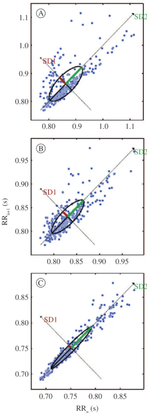
Figure 2 demonstrates the Poincaré plot during
the three study conditions, which revealed lower chaotic behavior for autonomic cardiac sinus node
modulation at 30% of MIP. Signiicant differences (p>0.05) in representative sympathetic heart rate
indices were not found between the three imposed loads.
Table 1. Demographic, anthropometric and clinical data.
n=25
Age (years) 66±4
Body mass (kg) 72±9
Height (cm) 167±5
BMI (kg/m2) 25.3±2 Clinical data
HR (bpm) 71±4
RR (cpm) 14±2
SBP (mmHg) 123±12
DBP (mmHg) 76±7
MIP (cmH2O) 97±30
MEP (cmH2O) 124±25
MIP expected (cmH2O) 105±5 MEP expected (cmH2O) 112±3
MIP % 92±38
MEP % 96±46
Values are expressed as the mean±SD. BMI=body mass index; HR=heart rate; RR=respiratory rate; SBP=systolic blood pressure; DBP=diastolic blood pressure; MIP=maximum inspiration pressure; MEP=maximum expiration pressure; MIP%=ratio MIP/MIP expected; MEP%=ratio MEP/MEPexpected.
Table 2. Heart rate variability at different inspiratory load levels during controlled respiratory breathing.
30% 60% 80% P value
Time domain HRV
Mean RR (ms) 781±23 789±19 768±24 0.24
Mean HR (1/min) 79±3 77±2 80±3 0.15
rMSSD (ms) 25±3 22±2 19±2* 0.02
SDNN (ms) 39±3 36±4 34±3 0.40
Frequency domain HRV
LF (ms2) 146±36 302±130 197±61 0.40
HF (ms2) 447±99 337±67 228±61* 0.004
LF/HF 1.2±0.4 1.2±0.3 1.8±0.7 0.07
Nonlinear HRV
SD1 18±2.3 16±1.6 13±1.7* 0.02
SD2 53±4.4 49±5 46±4 0.35
ApEn 0.91±0.02 0.97±0.01 0.93±0.02 0.08
Discussion
The purpose of this study was to investigate
acute HR modulation during different inspiratory resistive loading intensities (mild, moderate and high) in healthy elderly men. The main indings
of this study demonstrated that load increment
produced signiicantly lower values for representative
parasympathetic cardiac modulation indices in the
healthy elderly. Signiicantly reduced rMSSD, HF, and SD1 values were determined at 80% of MIP compared with 30% of MIP during inspiratory resistive loading. These indings suggest that lower
inspiratory resistive loading intensities promote
marked, positive improvement in parasympathetic
sinus node modulation.
Although inspiratory muscle training is the most
widely used method, cardiovascular adjustment during different inspiration intensities remains
unstudied. In a previous study on cardiovascular
adjustment during respiratory resistive loads in healthy subjects20, the authors analyzed HRV during
four different resistive loads that were applied
throughout the entire breathing cycle. In seven healthy subjects aged 19 to 55, resistive respiratory loads of 3.25 to 12.5 cmH2O were applied during an average Figure 1. HRV indices in (A) rMSSD, (B) LF – low frequency
and (C) HF - high frequency compared with spontaneous breathing (SB) and different maximal inspiratory load percentages. *p<0.05 compared with SB; # p<0.05 compared with 60 and 80%.
of 50 breaths and the HRV was analyzed. These authors observed a progressive increase in both LF and HF spectral components. Furthermore, the LF/ HF demonstrated a progressive load intensity increase that exceeded a sympathovagal balance of 1 at 8.25 and 12.5 cmH2O. These results by Calabrese et al.20
can only be partially compared with ours.
McConnell and Griffiths21 also evaluated the
inluence of different MIP percentages (50, 60, 70, 80 and 90%) in eight healthy males and observed that beginning at 60% MIP, HR and blood pressure increased. In addition, Sheel et al.22 demonstrated that HR increased during an MIP protocol, which
was time-dependent. These authors23 also observed that the diaphragm metaboreflex is intensively
activated during loaded breathing until task failure,
which induces sympathetic activation and peripheral vasoconstriction that may limit exercise performance.
In this context, important cardiovascular behavior
alterations have been attributed to local metabolic
changes during loaded breathing. Afferent receptors,
mechanical deformation, temperature elevation and vascular distension during muscular contractions may be stimulated by metabolic products24. Respiratory
muscle contraction causes changes in local metabolic homeostasis; consequently, myelinated group
III and unmyelinated IV afferent nerve ibers
trigger a cardiovascular adjustment with increased sympathetic activity to accompany the metabolic demand25. However, during an endurance protocol,
autonomic responses could indicate the best intensity at which to train inspiratory muscles because
of improved vagal activation. We demonstrated
that lower inspiratory resistive loading intensities promote parasympathetic modulation compared with
higher intensities. In agreement with these results,
Callegaro et al.26 observed that RMT at 60% of
MIP signiicantly attenuated the inspiratory muscle metaborelex.
One experimental study8 involving rats with
heart failure demonstrated improved cardiovascular
function after 6 weeks of RMT as assessed by
decreased left ventricular end-diastolic pressure, sympathetic tone, right ventricular hypertrophy and lung and hepatic congestion as well as increased vagal effects and arterial baroreceptor sensitivity.
N o b r e e t a l .2 7 o b s e r v e d t h a t E M G
(electromyography) activity in lower rib cage muscles increased during progressive respiratory workloads.
This may be directly related to high respiratory muscle recruitment because at intensities closer
to the MIP, the diaphragm and accessory muscles (scalene muscles, sternocleidomastoid, pectoralis
minor, external intercostals) increased intrathoracic
pressure and induced intense sympathetic activation.
Another plausible explanation for these results is
that because this intrathoracic pressure increase is generated at high intensities during each breathing cycle, it may reduce venous return to the right side of heart and act directly on the sinus node to reduce right atrial wall stretch28. Blood damming in the pulmonary
vascular bed that occurred because of the sudden intrathoracic pressure increase may also have reduced the blood volume that was sent to the left ventricle with a consequent instantaneous blood pressure
drop, leading to barorelex vagal withdrawal29,30.
Another explanation could be that the reduced vagal
modulation during the higher inspiratory resistive loading in this protocol was induced by the intense
effort that was required to sustain 80% of MIP
for the pre-determined time. These results can be
compared with those occurring in HRV in inspiratory
maneuvers at high volumes such as the respiratory
sinus arrhythmia accentuation maneuver (RSA-M). In contrast with the RSA-M (in which the inspired volume increased slowly over 5 seconds), the inspired volume increased rapidly at 80% of MIP because the inspiratory time was 2 s. Additionally, in the present study, the R-Ri was also collected differently than the RSA-M (which is standardized at 6 breaths per minute). Thus, even at higher exercise intensities and inspiratory volumes, the lung inlation-mediated
vagal response was reduced in relation to higher loads.
Finally, our results demonstrated that lower
inspiratory resistive loading intensities provided greater safety for the study population because it promoted a greater vagal response, which indicates
a powerful cardioprotective effect. Furthermore,
we emphasize the importance of our results for the
elderly population because the RMS is decreased
in these individuals. Thus, although our sample did
not present respiratory muscle weakness compared
to the reference values, we believe that future studies involving this population with inspiratory
muscle weakness will be worthwhile for greater understanding of the autonomic HR responses.
Some study limitations should be considered. First, control of tidal volume measurement during the
inspiratory control, which was not performed in this study, could have contributed to result consolidation
and interpretation. Second, barorelex and/or chemorelex were not evaluated, which could also
have contributed to the interpretation of our results.
In conclusion, our results suggest that lower inspiratory efforts produce higher HRV. These indings represent important clinical applications because low RMT intensities can produce greater parasympathetic HR modulation in this population.
Thus, we should choose the most appropriate load
for achieving the most beneicial autonomic effects,
which are associated with reduced cardiovascular event and morbidity incidence.
However, future studies are needed to conirm whether the RMT can enhance vagal modulation at low inspiratory muscle training intensity. Moreover, other studies are needed to conirm these indings
in other populations such as in chronic obstructive pulmonary disease and cardiac heart failure, where
respiratory muscle weakness is commonly present.
Acknowledgements
We acknowledge the UFSCar Cardiopulmonary Laboratory staff for their enthusiastic help. Special thanks are given to the subjects for their participation. Support for this work was provided by grants from Conselho Nacional de Desenvolvimento Cientíico e Tecnológico (CNPq) and Fundação de Amparo a Pesquisa do Estado de São Paulo (FAPESP) n° 2009/01842-0.
References
1. Westerdahl E, Lindmark B, Almgren SO, Tenling A.
Chest physiotherapy after coronary artery bypass graft surgery: a comparison of three different deep breathing
techniques. J Rehabil Med. 2001;33(2):79-84. http://
dx.doi.org/10.1080/165019701750098920
2. Westerdahl E, Lindmark B, Eriksson T, Hedenstierna
G, Tenling, A. The immediate effects of deep breathing
exercises on atelectasis and oxygenation after cardiac
surgery. Scand Cardiovasc J. 2003;37(6):363-7. http://
dx.doi.org/10.1080/14017430310014984
3. Stein R, Maia CP, Silveira AD, Chiappa GR, Myers J, Ribeiro JP. Inspiratory muscle strength as a determinant of
functional capacity early after artery bypass graft surgery.
Arch Phy Med Rehabil. 2009;90(10):1685-91. http://
dx.doi.org/10.1016/j.apmr.2009.05.010
4. Dall’ago P, Chiappa GR, Güths H, Stein R, Ribeiro JP. Inspiratory muscle training in patients with heart failure and inspiratory muscle weakness: a randomized trial. J Am Coll Cardiol. 2006;47(4):757-63. http://dx.doi.
org/10.1016/j.jacc.2005.09.052
5. Neder JA, Andreoni S, Lelario MC, Nery LE. References values for lung function tests. II. Maximal respiratory pressures and voluntary ventilation. Braz J Med Biol Res. 1999;32(6):719-27. http://dx.doi.org/10.1590/ S0100-879X1999000600007
6. Meyer FJ, Borst MM, Zugck C, Kirsche A, Schellberg D, Kubler, et al. Respiratory muscle dysfunction in
congestive heart falure: clinical correlation and prognostic
signiicante. Circulation. 2001;103(17):2153-8. http://
dx.doi.org/10.1161/01.CIR.103.17.2153
7. Ribeiro JP, Chiappa GR, Neder JA, Frankestein L. Respiratory muscle function and exercise intolerance in heart failure. Curr Heart Failure Rep. 2009;6(2):95-101. http://dx.doi.org/10.1007/s11897-009-0015-7
8. Jaenisch RB, Hentschke VS, Quagliotto E, Cavinato PR, Schmeing LA, Xavier LL, et al. Respiratory muscle
training improves hemodynamics, autonomic function, baroreceptor sensitivity, and respiratory mechanics in rats
with heart failure. J Appl Physiol. 2011;111(6):1664-70. http://dx.doi.org/10.1152/japplphysiol.01245.2010 9. Grossman P, Wilhelm FH, Spoerle M. Respiratory sinus
arrhythmia, cardiac vagal control and daily activity. Am J Physiol. 2004;287:H728-H34.
10. European Society of Cardiology, North American Society of Pacing Electrophysiology. Task force. Heart rate variability. Standards of measurement, physiological interpretation, and clinical use. Circulation. 1996;93:1043-65. http://dx.doi.org/10.1161/01.CIR.93.5.1043 11. Bainbridge FA. The inluence of venous illing upon the
rate of the heart. J Physiol. 1915;50:65-84.
12. Bernardi L, Porta C, Gabutti A, Spicuzza L, Sleight P. Modulatory effects of respiration. Auton Neurosci. 2001;90(1-2):47-56. http://dx.doi.org/10.1016/ S1566-0702(01)00267-3
13. Reis MS, Arena R, Deus AP, Simões RP, Catai AM, Borghi-Silva A. Deep Breathing and respiratory muscle weakness in COPD. Clinics. 2010;65(4):369-75. http://
dx.doi.org/10.1590/S1807-59322010000400004 14. Simões RP, Deus APL, Auad MA, Dionisio J, Mazzonetto
M, Borghi-Silva A. Maximal respiratory pressure in healthy 20 to 89 year-old sedentary individuals of central São Paulo State. Rev Bras Fisioter. 2010;14(1):60-7. http://
dx.doi.org/10.1590/S1413-35552010000100010 15. Parreira VF, Bueno CJ, França DC, Vieira DS, Pereira DR,
Britto RR. Breathing pattern and thoracoabdominal motion in healthy individuals: inluence of age and sex. Rev Bras Fisioter. 2010;14(5):411-6. http://dx.doi.org/10.1590/ S1413-35552010000500010
16. Simões LA, Dias JMD, Marinho KC, Pinto CLLR, Britto RR. Relationship between functional capacity assessed by walking test and respiratory and lower limb muscle function in community-dwelling elders. Rev Bras Fisioter. 2010;14(1):24-30. http://dx.doi.org/10.1590/ S1413-35552010000100005
17. American Thoracic Society, European Respiratory Society. ATS/ERS Statement of Respiratory Muscle Testing Guidelines. Am J Respir Crit Care Med. 2002;166(4):518-624. http://dx.doi.org/10.1164/rccm.166.4.518
18. Nunan D, Donovan G, Jakovljevic DG, Hodges LD, Sandercock GR, Brodie DA. Validity and reliability of short-term heart-rate variability from the Polar S810. Med Sci Sports Exerc. 2009;41(1):243-50. http://dx.doi.
19. Stein PK, Reddy A. Non-linear heart rate variability and risk stratiication in cardiovascular disease. Indian Pacing Electrophysiol J. 2005;5(3):210-20.
20. Calabrese P, Perrault H, Dinh TP, Eberhard A, Benchetrit G. Cardiorespiratory interactions during resistive load breathing. Am J Physiol Regul Integr Comp Physiol. 2000;279(6):R2208-13.
21. McConnell AK, Grifiths LA. Acute cardiorespiratory responses to inspiratory pressure threshold loading. Med Sci Sports Exerc. 2010;42(9):1696-703. http://dx.doi.
org/10.1249/MSS.0b013e3181d435cf
22. Sheel AW, Derchak PA, Morgan BJ, Pegelow DF, Jacques AJ, Dempsey JA. Fatiguing inspiratory muscle work causes relex reduction in resting leg blood low in humans. J Physiol 2001;537(Pt 1):277-89. http://dx.doi.
org/10.1111/j.1469-7793.2001.0277k.x
23. Sheel AW, Derchak PA, Pegelow DF, Dempsey JA. Threshold effects of respiratory muscle work on limb vascular resistance. Am J Physiol Heart Circ Physiol. 2002;282(5):H1732-8.
24. Haouzi P, Chenuel B, Huszczuk A. Sensing vascular distension in skeletal muscle by slow conducting afferent
fibers: neurophysiological basis and implication for
respiratory control. J Appl Physiol. 2004:96:407-18. http://
dx.doi.org/10.1152/japplphysiol.00597.2003
25. Roseguini BT, Alves CN, Chiappa GR, Stein R, Knorst
MM, Ribeiro JP. Attenuation of muscle metaborrelex in chronic obstrutive pulmonary disease. Med Sci Sports Exerc. 2008;40(1):9-14.
26. Callegaro CC, Ribeiro JP, Tan CO, Taylor JA. Attenuated inspiratory muscle metaborelex in endurance-trained individuals. Respir Physiol Neurobiol. 2011;177(1):24-9. http://dx.doi.org/10.1016/j.resp.2011.03.001
27. Nobre ME, Lopes F, Cordeiro L, Marinho PE, Silva TN, Amorim C, et al. Inspiratory muscle endurance testing:
pulmonary ventilation and electromyographic analysis.
Respir Physiol Neurobiol. 2007;155(1):41-8. http://dx.doi.
org/10.1016/j.resp.2006.04.005
28. Tulppo MP, Makikallio TH, Takala TE, Seppanen T, Huikuri HV. Quantitative beat-to-beat analysis of heart rate dynamics during exercise. Am J Physiol Heart Circ Physiol. 1996;271:H244-52.
29. Tulppo MP, Makikallio TH, Takala TE, Seppanen T, Huikuri HV. Vagal modulation of heart rate during exercise: effects of age and physical itness. Am J Physiol. 1998;274(2):H424-9.
30. Hirsch JA, Bishop B. Respiratory sinus arrhythmia in humans: how breathing patterns modulates heart rate. Am J Physiol. 1981;10:620-9.
Correspondence
Audrey Borghi-Silva
Universidade Federal de São Carlos Departamento de Fisioterapia
Rodovia Washington Luis, Km 235, Monjolinho CEP 13565-905, São Carlos, SP, Brazil