ORIGIN
AL RESEAR
CH
Corresponding address: Mauricio Jamami – Laboratório de Espirometria e Fisioterapia Respiratória (DFisio). Rodovia Washington Luiz, Km 235 – São Carlos (SP), Brazil – Zip Code: 13565-905 – Email: [email protected] – Phone number: (+55) (16) 3351-8343 – Financing source: Scientiic Research Scholarship by CNPq – Conlict of interest: Nothing to declare – Presentation: Nov. 2016 – Accepted for publication: Mar. 2017 – Approved by Ethics Committee: Opinion no. 042/2011.
Study developed in the Laboratory of Spirometry and Respiratory Physical Therapy of Universidade Federal de São Carlos (UFSCar).
1Graduate in Physical Therapy by Universidade Federal de São Carlos (UFSCar), São Carlos, SP, Brazil.
2Doctor in Physical Therapy by Universidade Federal de São Carlos (UFSCar); Professor of Universidade do Sagrado Coração, Bauru,
SP, Brazil.
3Masters in Physical Therapy by Universidade Federal de São Carlos (UFSCar), São Carlos, SP, Brazil.
4Doctor in Physiological Sciences; Associate Professor of Universidade Federal de São Carlos (UFSCar), São Carlos, SP, Brazil. ABSTRACT | Manovacuometry is a simple, fast, and
non-invasive test, with maximal inspiratory pressure (MIP) and maximal expiratory pressure (MEP) obtained to assist respiratory muscle assessment. Currently, there is a wide variety of models and brands of manovacuometers with diferent trachea diameters and lengths. However, the interference of these models in the measurements obtained by these equipments needs to be investigated. Thus, this study mainly aimed to verify the inluence of tracheal length on maximal respiratory pressures (MRP), obtained by an analog manovacuometer, in healthy individuals. Our secondary objective was to verify the correlation between measurements. Fifty individuals, aged 18 to 30, of both sexes, were evaluated by spirometry and manovacuometry. MIP and MEP were performed using tracheas with same internal diameter (0.5 cm) and 30 cm, 60 cm, and 90 cm length. Signiicantly lower MIP values were observed when comparing a 90 cm trachea to 30 and 60 cm tracheas (Friedman’s ANOVA test and Wilcoxon test with Bonferroni adjustment). Tracheas with 30, 60, and 90 cm length and same diameter did not afect MIP and MEP values, except the 90 cm trachea for MIP values, which may interfere in the physical therapy clinical practice. Further studies are required to analyze the need for standardizing the trachea length used in manovacuometers.
Keywords | Respiratory Muscles; Healthy Volunteers; Physical Therapy Modalities.
9
RESUMO | A manovacuometria é um teste simples, rápido e não invasivo por meio do qual a pressão inspiratória máxima (PImáx) e a pressão expiratória máxima (PEmáx) são obtidas, a im de auxiliar na avaliação muscular respiratória. Atualmente, há grande variedade de modelos e marcas de manovacuômetros, com diferentes diâmetros e comprimentos de traqueias, no entanto, a interferência desses modelos nas medidas obtidas por esses equipamentos necessita de investigação. Desta forma, o objetivo primário deste estudo foi veriicar a inluência do comprimento de traqueias nas pressões respiratórias máximas, obtidas por meio de manovacuômetro analógico, em indivíduos saudáveis e, secundariamente, se há correlação entre as medidas. Foram avaliados 50 indivíduos, de 18 a 30 anos, de ambos os sexos, por meio da espirometria e manovacuometria. As PImáx e PEmáx foram realizadas com uso de traqueias de mesmo diâmetro interno (0,5 cm) e comprimentos de 30, 60 e 90 cm. Foram observados valores signiicativamente menores de PImáx obtidos com a traqueia de comprimento de 90 cm comparados às PImáx obtidas com as traqueias de 30 e 60 cm (teste de Friedman’s ANOVA com teste de Wilcoxon com ajuste de Bonferroni). As traqueias de 30, 60 e 90 cm de comprimento e mesmo diâmetro não inluenciaram os valores de PEmáx e PImáx, exceto a traqueia de 90 cm para os valores de PImáx, o que pode interferir na prática clínica isioterapêutica. Novos estudos são necessários
Manovacuometry performed by diferent length
tracheas
Manovacuometria realizada por meio de traqueias de diferentes comprimentos
La realización de la manovacuometría con tráqueas de distintas longitudes
Roberta Magalhães Guedes dos Santos1, Bruna Varanda Pessoa-Santos2, Ivanize Mariana Masselli dos
para analisar a necessidade de padronização do comprimento da traqueia utilizada em manovacuômetros.
Descritores | Músculos Respiratórios; Voluntários Saudáveis; Modalidades de Fisioterapia.
RESUMEN | La manovacuometría es una prueba sencilla, rápida y no invasiva por la cual se obtienen la presión inspiratoria máxima (PImax) y la presión espiratoria máxima (PEmax), con el objetivo de ayudar en el examen muscular respiratorio. Hoy día se encuentran una gran variedad de modelos y marcas de manovacuometros, con diferentes diámetros y longitudes de las tráqueas, pero hacen falta estudios sobre la interferencia de estos modelos en las mediciones por este instrumento. En este texto se propone examinar en sujetos sanos, en primer lugar, la inluencia en la longitud de las tráqueas en las presiones respiratorias máximas, obtenidas por manovacuometros analógicos, y en segundo lugar comprobar la existencia de
correlación entre las mediciones. Se evaluaron a cincuenta sujetos entre 18 y 30 años de edad, tanto varones como mujeres, empleando la espirometría y la manovacuometría. Se midió la PImax y la PEmax empleando tráqueas de mismo diámetro interno (0,5 cm) y con longitudes de 30, 60 e 90 cm. Se observaron valores signiicativamente menores de PImax con la tráquea de longitud de 90 cm en comparación con las PImax con las tráqueas de 30 y 60 cm (prueba de Friedman’s ANOVA, la de
Wilcoxon con ajustes de Bonferroni). Las tráqueas de 30, 60 y 90 cm de longitud y mismo diámetro no inluyeron en los valores de la PEmax y de la PImax, con excepción de la tráquea de 90 cm en los valores de la PImax, lo que puede interferir la práctica clínica isioterapéutica. Se necesitan más estudios para evaluar la necesidad de estándares de la longitud de tráqueas empleadas en manovacuometros.
Palabras clave | Músculos Respiratorios; Voluntarios Sanos; Modalidades de Fisioterapia.
INTRODUCTION
Manovacuometry, also known as maximal respiratory pressures (MRP), consists of measuring maximum static respiratory pressures by a classic and reliable equipment, named manovacuometer1-4. his
is a simple, fast, non-invasive, volunteer, and efort-dependent test, in which the maximal inspiratory pressure (MIP) and the maximal expiratory pressure (MEP) are obtained5,6. hese are indexes
of inspiratory and expiratory muscle force and their respective values represent the force generated by the set of inspiratory and expiratory muscles, obtained at mouth level3,5,6.
Its applicability is large and aims to identify clinical changes, such as muscle weakness7 and ability
to cough and expectorate (relected by the MEP). hus, it helps the diagnosis of neuromuscular and progressive diseases, the prescription of respiratory muscle training programs3,7,8, the weaning from
mechanical ventilation9, and the assessment of
responsiveness to interventions2,5,6,10.
MIP and MEP are generated during maximum inspiration and expiration against an occluded airway11, respectively, and the values obtained depend
on the elastic retraction strength of the pulmonary system, on the respiratory muscle itself, on the instructions provided, and on the collaboration of
the individual to perform the maneuver11. herefore,
procedure standardization is necessary3,11,12. Studies
have investigated other variables able to afect the values obtained, such as, for example, types of equipment, buccal pieces8,10, tracheas, manometers,
air-escape oriice, use of nose clip, volunteer’s posture when performing the tests, rest time between repetitions and between tests, maximum pressure deinition, and lung (that in which the maneuver is carried out) volume determination4-6.
In their studies, Onaga et al.8, Koulouris et al.13, and
Gibson10 concluded that diferent buccal types strongly
inluence measures of respiratory muscle pressures. Currently, there is a wide range of models and brands of manovacuometers with diferent diameters and lengths of tracheas. However, the inluence of these models on the measures obtained by such equipment is not clear.
he existing standardization refers to the presence of air-escape oriice (1-2 mm diameter) and a maximum of eight eforts for each test (with at least three acceptable and two reproducible)5-6. herefore,
given the small number of studies on the topic, it is important to compare the data obtained by diferent lengths of tracheas, which justiies our position of assisting the standardization method of such measures.
pressures, obtained by analog manovacuometers, in healthy individuals. Secondarily, it aimed to verify the correlation between the maximal respiratory pressure (MRP) measures obtained with diferent lengths of tracheas.
METHODOLOGY
Sample
his study’s sample consisted of 50 healthy individuals, of both sexes, aged 18 to 30, with body mass index between 18 and 29.9 kg/m²14, and
who belonged to the community of São Carlos, SP, Brazil and surroundings. Individuals in the following conditions were excluded: respiratory and neurological diseases and/or temporomandibular joint syndrome; use of any type of medicines that could interfere and change MRP values; smokers and ex-smokers.
To determine the sample size, the previous study of Onaga et al.8 was used, considering the MEP
variable as primary outcome. Calculation was carried out by the GPower software, version 3.1, adopting 95% conidence level and 80% study power. A number of 38 individuals was suggested to detect a 0.42 efect size. However, 50 individuals were included in this research.
his study was approved by the Research Ethics Committee of Universidade Federal de São Carlos (UFSCar) (protocol number 042/2011). All participants were informed about the experiment characteristics and signed the Informed Consent Form.
Experimental procedure
Individuals who agreed to participate in the study illed out a standardized assessment form containing personal data. All of them underwent anamnesis and physical examination that collected anthropometric data, medications used, and smoking habit information. he short version of the International Physical Activity Questionnaire (IPAQ) was applied to evaluate physical activity level15.
Height and body mass measures were obtained using biometric scale (Welmy®, 110FF model, São Paulo, SP, Brazil), and then the body mass index (BMI) was calculated. Subjects were submitted to spirometry
and manovacuometry tests. Data collection was carried out in a single day by the same evaluator.
Spirometry: A portable spirometer (NDD
EasyOneTM, Zurich, Switzerland) was used,
following the standards of the American horacic Society/European Respiratory Society (ATS/ ERS)16. he values obtained were compared to those
predicted by Knudson et al.17.
Respiratory muscle pressures: they were
measured with the subject on standing position using a nose clip, by an analog manovacuometer (Ger-Ar, São Paulo, Brazil) calibrated in cmH2O, with a –300 to +300 cmH2O operational limit, scale ranging each 10 cmH2O, equipped with a buccal adapter with an approximate 2 mm diameter oriice, aiming to prevent contraction of facial muscles10,18-22. All
individuals received standardized verbal stimuli23.
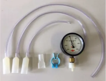
Measures were registered using tracheas with the same internal diameter (0.5 cm) and 30, 60, and 90 cm lengths (Ger-Ar, São Paulo, Brazil). hese trachea lengths were determined according to the manovacuometer models that are commonly available for sale on the market. A rectangle type buccal device was used (Ger-Ar, São Paulo, Brazil), since it is considered more anatomical, allowing less air escape during the execution of maneuvers8.
Figure 1. Analog manovacuometer (Ger-Ar) and different tracheas’ lengths with respective buccal device adapter and rectangular buccal device used
MIP was obtained by a maximal inspiratory efort maneuver after a maximal expiration, close to the residual volume (RV)2,21. MEP was obtained by a
of MIP and MEP maneuvers and tracheas lengths (30 cm, 60 cm, and 90 cm) to be used were randomly determined through lots, for each individual.
Maneuvers were performed at least three times and, at most, ive times, in case there was more than 10% variation between the values obtained21,
and the efort was held by at least three seconds2,24.
he following intervals were adopted: 15 seconds between measurements, 30 seconds between maneuvers, and one minute between change of tracheas8. For statistical analysis, maximum values
were considered. he predicted values of MIP and MEP were calculated according to Neder et al.25.
Statistical Analysis
Data of this study were analyzed by the Statistical Package for the Social Sciences (SPSS) software for Windows, version 20.0. Data normality was veriied using the Shapiro–Wilk test. For sample characterization, descriptive statistics was expressed as median (interquartile range). For analysis of MIP and MEP values, Friedman’s ANOVA test and Wilcoxon test with Bonferroni adjustment were used. he correlation between the values obtained with diferent trachea lengths for MIP and MEP values was obtained by Spearman’s correlation coeicient. he signiicance level adopted was 5%.
RESULTS
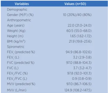
Table 1 shows demographic, anthropometric, and spirometric characteristics of the individuals studied. Regarding the level of physical activity of individuals, veriied by the IPAQ14, 2% of them
were classiied as very active; 42% as active; 50% as irregularly active (24% irregularly active A and 26% irregularly active B), and 6% as sedentary.
Table 1. Demographic, anthropometric, and spirometric variables of the volunteers
Variables Values (n=50)
Demographic
Gender (M/F) (%) 10 (20%)/40 (80%) Anthropometric
Age (years) 22.0 (21.0–24.0) Weight (Kg) 60.5 (55.0–68.0) Height (m) 1.65 (1.62–1.72) BMI (kg/m²) 21.9 (19.8–23.6) Spirometric
FEV1 (predicted %) 94.9 (86.8–102.6)
FEV1 (L) 3.2 (2.9–3.8)
FVC (predicted %) 97.0 (88.8–104.3)
FVC (L) 3.7 (3.2–4.7)
FEV1/FVC (%) 97.8 (92.0–101.3) FEV1/FVC (L) 0.9 (0.8–0.9) MVV (predicted %) 97.0 (86.7–106.5)
MVV (L/min) 124.9 (108.2–147.5)
The data were expressed as median (interquartile range); M: Male; F: Female; BMI: body mass index; FEV1: forced expiratory volume in one second; FVC: forced vital capacity; FEV1/
FVC: relation FEV1/FVC; MVV: maximum voluntary ventilation.
Table 2 presents MIP and MEP values obtained by tracheas with diferent lengths. No statistically signiicant diferences were found among the three types of trachea lengths for the MEP. However, signiicantly lower MIP values were obtained with a 90 cm trachea length compared to the MIP values obtained with 30 cm and 60 cm tracheas.
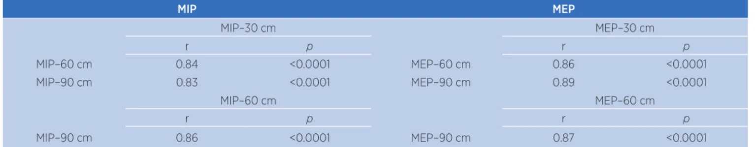
Strong positive and statistically signiicant correlations were observed between MIP values with tracheas of all lengths (30 cm, 60 cm, and 90 cm). he same occurred with MEP values, as shown in Table 3.
Table 2. MIP and MEP values with diferent length tracheas
Predicted Values Obtained Value % Predicted Value Obtained Value % Predicted
Value Obtained Value
% Predicted Value
MIP MIP–30 cm MIP–30 cm MIP–60 cm MIP–60 cm MIP–90 cm MIP–90 cm p-value* 100.1 (98.6–100.7) 100 (90–110) 96.3 (83.9–105.3) 100 (85–110) 91.0 (80.2–109.0) 90 (80–110) § ¥ 88.9 (79.1–101.4) 0.0001
MEP MEP–30 cm MEP–30 cm MEP–60 cm MEP–60 cm MEP–90 cm MEP–90 cm
102.8 (101.0–103.6) 110 (95–125) 97.3 (84.3–117.3) 110 (90–125) 100.0 (82.6–117.1) 110 (90–120) 97.3 (83.7–116.3) 0.076
Data expressed as median (interquartile range).
among individuals, by standardizing verbal encouragement and body positioning. he evaluator remained the same during measurements, in such a way that, besides the physical characteristics of each individual, no other factor could afect the acquisition of MIP and MEP values.
In this study, the tracheas’ length was the only factor relevant to the equipment that could afect the inal pressure value obtained, since we assured diameter and roughness of tracheas were the same. hus, considering all these factors, we found that the pressure obtained sufered no signiicant inluence from the tracheas’ lengths, as there was no signiicant diference between MEP values, with strong association between the diferent lengths of tracheas. However, we found that the 90 cm length trachea resulted in lower MIP values when compared to the values obtained with the 30 cm and 60 cm tracheas, suggesting that, from that length, a greater inspiratory efort is required to overcome the resistance of the circuit, which can compromise a reliable assessment of individuals. Even noticing the lower MIP values obtained with the 90 cm trachea, we considered the association between values strong.
Our sample was predominantly formed by females, a factor that may have afected our results and constitute a limitation of the study. Another factor considered as a limitation is the impossibility of identifying the measurement time and the non-visualization of MIP and MEP curve of measures, constituting a disadvantage of the analog manovacuometer. In addition, the 15-second interval established between measurements, although used in a previous study, is diferent from the most commonly used in the literature, which is close to one minute27,28.
DISCUSSION
Our main result is that we found no signiicant diferences on MEP values between 30, 60, and 90 cm trachea lengths, with positive correlation between them. However, we observed the 30 cm and 60 cm tracheas provided higher MIP values than the 90 cm trachea.
In this study, an analogic manovacuometer calibrated in cmH2O was used. his choice was made because this type is the most used in clinical practice.
Regarding the buccal device, we have chosen the rectangular format, since, according to Gibson10, this type
has great inluence on the measurement of respiratory pressure values. For Onaga et al.8, the rectangular buccal
device guarantees a minor air escape for MEP measures. However, Montemezzo et al.4 mostly used the tubular
type buccal device, while Souza21 considers the diver
type the most indicated one.
In the analysis of some aspects of luid mechanics, it is possible to better understand the results of this research. According to Munson et al.26, the inal
pressure is inluenced by three main factors: luid characteristics (speciic mass and viscosity); tube characteristics (diameter, length, and roughness); and user performance (speed and pressure with which the air is propelled at the tube entrance). However, the diferent lengths of tracheas established in this study were not suicient to provide diferences in the MEP assessment. Nevertheless, this can be veriied in the MIP values obtained with the 90 cm trachea, which are lower when compared to the 30 cm and 60 cm tracheas.
hus, once two of these major factors are guaranteed, such as luid characteristics and user performance, the only variable factor relates to the tube characteristics. We were careful to minimize performance diferences
Table 3. MIP and MEP values with diferent length tracheas
MIP MEP
MIP–30 cm MEP–30 cm
r p r p
MIP–60 cm 0.84 <0.0001 MEP–60 cm 0.86 <0.0001
MIP–90 cm 0.83 <0.0001 MEP–90 cm 0.89 <0.0001
MIP–60 cm MEP–60 cm
r p r p
MIP–90 cm 0.86 <0.0001 MEP–90 cm 0.87 <0.0001
CONCLUSION
his study showed that 30, 60, and 90 cm tracheas with same diameter did not afect MIP and MEP values, except the 90 cm trachea for MIP values, which may interfere in the physical therapy clinical practice.
REFERENCES
1. Cook CD, Mead J, Orzalesi MM. Static volume-pressure characteristics of the respiratory system during maximal eforts. J Appl Physiol. 1964;19:1016-22.
2. Black LF, Hyatt RE. Maximal respiratory pressures: normal values and relationship to age and sex. Am Rev Respir Dis. 1969;99(5):696-702.
3. Parreira VF, França DC, Zampa CC, Fonseca MM, Tomich GM, Britto RR. Pressões respiratórias máximas: valores encontrados e preditos em indivíduos saudáveis. Rev Bras Fisioter. 2007;5(11):361-8.
4. Montemezzo D, Velloso M, Britto RR, Parreira VF. Pressões respiratórias máximas: equipamentos e procedimentos usados por isioterapeutas brasileiros. Fisioter Pesqui. 2010;17(2):147-52.
5. Montemezzo D, Lages AC, Tierra-Criollo CJ, Veloso M, Britto RR, Parreira VF. Relationship between maximum mean pressure and peak pressure obtained by digital manometer during maximal respiratory pressure. J Resp Cardiov Phy Ther. 2012;1(1):9 15.
6. Montemezzo D, Vieira DSR, Tierra-Criollo CJ, Britto RR, Velloso M, Parreira VF. Inluence of 4 interfaces in the assessment of maximal respiratory pressures. Respir Care. 2012;57(3):392-8.
7. Decramer M, Scano G. Assessment of respiratory muscle function. Eur Respir J. 1994;7:1744-5.
8. Onaga FI, Jamami M, Ruas G, Di Lorenzo VAP, Jamami LK. Inluência de diferentes tipos de bocais e diâmetros de traqueias na manovacuometria. Fisioter Mov. 2010;23(2):211-9. 9. Bethlem N. Pneumologia. 4ª ed. São Paulo: Atheneu; 2002. 10. Gibson GJ. Measurement of respiratory muscle strength.
Respir Med. 1995;89:529-35.
11. Evans JA, Whitelaw WA. The assessment of maximal respiratory mouth pressures in adults. Respir Care. 2009;54(10):1348-59.
12. Brunetto AF, Fregonezi GAF, Paulin E. Comparação das medidas de pressões respiratórias máximas (PImáx, PEmáx) aferidas através de Manuvacuômetro e Sistema de
Aquisição de dados (SAqDados). Rev Bras Ativ Fís Saúde. 2000;5(1):30-7.
13. Koulouris N, Mulvey DA, Laroche CM, Green M, Moxham J. Comparison of two diferent mouthpieces for the measurement of PImax and PEmax in normal and weak subjects. Eur Respir J. 1988;1:863-7.
14. Coutinho WF. Consenso latino-americano de obesidade. Arq Bras Endocrinol Metab. 1999;43(1):21-67.
15. Matsudo S, Araújo T, Matsudo V, Andrade D, Andrade E, Oliveira LC, et al. Questionário internacional de atividade física (IPAQ). Estudo de validade e reprodutibilidade no Brasil. Rev Bras Ativ Fís Saúde. 2001;6(2):5-18.
16. Miller MR, Hankinson J, Brusasco V, Burgos F, Casaburi R, Coates A, et al. Standardisation of spirometry. Eur Respir J. 2005;26(2):319-38.
17. Knudson RJ, Lebowitz MD, Holberg CJ, Burrows B. Changes in the normal maximal expiratory low-volume curve with growth and aging. Am Rev Respir Dis. 1983;127:725-34. 18. Camelo JS, Terra Filho J, Manço JC. Pressões respiratórias
máximas em adultos normais. J Pneumol 1985;11(4):181-4.
19. Green M, Road J, Sieck GC, Similowski T. ATS/ERS Statement on Respiratory Muscle Testing. Am J Respir Crit Care Med. 2002;166(4):518-624.
20. Sobush DC, Dunning M. Assessing maximal static ventilatory muscle pressures using the bugle dynamometer. Suggestion from the ield. Phys Ther. 1984;64(11):1689-90.
21. Souza RB. Pressões respiratórias estáticas máximas. J Pneumol. 2002;28(Suppl 3):S155-65.
22. Badr C, Elkins MR, Ellis ER. The efect of body position on maximal expiratory pressure and low. Aust J Physiother. 2002;48(2):95-102.
23. Hautmann H, Hefele S, Schotten K, Huber RM. Maximal inspiratory mouth pressure (PIMAX) in healthy subjects: what is the lower limit of normal? Respir Med. 2000;94:689-93. 24. Neder JA, Andreoni S, Castelo-Filho A, Nery LE. Reference
values for lung function tests. I. Static volumes. Braz J Med Biol Res. 1999;32(6):703-17.
25. Neder JA, Andreoni S, Lerario MC, Nery LE. Reference values for lung function tests. II. Maximal respiratory pressures and voluntary ventilation. Braz J Med Biol Res. 1999;32(6):719-27. 26. Munson BR, Young DF, Okiishi TH. Fundamentos da Mecânica
dos Fluidos. 4ª ed. São Paulo: Edgar Blücher, 2004.
27. Aldrich T, Spiro P. Maximal inspiratory pressure: does reproducibility indicate full efort? Thorax. 1995;50:40-3. 28. Rochester DF. Tests of respiratory muscle function. Clin Chest