Review Article
Coccidioidomycosis and Histoplasmosis in Equines: An
Overview to Support the Accurate Diagnosis
Raimunda Sâmia Nogueira Brilhante
a,*, Paula Vago Bittencourt
b, Rita Amanda Chaves Lima
a,
Débora Castelo-Branco
a, Jonathas Sales Oliveira
a, Adriana Pinheiro
b, Rossana Cordeiro
a,
Zoilo Pires Camargo
c, José Júlio Costa Sidrim
a, Marcos Fábio Gadelha Rocha
a,baDepartment of Pathology and Legal Medicine, School of Medicine, Specialized Medical Mycology Center, Postgraduate Program in Medical Microbiology, Federal University of Ceará, Fortaleza, Ceará, Brazil
bSchool of Veterinary, Postgraduate Program in Veterinary Science, State University of Ceará, Fortaleza, Ceará, Brazil cDepartment of Microbiology, Immunology and Parasitology, São Paulo Federal University, São Paulo, Brazil
a r t i c l e
i n f o
Article history:
Received 15 September 2015
Received in revised form 9 February 2016 Accepted 9 February 2016
Available online 21 February 2016
Keywords:
Coccidioidomycosis Histoplasmosis Equines Epidemiology
Pathogenesis and diagnosis
a b s t r a c t
Fungal infections of the respiratory tract of horses are not as frequent as those of bacterial and viral origin, often leading to worsening of clinical conditions due to misdiagnosis and incorrect treatment. Coccidioidomycosis and histoplasmosis are systemic mycoses caused by the dimorphic fungiCoccidioidesspp. andHistoplasma capsulatum, respectively, which affect humans and a variety of other animals, including equines. These systemic mycoses of chronic and progressive nature can exhibit clinical manifestations similar to other microbial infections. Thus, this article broadly discusses the epidemiology, etiology, viru-lence, pathogenesis, clinical presentation, treatment, and diagnostic strategies of coccidi-oidomycosis and histoplasmosis, to support accurate diagnosis.
Ó2016 Elsevier Inc. All rights reserved.
1. Introduction
Coccidioidomycosis and histoplasmosis, respectively caused by Coccidioidesspp. and Histoplasma capsulatum, share some similarities: both etiologic agents are sapro-phytic dimorphic fungi that can infect humans and several other mammal species, including horses, mainly through the inhalation of fungal propagules that are present in the environment, and both diseases have diverse clinical presentations[1–3]. Throughout the last decades, several researches focusing on novel diagnostic strategies for human coccidioidomycosis and histoplasmosis have been
published, which have contributed for better understand-ing these diseases in human hosts. However, literature on equine coccidioidomycosis and histoplasmosis is still scarce, as it is mainly limited to a few case reports. Curi-ously, in some regions where these mycoses seem to be endemic, according to the notification of human cases, the diagnostic resources in veterinary practice are still insuffi -cient. In fact, the broad spectrum of clinical presentations of coccidioidomycosis and histoplasmosis in horses can lead to misdiagnosis. Besides the difficulty of establishing the clinical diagnosis of systemic mycoses, these diseases are not of compulsory notification[4], which contributes for the little epidemiologic data on coccidioidomycosis and histoplasmosis in horses. Furthermore, it is important to highlight the technical limitations associated with the manipulation of the culture of these fungal pathogens, which are restricted to biosafety level 3 laboratories[5]. All these issues justify the development of this work that *Corresponding author at: Raimunda Sâmia Nogueira Brilhante,
Department of Pathology and Legal Medicine, Federal University of Ceará, School of Medicine, Specialized Medical Mycology Center, Postgraduate Program in Medical Microbiology, 60430275 Fortaleza, Brazil.
E-mail address:[email protected](R.S.N. Brilhante).
Contents lists available atScienceDirect
Journal of Equine Veterinary Science
j o u r n a l h o m e p a g e : w w w . j - e v s . c o m
0737-0806/$–see front matterÓ2016 Elsevier Inc. All rights reserved.
http://dx.doi.org/10.1016/j.jevs.2016.02.230
makes a parallel approach of these mycoses in humans and horses, mainly considering the great number of scientific papers on human medical literature addressing this theme, but at the same time highlighting peculiar aspects of these mycoses in horses. Initially, taxonomic, morphologic, and eco-epidemiologic aspects ofCoccidioidesspp. andH. cap-sulatumvar.capsulatumwere quickly presented, followed by the discussion of the virulence and pathogenesis of these fungal pathogens and a description of the clinical presentations and treatment of coccidioidomycosis and histoplasmosis. Finally, different strategies for the diag-nosis of these mycoses are thoroughly discussed and pre-sented in this article.
2. Taxonomy, Morphology, and Eco-epidemiologic Aspects ofCoccidioidesspp. andHistoplasmaspp.
The generaCoccidioidesandHistoplasmaare classified in the Onygenales order, which is contained in the Euro-tiomycetes class, in turn included in the Ascomycota phylum, belonging to the Fungi kingdom[6].Coccidioides immitisandCoccidioides posadasiiare the only species in the genusCoccidioides. Thefirst species is native and restricted to San Joaquin Valley, California, United States, whereas the second presents larger geographic distribution, occurring in other states in the United States, and other countries, such as Mexico and Central and South America[7,8]. This genus belongs to the Onygenacea family[9].
In turn, theHistoplasmagenus is part of the Ajellomy-cetaceae family and presents the speciesHistoplasma
cap-sulatum, whose teleomorph is named Ajellomyces
capsulatus[6]. This species has three varieties:H. capsu-latumvar.capsulatum, which is pathogenic to humans and a variety of mammals, including equines causing classic histoplasmosis [10–13]; H. capsulatum var. duboisii, restricted to Central Africa, which causes African histo-plasmosis, mainly in humans; andH. capsulatumvar.
far-ciminosum, endemic to western, northern, and
northeastern countries in Africa and Asia, including India, Pakistan, and Japan, causing epizootic lymphangitis in horses[2,14].
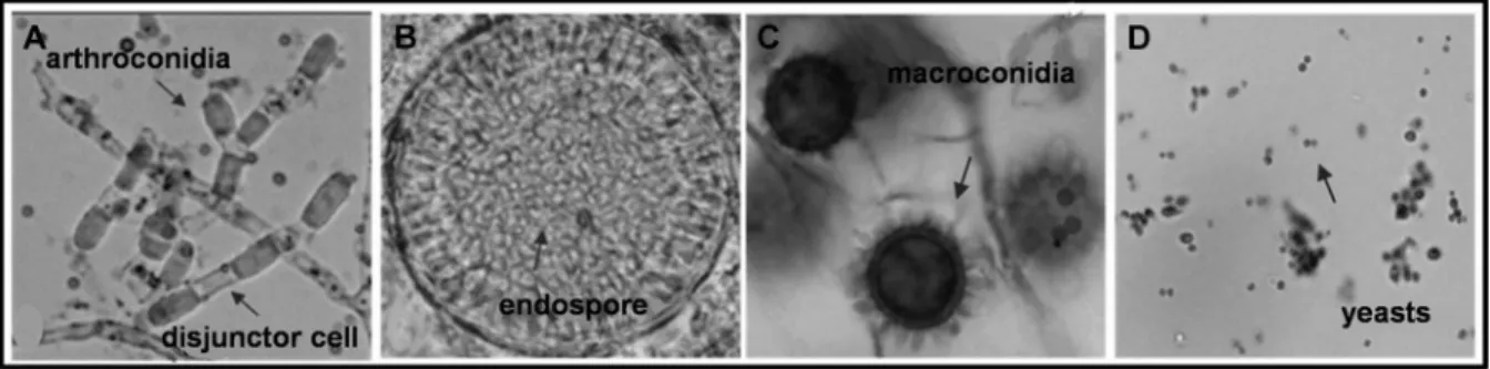
Coccidioides spp. have a mycelium formed by hyaline
and septate hyphae that originate arthroconidia, which are asexual reproduction structures, measuring 2–4
m
m by 3–6
m
m, intercalated with sterile cells devoid of cytoplasmicmaterial known as disjunctor cells (Fig. 1A). When reaching maturity, the infective arthroconidia are released, and the remnants of the disjunctor cells are seen at the end of in-dividual arthroconidia. This characteristic is responsible for their easy aerial propagation. The yeast form is named spherule, which are large rounded structures with thick walls that measure 20–200
m
m in diameter. Each spheruleproduces a large number of small endospores inside, measuring 2–4
m
m in diameter (Fig. 1B)[15].Thefilamentous form ofH. capsulatumshows a myce-lium composed of hyaline, septate, and ramified hyphae, with two types of conidia. Thefirst, microconidia, are oval structures with smooth walls and a diameter of about 2– 4
m
m, which are easily dispersed in the air due to theirsmall size and are considered the main infective propagules of this fungus [16]. The second type, macroconidia, also called tuberculate macroconidia, are round large struc-tures, measuring 8–15
m
m, with a rough wall covered withdistinctive projections (Fig. 1C) [16–18]. The yeast form presents small oval uninucleate cells, measuring about 3– 5
m
m in diameter (Fig. 1D)[17,18].Coccidioidesspp. are geophilic fungi that develop in soils with high salinity and alkaline pH, usually found at a depth of 10–50 cm[7]. They are associated with arid and semi-arid regions, which have an average temperature above 30C, recurrent droughts, and poor and sparse xerophytic vegetation [19]. The survival of Coccidioides species is substantially reduced by competition with other microor-ganisms in the soil[15]. Several studies have confirmed the importance of armadillo burrows in the ecology of Cocci-dioidesspp. especially in Northeastern Brazil[20,21]. There are also reported cases of coccidioidomycosis associated with contact with rodent burrows[22].
The saprophytic phase ofH. capsulatumis also geophilic, but it has been associated with soils rich in nitrogen com-pounds, with acidic pH and high moisture, which are characteristics found in soils enriched with the feces of birds and bats[23,24]. Thus, places like caves, hollow trees, chicken pens, and abandoned houses and parks are likely sites of colonization by this fungus[25]. Bats are suscepti-ble to infection caused by Histoplasma capsulatum var. capsulatumand actively participate in the epidemiology of histoplasmosis. By their migration, these animals can disseminate the pathogen from one location to another, through the elimination of viable forms of the fungus in
Fig. 1.Micromorphologic aspect ofCoccidioides spp.(A and B) andH. capsulatum(C and D). (A) Filamentous phase, showing arthroconidia and disjunctor cells; (B) Yeast phase, demonstrating a mature spherule with numerous endospores; (C) Filamentous phase, showing tuberculate macroconidia; (D) Yeast phase, showing oval and rounded yeasts.
guano[26,27]. Birds are rarely affected, probably because of their high body temperature, around 41C, but it is believed that they carry the organism on their wings, feathers, and beak[28,29]. The epizootic lymphangitis, disease caused by Histoplasma capsulatum var. farciminosum, rarely affect humans, cattle, and camels; however, its occurrence pre-dominates in horses, donkeys, and mules[30].
Considering the reports of human cases and environ-mental isolation of both fungal agents, it is estimated that the prevalence of coccidioidomycosis and classic histo-plasmosis is high in the Americas (Fig. 2). However, coccidioidomycosis has a limited geographic distribution, with the largest incidence of the disease being described in the United States, where several endemic areas have been identified, such as in Nevada, Colorado, Arizona, California, New Mexico, Texas, and Utah[5]. Outbreaks have also been reported in northeastern Mexico[31]. Additionally, several countries in Central and South America, such as Guatemala, Honduras, Argentina, Paraguay, Bolivia, Colombia, Venezuela, and Brazil, are also considered endemic areas [8,32]. Although human coccidioidomycosis has been known since 1889[33], in equids, this disease was initially described in mules[34]. The oldest reports of this disease in horses were described in areas where coccidioidomycosis is endemic [35]. Since then, several researches have been published approaching epidemiologic surveys with the intradermic use of coccidioidin and isolated clinical cases of this mycosis in equids [36]. Cases of equine coccidioido-mycosis have been reported by Zontine[37], Crane[38], Langham et al.[39], Reed et al.[40], De Martini and Riddle [41], Hodgin et al. [42], Stoltz et al. [43], Kramme and Ziemer[44], Ziemer et al.[36], and Walker et al.[45].
In contrast,H. capsulatumvar. capsulatumhas cosmo-politan distribution. Thus, classical histoplasmosis, although more prevalent in the Americas, has a wide geographic distribution, having been reported in more than 50 countries. There are several endemic areas in the central and southern regions of the United States in the valleys of the Mississippi and Ohio rivers. It is also found on several Caribbean islands and countries such as Mexico, Honduras, Guatemala, Nicaragua, and Panama. In South America, Venezuela, Colombia, Peru, Argentina, Uruguay, and Brazil also have high prevalence of histoplasmosis. Sporadic autochthonous cases of histoplasmosis have also been re-ported in Africa, Asia, and Europe[18,23]. Although this disease is considered to be the most common systemic mycoses in humans, the reports of histoplasmosis in horses are rare[46]. It is estimated that 50%–73% of horses from endemic areas are positive for the intradermic tests with histoplasmin. Moreover, cases of equine classic histoplas-mosis have been reported by Hall[47], Goetz and Coffman [48], Johnston et al.[11], Richter et al.[12], and Nunes et al. [13].
Cases of infections caused by H. capsulatumvar.
far-ciminosum have been reported in Iraq, Egypt, Sudan,
Central African Republic, Nigeria, Italy, Russia, UK and Ireland, Japan, China, and India[49]. Most of the recent reports are from Ethiopia[50–53], with high prevalence in towns in mid-altitude regions between 1500 and 2300 m above sea level. The Office International des Epizooties (OIE) previously listed epizootic lymphangitis as a list B disease; however, the infection is no longer listed as notifiable, although still prevalent in some countries[54].
Fig. 2.Endemic areas for coccidioidomycosis and histoplasmosis in the Americas, considering the notifications of human cases, environmental recovery, and reports of cases of these mycoses in horses.
3. Virulence and Pathogenesis ofCoccidioidesspp. and H. capsulatum
Coccidoides spp.andH. capsulatumvar.capsulatumare able to cause disease even in immunocompetent in-dividuals. They have biochemical and morphologic char-acteristics that allow them tofind a microenvironmental niche with sufficient nutritional substrates to survive and multiply in the host. Moreover, these two pathogens have virulence factors that allow them to avoid, subvert and/or rupture the immunologic defenses of the host. Therefore, they are classified as primary pathogens and considered to be the most virulent and infective fungal agents[55,56]. The dimorphism presented byCoccidoidesspp andH. cap-sulatumis considered one of the most relevant virulence factors in the pathogenesis of these fungi. The establish-ment of these. pathogens in the host directly depends on the conversion of the filamentous phase to the parasitic yeast phase[57]. This phase transition results not only in a morphologic change of the cell but also in an alteration in the antigenic composition of the cell wall, as well as in the expression of virulence related genes. Thus, in response to high temperatures and other adverse conditions found in the host environment, these pathogens express several genes that are specific for the yeast phase, some of which seems to contribute for the virulence of these fungal pathogens[58].
One of the major virulence factors specific for
Cocci-dioidesspp. is the production of an immunodominant
an-tigen, the glycoprotein SOWgp, which is deposited on the external wall of the spherules. The exposure of the host to the SOWgp modulates the immune response to the Th2 pathway. The persistence of a Th2 immune response is advantageous to the pathogen because it impairs the cellular immunity of the host [59]. Moreover, a metal-loproteinase (Mep1) secreted during differentiation of en-dospores digests the cell surface of the spherules, thus resulting in a depletion of the immunodominant antigen SOWgp, preventing recognition of free endospores by the host’s immune system during their development, when they are most vulnerable to phagocytic cells[60].
Histoplasma capsulatum, in the parasitic phase, modifies the biosynthesis of the glucans that makes up its cell wall, leading to the production of a layer of
a
-1,3 glucan. Thepresence of
a
-1,3 glucan is related to pathogenicity andvirulence of the strain as it prevents the detection of the fungus by the innate immune system, hence, interfering with the activation of cellular immunity and allowing them to remain inside the phagolysosomes [55]. The YPS3p protein encoded by theyps-3gene is also found as a con-stituent of the wall of theH. capsulatumyeast. It is believed that this protein participates in the dissemination of the fungus to extrapulmonary sites[61]. Moreover, the ability ofH. capsulatumto survive and proliferate within macro-phages is mediated by factors such as siderophores asso-ciated with the S1D1 gene [62], responsible for the acquisition of iron and calcium binding protein (CBP1), encoded by the CBP1gene, responsible for binding and transporting Ca2þto the intracellular environment[63].
Human and animal coccidioidomycosis and classical histoplasmosis are acquired through inhalation of
infectious conidia produced in the saprophyticfilamentous phase of the fungi Coccidioides spp. and H. capsulatum, respectively. In soil, the conidia of these pathogens can be easily dispersed in the air, after which they can follow one of two paths: germinate and perpetuate the saprobic cycle or be inhaled by a susceptible host and start the parasitic cycle, converting to the yeast phase (Fig. 3)[64].
Histoplasma capsulatum var. farciminosum is a soil
saprophyte fungus. Contact of injured skin with material contaminated by the mycelial form of the pathogen is the main via of infection[51]. A study showed the reproduc-ibility of epizootic lymphangitis after experimental infec-tion of two horses with yeast and mycelial form of the fungus[65]. Bitingflies of the generaMuscaorStomoxysare believed to spread the conjunctival form of the disease[66]. Also, pulmonary infection can be acquired by inhalation of fugal conidia[54]however is uncommon. In endemic areas, the seasonal dusty winds expose horses to the inhalation of dust and spores, leading to pneumonia[30].
In coccidioidomycosis, the initial response of the host targets the arthroconidia and is characterized by influx of polymorphonuclear leukocytes that respond to chemo-tactic substances generated in response to the activation of the complement system. In thefirst 72 hours, the arthro-conidia are converted to spherules. The spherules form many endospores inside their structures through cell wall thickening and fragmentation of the cytoplasm. Afterward, the inflammatory response shifts to infiltration of mono-nuclear cells, which persists throughout infection, leading to formation of granulomas[1]. The spherules rupture at maturity, releasing endospores, which in turn undergo isotropic growth and progressively mature. Thus, each endospore acts as a new infectious agent, growing and differentiating into a new spherule, which is again able to produce 200 to 300 endospores, restarting the parasitic cycle[67]. Mature spherules can easily evade phagocytosis because they are too large to be ingested by neutrophils, macrophages, and dendritic cells. Endospores released from immature spherules are susceptible to phagocytosis; however,in vitrostudies have shown that some endospores may survive. Therefore, the participation of T cells is highly effective againstCoccidioidesspp., as they potentiate the action of phagocytes[67].
In classical histoplasmosis, inhaled spores reach the pulmonary alveoli, stimulating an inflammatory response in the host, composed of mononuclear cells and macro-phages. After conversion to the yeast phase, the fungal cells bind to receptors (CD11/CD18) on the surface of resident alveolar macrophages and are, then, engulfed. Afterward, phagosomes containing the blastoconidia are fused with lysosomes. Thus, the yeast cells survive and multiply within phagolysosomes [68]. Activation of macrophages by the immune system results in a granulomatous reaction fol-lowed by scarring, fibrosis, and calcification[23]. In the absence of an immunologic barrier, the causative agent of histoplasmosis can be transported via the lymphatic sys-tem to the mediastinal lymph nodes and disseminate to other organs and systems through the blood, producing new inflammatory foci in the liver, spleen, and bone marrow. Immunocompromised patients present the disseminated form of the disease, as there is no
inflammatory reaction or efficient formation of compact granulomas to prevent the spread of the fungus. Moreover, the pathogen H. capsulatum can remain latent for many years before reactivating in cases of immunosuppression [18,23,24].
4. Clinical Presentation of Coccidioidomycosis and Histoplasmosis
Coccidioidomycosis and classical histoplasmosis are primarily pulmonary diseases but can present a wide spectrum of clinical manifestations. The severity and pro-gression of these mycoses will be determined by the amount of inhaled particles, immune status of the host, and virulence of the infecting strain [17,69]. Thus, these
infections can be merely asymptomatic or acute but self-limited in most hosts. However, in susceptible individuals, the infection can also progress to a chronic disease and lead to dissemination, infecting other organs and systems [17,69].
In this context, coccidioidomycosis has three main clinical forms: primary pulmonary, progressive pulmonary, or disseminated [70]. In humans, primary pulmonary infection is asymptomatic in 60% of cases. Infected in-dividuals have no symptoms or exhibit common symptoms of upper respiratory tract infection [71], ranging from flu-like to severe nonspecific respiratory infection in more severe cases. Primary pulmonary coccidioidomycosis usu-ally resolves spontaneously within 30–60 days, even without antifungal treatment [72]. In some cases, acute Fig. 3.Biological cycle ofCoccidioides spp.(A) andH. capsulatum(B), showing the saprophytic phase in the environment (on the left) and parasitic phase after being inhaled by a susceptible host (on the right). Image created by the authors, based on an active search through the internet (www.medicalook.com/Lung_ diseases/Histoplasmosis.html; acxmassa.blogspot.com/2012/02/vegetacoes-da-caatinga-ne-brasil.html; pt.dreamstime.com; fungalinfections.wordpress.com;
www.cdc.gov;www.thinklikeahorse.org).
pulmonary coccidioidomycosis regresses, without pro-gressing to chronic pneumonia. This progressive pulmo-nary form is usually chronic and evolves from infections that do not resolve after 2 months. These forms can cause nodular or cavitary lesions, lung disease with fibrosis or miliary pulmonary dissemination with nonspecific clinical and radiological manifestations[72]. The most severe form of the disease is disseminated and can quickly reach several organs and systems, leading to death if not diagnosed and treated. However, only approximately 1%–5% of patients with primary pulmonary evolve to dissemination, with skin lesions as the most common extrapulmonary location. Disseminated lesions are also usually observed in bones, joints, central nervous system, and genitourinary system[69].
In equines, the cases described traditionally are severe and often fatal[73]. In general, when clinical symptoms are present, the disease is already severe. The animals may have fever, chronic weight loss, cough and/or tachypnea, and leukogram results show inflammatory characteristics, such as hyperfibrinogenemia, nonregenerative anemia, leukocytosis with mature neutrophilia and monocytosis, and hyperglobulinemia[74,75]. The clinical presentations include interstitial pneumonia with or without pleural or pericardial effusion, cutaneous lesions, and disseminated osteomyelitis [43,45,67]. Miscarriages accompanied by placentitis, fetal infection, and osteomyelitis with or without respiratory signs have also been reported[43,76]. Mastitis can also occur as a result of disseminated in-fections [45]. Apparently, horses have greater risk of developing the disseminated form of the disease, with unfavorable prognosis [46]. Risk factors for severity of infection include the breed, with Arabians being more susceptible than Quarter Horses and thoroughbreds, as well as mares during pregnancy[2]. In Arizona, about 4% of the horses in endemic areas show subclinical infections[4]. Classical histoplasmosis, in turn, can be clinically clas-sified as acute pulmonary, chronic pulmonary, or dissemi-nated [77]. In humans, the acute pulmonary disease is observed in most patients, of which about 90%–95% are asymptomatic, showing no clinical signs. Symptomatic cases generally show symptoms similar to a viral respira-tory infection. A more serious acute pulmonary condition can also be observed, especially when there is massive aspiration of infective particles of the fungus. Symptoms disappear after 2 to 4 weeks, by spontaneous regression without treatment [17,23,24]. Chronic pulmonary histo-plasmosis, in turn, affects people>50 years, smokers and patients with chronic obstructive pulmonary disease. The clinical signs are quite similar to those observed in chronic pulmonary tuberculosis, though, less severe[78]. Dissem-inated histoplasmosis is the most serious form of the dis-ease, occurring extrapulmonary and extramediastinal foci of the infection, with a progressive course. The dissemi-nation commonly results in infiltration of lymph nodes, liver, spleen, bone marrow, adrenal glands, skeletal system, and integument. The clinical signs vary according to the organs and systems involved[18,79]. About 90% of cases of dissemination are fatal when not properly treated[18,77]. Immunocompromised patients, especially HIV positive in-dividuals, are the most affected, although a small number
of people who are healthy or have transient immunosup-pression develop disseminated histoplasmosis[80].
In equines, there are few classical histoplasmosis cases reported in the literature when compared to those described in humans, dogs, and cats. However, reports indicate that these animals are commonly exposed to the fungal propagules in the environment, most likely resulting in asymptomatic infections. This hypothesis is based on researches performed in endemic areas which showed that 50% of 467 healthy horses raised in Central Kentucky (USA) and 73% of 44 horses from Missouri (USA) were positive for histoplasmin intradermic test, as were 8.7% of 51 horses from Uruguay and 7.9% of 2221 horses from Mexico[46]. Histoplasmosis can be presented as localized infections in different organs and tissues, such as lungs, bone marrow, placenta, eyes, colon, cecum, and mesenteric lymph nodes [10,12,13,48]. The disseminated form is less frequent and can involve the lungs, pleura, spleen, kidneys, liver, small intestine, and colon[10,11]. In general, the described clin-ical signs include dyspnea, weight loss, diarrhea, granulo-matous colitis, placentitis, miscarriage, and keratitis[2]. In the disseminated form, generalized lymphadenopathy as well as pleural and peritoneal effusion have also been re-ported[11]. It is believed that the systemic infection is the result of immunologic compromise[11]. Blood work usu-ally reveals nonregenerative anemia and thrombocyto-penia. Several cases of disseminated histoplasmosis were described by Razabek et al.[10] in fetuses and newborn foals. It is noteworthy that the absence of generalized clinical signs in mares with infected foals or miscarried fetuses has been reported, indicating that ascending in-fections of the reproductive tract can occur[10]. Similarly, cases of gastrointestinal infection in horses, with no pul-monary involvement, have been described, suggesting that in these cases, the acquisition of classical histoplasmosis may have occurred by the oral route[13].
Histoplasma capsulatumvar.farciminosumis the causa-tive agent of epizootic lymphangitis, a fungal disease occurring manly in equids. The disease can be grouped into four different forms, namely: cutaneous, respiratory, ocular, and asymptomatic. The cutaneous form of the disease, after which has a tendency to ulcerate, or to undergo alternating periods of discharge and closure for some weeks before healing which residual scar formation[30]. It is character-ized by multi-focal pyo-granulomatous sub-cutaneous nodules caused by the dissemination via the lymphatic system[49]. Thickening of lymphatics with the formation of purulent nodes can occur, and regional lymph nodes may be enlarged and inflamed[51]. Lesions are most common in the forelimbs, the chest wall, and the neck. In severe cases, skin over the entire body may be affected. The lesions begin as indolent, chancre-like papules, becoming larger over the course of weeks, and eventually form irregular pyogranu-lomatous nodules [30]. Severe debilitation caused by chronic disease may lead to multisystemic involvement of the organism[66]. Mortality does not usually exceed 10%– 15%, and the main loss results from the inability of animals to work for several weeks because of extremely painful lesions[30].
Histoplasma capsulatumvar.farciminosumalso can cause less frequent infections as ulcerating conjunctivitis or
multifocal pneumonia[54]. Respiratory form is character-ized by pyo-granulomatous lesions within the nasal mu-cosa that can extend throughout the respiratory tract to the lung parenchyma[49]. This form usually occurs as a late development in the cutaneous form of the disease. On the nasal mucosa, the lesions begin as yellowish papules or nodules and these soon form crater-like granulating ulcers that bleed easily. The lesions are usually found near the external nares. These lesions may also occur in the lungs. Asymptomatic carriers can be identified clinically by the identification offibrocalcific skin lesions at previous sites of infection. Such horses will give positive reactions to sero-logical tests[30].
5. Treatment
To start the treatment of coccidioidomycosis and his-toplasmosis, it is necessary to know the extent of the infection and identify factors that predispose the host to the disease. In both cases, patients with localized pulmo-nary infections and no risk factors for complications often require only periodic reevaluation to demonstrate sponta-neous resolution of their infection, with rest and clinical observation as the most effective measures. On the other hand, patients with extensive spread of infection or who are at high risk of complications due to immunosuppres-sion or other preexisting factors require appropriate treatment strategies [69,78]. In this context, the existing treatments for acute, chronic, or disseminated forms of coccidioidomycosis and histoplasmosis can be prolonged and difficult to tolerate, because the therapy takes months to years. In some patients, suppressive antifungal therapy is needed throughout life to prevent relapse[69,78]. Among the therapeutic options currently available, azoles such as itraconazole,fluconazole, and more recently voriconazole have replaced amphotericin B in the treatment of most infections. They are used especially in patients with mild or moderate symptoms. Azoles are also commonly used as maintenance therapy after initial treatment with ampho-tericin B in patients with the most severe forms of the diseases[17,69]. Thus, although the use of amphotericin B is considered the gold standard for the treatment of these systemic mycoses, this drug has been reserved only for the most serious cases, such as rapidly progressive infection, severe immunosuppression, respiratory failure, or preg-nancy, due to its high toxicity[69,78,81].
In animals, according to the Infectious Disease Com-mittee of the American Association of Zoo Veterinarians [82], the treatment of these mycoses traditionally includes the use of amphotericin B, itraconazole, andfluconazole as the drugs of choice, with the latter being indicated for ocular and central nervous system involvement. Pos-aconazole and voriconazole are newer and effective drugs, but are expensive and have little information in veterinary medicine literature. For coccidioidomycosis, itraconazole may be more effective for skeletal lesions, but bone in-fections may be incurable. In addition, the treatment for this mycosis is recommended for 1–6 months after reso-lution of clinical signs, however, relapses may occur after treatment. For histoplasmosis, treatment should be done for 4–6 months and at least 1 month after resolution of
clinical signs and/or afterH.capsulatum antigen concen-trations reach levels<2 ng/mL[82].
In equines, therapeutic success of coccidioidomycosis was reported for pulmonary forms in horses treated with fluconazole, which showed resolution of clinical signs in just 2 weeks[72]. Itraconazole was also effective in treating a foal with vertebral osteomyelitis during 9 months, without showing signs of toxicity[75]. In histoplasmosis, there are reports of effective treatment with the use of a 5-week regimen of amphotericin B against the pulmonary form [83] and a 10-day regimen of topical fluconazole against corneal Histoplasma infection [12]. Moreover, therapy with itraconazole should also be considered against histoplasmosis, despite the lack of data in equine veterinary practice[84].
6. Diagnostic Methods
The diagnosis of coccidioidomycosis and histoplasmosis is determined through a combination of clinical, epidemi-ologic, and laboratory data, with the latter being essential for a definitive diagnosis. Currently, the laboratory tests used are based on mycologic, histopathologic, immuno-logic, and molecular techniques[23,85].
6.1. Mycologic Detection
The mycologic diagnosis of coccidioidomycosis and histoplasmosis includes direct examination of clinical specimens through optical microscopy to detect parasitic structures that are characteristic ofCoccidioidesspp. andH. capsulatum[23,55]. The most common clinical samples in coccidioidomycosis are fresh sputum obtained by bronchial lavage, bone marrow aspirates, biopsy material from bone lesions and joints, urine, lymph node aspirates or biopsy, and other similar clinical specimens. Clinical samples should be evaluated under the microscope in wet mounts with 10% KOH or calcofluor white or[86,87]. The sample is considered positive when intact or ruptured spherules are observed (Fig. 4A). In histoplasmosis, the most frequent samples are buffy coat smears and bone marrow aspirates, which can be stained with Giemsa. Positivity is indicated by the presence of small ovoid structures measuring about 3– 5
m
m, surrounded by a clear halo, inside the macrophagesand monocytes (Fig. 4B)[23,88].
Concomitant to the direct examination, fungal culture continues to be the gold standard for definitive diagnosis of these mycoses.Coccidioidesspp. andH. capsulatumare not particularly demanding, so a variety of primary isolation media can be used to obtain these cultures. Mycological media commonly used include BHI (brain heart infusion) agar, potato dextrose agar, and Sabouraud dextrose agar, with or without chloramphenicol and cycloheximide [88,89]. Generally, the colonies ofCoccidioidesspp. grow at 30C, within 3–5 days. It is rarely necessary to maintain the cultures for >3 weeks, except when animals are under antifungal treatment at the time of sample collection. The colonies are initially white to cream, with glabrous texture, growing adhered to the medium. As they mature, they exhibitfilamentous areas with cottony aspect (Fig. 5A). In general, the colonies are white to off-white, although other
colors have been observed, such as variations of yellow, brown, gray, and lavender[56]. Thein vitroreversion of the filamentous phase of Cocdidioides spp. to the parasitic phase is not routinely performed, not only because it re-quires specific growth conditions (high concentrations of CO2at 37C in a rich medium), but mainly because of the biological risk of handling this fungal agent[90]. As forH.
capsulatum, colonies grow well at room temperature,
around 22–30C, after 1–3 weeks of incubation, showing
filamentous aerial mycelium with suede-like or cottony texture and white color that tends to darken over time (Fig. 5B)[88]. Thein vitroconversion offilamentous phase to the yeast phase has also been used for a precise diagnosis of histoplasmosis. In this case, enriched nutrient media are required, such as BHI agar or Sabouraud agar supplemented with sheep blood, and the strains are incubated at 35–37C. However, the dimorphic transition is a laborious process and requires 2–4 weeks, increasing the period to obtain diagnosis[23,88]. Macroscopically, the yeast colonies ofH. capsulatumpresent white to brown color, creamy texture, and smooth or rough surface (Fig. 5C). Microscopically, small (3–5
m
m) and oval yeast cells can be observed[18].6.2. Histopathologic Detection
For proper analysis of histologic samples, special stains for fungi are generally used, such as Gomori-Grocott and periodic acid-Schiff (PAS). However, other stains such as hematoxylin-eosin and Giemsa have also been successfully used[15,17]. In coccidioidomycosis, histopathology is per-formed with tissue orfluid samples from cutaneous lesions, lungs, musculoskeletal system or brain, or with other sus-picious material, including samples obtained from nec-ropsy. In positive cases, spherules are visualized in different stages of maturation, with endospores inside (Fig. 6A)[15]. The inflammatory tissue consists of granulomas, with the presence of Langhans giant cells, plasmocytes, and lym-phocytes[68]. In histoplasmosis, common biopsy samples include material from lungs, bone marrow, liver, and lymph nodes. The histopathologic examination is positive when macrophages containing spherical to oval yeast cells, sur-rounded by a very thin and hyaline cell wall, are visualized (Fig. 6B) [17,88,91]. The inflammatory tissue reveals the presence of granulomas with or without caseous necrosis
in immunocompetent individuals, whereas in
Fig. 4.(A) Direct examination of sputum in preparation with KOH 30%, showing spherules ofC. posadasiiin different stages of maturation; (B) Buffy coat smear stained with Giemsa, showing intracellular yeasts ofH. capsulatum.
Fig. 5.(A) and (B) Macromorphological aspect ofCoccidioides spp.(A) andH. capsulatum(B) after 15 days of incubation at 28
C on potato agar, demonstrating colonies with white color and cotton texture; (C) Macroscopic aspect ofH. capsulatumin the yeast phase, after 15 days of incubation at 35
C on Sabouraud agar (on the right) and BHI agar (on the left) supplemented with 10% sheep blood, showing colonies with beige color and creamy texture, with smooth or rough surface.
immunocompromised individuals, loose granuloma, lympho-histiocytic aggregates, or mononuclear diffuse in-filtrates are often found[17].
6.3. Immunologic Detection
Serological tests are ancillary tools for the diagnosis of coccidioidomycosis and histoplasmosis, besides revealing important information on the epidemiology of the disease
[78,92,93]. The immunologic techniques used have
different levels of sensitivity and specificity[15].
In coccidioidomycosis, IgM antibodies become measur-able early in the acute phase, between thefirst and third weeks of infection. IgG antibodies react at the end of the infectious process and remains for several months, so it can be used for the prognosis of infection[89]. The technique of complement fixation detects IgG antibodies in the pro-gressive and disseminated forms of coccidioidomycosis and antibody titers are usually directly correlated with the severity of the disease. Immunodiffusion (ID) (Fig. 7A) can be applied to detect IgM and IgG antibodies, but they require longer incubation period (4 days) to reduce the occurrence of false negatives[89]. Enzyme immunoassays (EIA) are also available for the detection of IgM and IgG and apparently are more sensitive than the other methods. However, they may be less specific, especially if only the
IgM test is positive. For instance, a retrospective analysis of EIA FC and ID of sera from human patients during the acute phase of the disease showed that the overall sensitivity of immunoassays is only 82%. Therefore, negative serology does not rule out coccidioidomycosis, especially in the early phase of infection[15]. A study conducted in the United States indicated that the number of animals with positive serology in endemic areas was low: only 4.1% (8 of 197 samples) of healthy horses had positive titers (IgG) for coccidioidomycosis[4].
In histoplasmosis, the serological diagnosis is based on the identification of anti-H and anti-M antibodies. Sero-conversion occurs late in the course of infection, and 2 to 6 weeks are needed for the detection of antibodies. M and H antigens are the building standards used in double immunodiffusion (Fig. 7B) and complementfixation tests. The presence of the M and H precipitins is considered conclusive for diagnosis of histoplasmosis[88,94]. When properly executed, the serological tests assist the diagnosis of histoplasmosis, with sensitivity>90%[17]. Apart from immunodiffusion, the main techniques used for the sero-logical diagnosis of histoplasmosis are complementfi xa-tion and radioimmunoassay. However, these methods have some limitations, mainly the occurrence of cross-reactions with other fungi that cause systemic mycoses such as blastomycosis, coccidioidomycosis, paracoccidioi-Fig. 6.Histopathologic aspect ofCoccidioides spp.in biopsies from mice, showing mature spherules containing a large number of endospores stained with Gomori-Grocott (A) and PAS (B).
Fig. 7.(A) and (B) The immunodiffusion test positive for coccidioidomycosis in A and for histoplasmosis in B, showing the precipitation bands H and M, where: (S) patient sera, (1) commercial antigen and (2) experimental antigen; (C) and (D) PCR amplification product of gene Ag2/PRA ofCoccidioides spp.in C and of gene 100 kDa-like protein ofH. capsulatumin D, where (M) molecular marker (1) the test sample and (2) positive control.
domycosis, and aspergillosis[17,94]. Intradermal skin tests with histoplasmin are widely used in epidemiologic sur-veys but have no diagnostic value[88].
Methods for detection of antigens have also been used, especially when the detection of antibodies is unlikely, as in immunocompromised patients, whose antibody titers may not be reliable as they are unable to mount an immune response. Another important advantage is the ability to identify cases early in infection before seroconversion can be detected. For coccidioidomycosis, promising results in detection of specific antigens ofCoccidoidesspp. in urine by EIA were demonstrated by Durkin et al.[95], resulting in 71% of positivity[95]. For histoplasmosis, detection of an-tigens ofH. capsulatumcan be performed by radioimmu-noassay or ELISA. These two methods are widely used in patients with acute histoplasmosis and/or severe dissemi-nated histoplasmosis[18].
6.4. Molecular Detection
Among the molecular tests performed directly on clin-ical samples (the same specimens that are used for myco-logical diagnosis), polymerase chain reaction (PCR)–based tests are the most widely used techniques [88,96–98]. Different targets have been used in PCR reactions to iden-tifyCoccidioidesspp. andH. capsulatum. Among the most used are the ITS region (internal transcribed spacer) of ri-bosomal DNA (rDNA) [99–102], followed by DNA sequencing for the identification of the fungal species. However, DNA sequencing-based techniques are not very useful in routine diagnostic laboratories, as they are costly and time demanding[103]. Other specific targets of each fungal genus have also been successfully explored. In Coc-cidioides spp., for instance,in situhybridization targeting variable regions of the 18S and 28S ribosomal DNA has been used to identifyC.immitisin formalin-fixed paraffi n-embedded tissue sections. These procedures have shown a sensitivity of 94.3% and specificity and positive predictive values of 100% [96]. In addition, a PCR assay for the amplification of fungal DNA from serum samples has been described[98], and a nested PCR and a real time PCR were developed targeting the genus-specific antigen2/proline rich antigen ofCoccidioides spp. (Fig. 7C)[97]. Both PCR assays correctly identified 120 clinical isolates, previously identified as C.posadasii. As for H. capsulatum, the gene encoding the 100-kDa protein HC100 (Fig. 7D), specific and essential for the survival of the pathogen in the host cells, as well as the gene sequence encoding the antigen M, a glycoprotein that activates the humoral and cellular im-mune response, have been used as targets, through tech-niques such as seminested and nested PCR[104,105]. This nested PCR targeting the Hcp100 gene has been used in a variety of clinical samples from humans, including respi-ratory specimens, fresh biopsies, and whole blood [106–109] reporting sensitivity values varying between 86% and 100% and specificity values between 92% and 100%. A real time PCR assay to detectH. capsulatumin formalin-fixed paraffin-embedded tissues that targeted the specific Hcp100 gene has also been described[110].
Although there are some available data on the use of
molecular techniques for the diagnosis of
coccidioidomycosis and histoplasmosis, these data are all from studies performed with human cases of these dis-eases. To our knowledge, there are no studies evaluating the performance of these techniques in the diagnosis of coccidioidomycosis and histoplasmosis in animals. Despite the significant and growing role of molecular techniques for the identification and detection of fungal agents in clinical mycology laboratories, there is a lack of standardi-zation of methods; hence, results vary greatly across labo-ratories. In addition, it is important to highlight that there are no Food and Drug Administration–approved nucleic acid–based assays for fungal diagnosis[103].
7. Final Considerations
Classical histoplasmosis is considered to be the most common endemic mycosis in humans. However, biblio-graphic data indicate that this mycosis is less frequent in horses than coccidioidomycosis. Although this premise can be stated, it is known that the prevalence of both systemic mycoses in nonhuman animal species is highly under-estimated, considering the total lack of epidemiological data from regions where microepidemics and epidemics of human coccidioidomycosis and histoplasmosis have been reported. This epidemiologic gap may be a consequence of the lack of diagnostic resources and the insufficient knowledge on the characteristics of these mycoses among animal health professionals, which may lead to diagnostic confusion with other infections, associated with the fact that these diseases are not of mandatory notification. Therefore, we believe that this review will draw the attention of the scientific community for the need of per-forming further studies focusing on the occurrence of coccidioidomycosis and histoplasmosis in horses.
Acknowledgments
This work was supported by National Scientific and Technological Research Council; Brazil (CNPq; process numbers 445670/2014-2; 552161/2011-0).
Conflict of interest statement: None of the authors of this article has afinancial or personal relationship with other people or organizations that could inappropriately influence or bias the content of the article.
References
[1] Dicaudo DJ. Coccidioidomycosis: a review and update. J Am Acad Dermatol 2006;55:929–42.
[2] Cafarchia C, Figueredo LA, Otranto D. Fungal diseases of horses. Vet Microbiol 2013;167:215–34.
[3] Cruz R.Histoplasma capsulatumvar.capsulatumDarling. Rev Chi-lena Infectol 2013;30:311–22.
[4] Higgins JC, Leith GS, Voss ED, Pappagianis D. Seroprevalence of antibodies againstCoccidioides immitisin healthy horses. J Am Vet Med Assoc 2005;226:1888–92.
[5] Dixon DM.Coccidioides immitisas a select agent of bioterrorism. J Appl Microbiol 2001;91:602–5.
[6] Untereiner WA, Scott JA, Naveau FA, Sigler L, Bachewich J, Angus A. The Ajellomycetaceae, a new family of vertebrate-associated Onygenales. Mycologia 2004;96:812–21.
[7] Laniado-Laborin R. Expanding understanding of epidemiology of coccidioidomycosis in the Western hemisphere. Ann N Y Acad Sci 2007;1111:19–34.
[8] Brown J, Benedict K, Park BJ, Thompson GR. Coccidioidomycosis: epidemiology. Clin Epidemiol 2013;5:185–97.
[9] Fisher MC, Koenig GL, White TJ, Taylor JW. Molecular and phenotypic description ofCoccidioides posadasiisp. nov., previ-ously recognized as the non-California population ofCoccidioides immitis. Mycologia 2002;94:73–84.
[10] Rezabek GB, Donahue JM, Giles RC, Petrites-Murphy MB, Poonacha KB, Rooney JR, et al. Histoplasmosis in horses. J Comp Pathol 1993;109:47–55.
[11] Johnston PF, Reams R, Jakovljevic S, Andrews DA, Heath SE, DeNicola D. Disseminated histoplasmosis in a horse. Can Vet J 1995;36:707–9.
[12] Richter M, Kauser B, Kaps S, Spiess BM. Keratitis due toHistoplasma spp. in a horse. Vet Ophthalmol 2003;6:99–103.
[13] Nunes J, Mackie JT, Kiupel M. Equine histoplasmosis presenting as a tumor in the abdominal cavity. J Vet Diagn Invest 2006;18:508–10. [14] Kasuga T, White TJ, Koenig G, Mcewen J, Restrepo A, Castañeda E, et al. Phylogeography of the fungal pathogenHistoplasma capsu-latum. Mol Ecol 2003;12:3383–401.
[15] Saubolle MA, Mckellar PP, Sussland D. Epidemiologic, clinical, and diagnostic aspects of coccidioidomycosis. J Clin Microbiol 2007;45: 26–30.
[16] Kauffman CA. Endemic mycoses: blastomycosis, histoplasmosis, and sporotrichosis. Infect Dis Clin North Am 2006;20:645–62. [17] Aidé MA. Histoplasmosis. J Bras Pneumol 2009;35:1145–51. [18] Ferreira MS, Borges AS. Histoplasmose. Rev Soc Bras Med Trop
2009;42:192–8.
[19] Paixão GC, Rocha MFG, Sidrim JJC. Coccidioidomicose e blastomi-cose. In: Sidrim JJC, Rocha MFG, editors. Micologia Médica à Luz de Autores Contemporâneos. Rio de Janeiro: Guanabara Koogan; 2004. p. 237–51.
[20] Eulálio KD, de Macedo RL, Cavalcanti MA, Martins LM, Lázera MS, Wanke B.Coccidioides immitisisolated from armadillos (Dasypus novemcinctus) in the state of Piauí, northeast Brazil. Mycopatho-logia 2001;149:57–61.
[21] Brilhante RS, Moreira Filho RE, Rocha MF, Castelo-Branco DS, Fechine MA, Lima RA, et al. Coccidioidomycosis in armadillo hunters from the state of Ceará, Brazil. Mem Inst Oswaldo Cruz 2012;107:813–5.
[22] Drutz DJ, Catanzaro A. Coccidioidomycosis: state of the art. Part II. Am Rev Respir Dis 1978;177:727–71.
[23] Rossini FT, Goulart LS. Classic histoplasmosis: review. Rev Bras Anal Clin 2006;38:275–9.
[24] Kauffman CA. Histoplasmosis. Clin Chest Med 2009;30:217–25. [25] Lyon GM, Bravo AV, Espino A, Lindsley MD, Gutierrez RE,
Rodriguez I, et al. Histoplasmosis associated with exploring a bat-inhabited cave in Costa Rica, 1998-1999. Am J Trop Med Hyg 2004; 70:438–42.
[26] Allton DR, Rivard RG, Connolly PA, Mccall S, Durkin MM, Boyd TM, et al. Detection of Latin American strains ofHistoplasmain a mu-rine model by use of a commercially available antigen test. Clin Vaccine Immunol 2010;17:802–6.
[27] Wanke B, Lazéra M. Histoplasmose clássica e africana. In: Sidrim JJC, Rocha MFG, editors. Micologia Médica à Luz de Autores Contemporâneos. Rio de Janeiro: Guanabara Koogan; 2004. p. 222–51.
[28] Vaughn S. What is your diagnosis? J Avian Med Surg 1996;10:37–9. [29] Quist EM, Belcher C, Levine G, Johnson M, Heatley JJ, Kiupel M, et al. Disseminated histoplasmosis with concurrent oral candidi-asis in an Eclectus parrot (Eclectus roratus). Avian Pathol 2011;40: 207–11.
[30] Al-Ani FK. Epizootic lymphangitis in horses: a review of the liter-ature. Rev Sci Tech 1999;18:691–9.
[31] Castañón-Olivares LR, Aroch-Calderón A, Bazán-Mora E, Córdova-Martínez E. Coccidioidomicosis y su escaso conocimiento en nuestro país. Rev Fac Med Univ Nac Auton Mex 2004;47:145–8. [32] Baptista-Rosas RC, Hinojosa A, Riquelme M. Ecological niche
modeling ofCoccidioidesspp. in western North American deserts. Ann N Y Acad Sci 2007;1111:35–46.
[33] Posadas A. Un nuevo vaso de micoses fungoidea com psor-ospermias. Anales Circulo Med Argentino 1892;15:585–94. [34] Wilding JL, Kimball AW, Whitaker MW, Trum BF, Rust JH. Some
blood values of the southwestern burro (Equus asinus asinus). Am J Vet Res 1952;13:509–13.
[35] Maddy KT. Ecological factors of the geographic distribution of Coccidioides immitis. J Am Vet Med Assoc 1957;130:475–6. [36] Ziemer EL, Pappagianis D, Madigan JE, Mansmann RA, Hoffman KD.
Coccidioidomycosis in horses - 15 cases (1975-1984). J Am Vet Med Assoc 1992;201:910–6.
[37] Zontine WJ. Coccidioidomycosis in the horse: a case report. J Am Vet Med Assoc 1958;132:490–2.
[38] Crane CS. Equine coccidioidomycosis, a case report. Vet Med 1962; 57:1073.
[39] Langham RF, Beneke SS, Whiteneck DL. Abortion in a mare due to coccidioidomycosis. J Am Vet Med Assoc 1977;170:178–80. [40] Reed SM, Boles CM, Dade AW, Gallagher KF. Localized equine
nasal coccidioidomycosis granuloma. J Equine Med Surg 1979;3: 119.
[41] De Martini JC, Riddle WE. Disseminated coccidioidomycosis in two horses and a pony. J Am Vet Med Assoc 1969;155:149. [42] Hodgin EC, Conaway DH, Ortenburger AI. Recurrence of
obstruc-tive nasal coccidioidal granuloma in a horse. J Am Vet Med Assoc 1984;184:339–40.
[43] Stoltz JH, Johnson BJ, Walker RL, Pappagianis D. Coccidioides immitisabortion in an arabian mare. Vet Pathol 1994;31:258–9. [44] Kramme PM, Ziemer EL. Disseminated coccidioidomycosis in a
horse with osteomyelitis. J Am Vet Med Assoc 1990;196:106–9. [45] Walker RL, Johnson BJ, Jones KL, Pappagianis D, Carlson GP.
Coccidioides immitismastitis in a mare. J Vet Diagn Invest 1993;5: 446–8.
[46] Sellon DC, Long MT. Miscellaneaus fungal diseases (histoplasmo-sis). In: Sellon DC, Long M, editors. Equine infectious diseases. Elsevier Health Sciences: St. Louis, Missouri; 2014. p. 433–48. [47] Hall AD. An equine abortion due to histoplasmosis. Vet Med Small
Anim Clin 1979;74:200–1.
[48] Goetz TE, Coffman JR. Ulcerative colitis and protein losing enter-opathy associated with intestinal salmonellosis and histoplasmosis in a horse. Equine Vet J 1984;16:439–41.
[49] Scantlebury CE, Zerfu A, Pinchbeck GP, Reed K, Gebreab F, Aklilu N, et al. Participatory appraisal of the impact of epizootic lym-phangitis in Ethiopia. Prev Vet Med 2015;120:265–76.
[50] Ameni G, Siyoum F. Study on Histoplasmosis (epizootic lym-phangitis) in cart-horses in Ethiopia. J Vet Sci 2002;3:135–40. [51] Ameni G. Epidemiology of equine histoplasmosis (epizootic
lym-phangitis) in carthorses in Ethiopia. Vet J 2006;172:160–5. [52] Ameni G, Terefe W, Hailu A. Histofarcin test for the diagnosis of
epizootic lymphangitis in Ethiopia: development, optimisation, and validation in thefield. Vet J 2006;171:358–62.
[53] Mekonnen N, Makonnen E, Aklilu N, Ameni G. Evaluation of berries of Phytolacca dodecandra for growth inhibition of Histoplasma capsulatumvar.farciminosumand treatment of cases of epizootic lymphangitis in Ethiopia. Asian Pac J Trop Biomed 2012;2:505–10. [54] Jones K. Epizootic lymphangitis: the impact on subsistence
econ-omies and animal welfare. Vet J 2006;172:402–4.
[55] Rappleye CA, Eissenberg LG, Goldman WE.Histoplasma capsulatum alpha-(1,3)-glucan blocks innate immune recognition by the beta-glucan receptor. Proc Natl Acad Sci U S A 2007;104:1366–70. [56] Sutton DA. Diagnosis of coccidioidomycosis by culture: safety
considerations, traditional methods, and susceptibility testing. Ann N Y Acad Sci 2007;1111:315–25.
[57] Nemecek JC, Wüthrich M, Klein BS. Global control of dimorphism and virulence in fungi. Science 2006;312:583–8.
[58] Klein BS, Tebbets B. Dimorphism and virulence in fungi. Curr Opin Microbiol 2007;10:314–9.
[59] Hung CY, Xue J, Cole GT. Virulence mechanisms ofcoccidioides. Ann N Y Acad Sci 2007;1111:225–35.
[60] Hung CY, Seshan KR, Yu JJ, Schaller R, Xue J, Basrur V, et al. A metalloproteinase of Coccidioides posadasii contributes to evasion of host detection. Infect Immun 2005;73:6689–703. [61] Holbrook ED, Rappleye CA.Histoplasma capsulatumpathogenesis:
making a lifestyle switch. Curr Opin Microbiol 2008;11:318–24. [62] Hilty J, George SA, Newman SL.Histoplasma capsulatumutilizes
siderophores for intracellular iron acquisition in macrophages. Med Mycol 2011;49:633–42.
[63] Murray PR. Patogênese das doenças fúngicas. In: Murray PR, editor. Microbiologia Médica. Rio de Janeiro: Elsevier; 2010. p. 661–9. [64] Laniado-Laborín R. Coccidioidomycosis and other endemic
myco-ses in Mexico. Rev Iberoam Micol 2007;24:249–58.
[65] Ameni G. Preliminary trial on the reproducibility of epizootic lymphangitis through experimental infection of two horses. Vet J 2006;172:553–5.
[66] Singh T, Vermani BML, Bhalla NP. Studies on epizootic lym-phangitis. II. Pathogenesis and histoplathology of equine histo-plasmosis. Indian J Vet Sci 1965;35:111–20.
[67] Drutz DJ, Huppert M. Coccidioidomycosis: factors affecting the host-parasite interaction. J Infect Dis 1983;147:372–90. [68] Newman SL. Macrophages in host defense against Histoplasma
capsulatum. Trends Microbiol 1999;7:67–71.
[69] Cox RA, Magee DM. Coccidioidomycosis: host response and vac-cine development. Clin Microbiol Rev 2004;17:804–39. [70] Galgiani JN, Ampel NM, Blair JE, Catanzaro A, Johnson RH, Stevens DA,
et al. Coccidioidomycosis. Clin Infect Dis 2005;41:1217–23. [71] Aviles-Salas A, Quintero-Cuadra Y, Cornejo-Juárez P.
Coccidioido-micosis extrapulmonar. Presentación de un caso y revisión de la literatura. Rev Chilena Infectol 2007;24:398–401.
[72] Deus Filho A. Chapter 2: coccidioidomycosis. J Bras Pneumol 2009; 35:920–30.
[73] Higgins JC, Pusterla N. Fungal pneumonia in horses. Clin Tech Equine Pract 2006;5:218–24.
[74] Long MT, Pappagianis D, Higgins J. Coccidioidomycosis. In: Sellon DC, Long M, editors. Equine infectious diseases. Elsevier Health Sciences: St. Louis, Missouri; 2014. p. 399–405.
[75] Higgins JC, Pusterla N, Pappagianis D. Comparison ofCoccidioides immitis serological antibody titers between forms of clinical coccidioidomycosis in horses. Vet J 2007;173:118–23.
[76] Foley JP, Legendre AM. Treatment of coccidioidomycosis osteo-myelitis with itraconazole in a horse. A brief report. J Vet Intern Med 1992;6:333–4.
[77] Seguro FS, Silveira PAA. Histoplasmose. Einstein 2008;6:378–9. [78] Mukherjee A, Tangri R, Verma N, Gautam D. Chronic disseminated
histoplasmosis bone marrow involvement in an immunocompe-tent patient. Indian J Hematol Blood Transfus 2010;26:65–7. [79] Wheat LJ, Freifeld AG, Kleiman MB, Baddley JW, Mckinsey DS,
Loyd JE, et al. Clinical practice guidelines for the management of patients with histoplasmosis: 2007 update by the Infectious Dis-eases Society of America. Clin Infect Dis 2007;45:807–25. [80] Brilhante RS, Fechine MA, Mesquita JR, Cordeiro RA, Rocha MF,
Monteiro AJ, et al. Histoplasmosis in HIV-positive patients in Ceará, Brazil: clinical-laboratory aspects and in vitro antifungal suscep-tibility ofHistoplasma capsulatumisolates. Trans R Soc Trop Med Hyg 2012;106:484–8.
[81] Johnson RH, Einstein HE. Amphotericin B and coccidioidomycosis. Ann N Y Acad Sci 2007;1111:434–41.
[82] Gamble KC, Clancy MM. Infectious diseases of concern to captive and free ranging animals in North America. 2nd ed. Yulee, Florida: Infectious Disease Committee, American Association of Zoo Vet-erinarians; 2013.
[83] Cornick JL. Diagnosis and treatment of pulmonary histoplasmosis in a horse. Cornell Vet 1990;80:97–103.
[84] Stewart J. Antifungal therapy for horses. Compend Contin Educ Pract Vet 2005;27:871.
[85] Blair JE, Coakley B, Santelli AC, Hentz JG, Wengenack NL. Serologic testing for symptomatic coccidioidomycosis in immunocompetent and immunosuppressed hosts. Mycopathologia 2006;162:317–24. [86] Galgiani JN, Ampel NM, Catanzaro A, Johnson RH, Stevens DA, Williams PL. Practice guideline for the treatment of coccidioido-mycosis. Clin Infect Dis 2000;30:658–61.
[87] Lacaz CS, Porto E, Martins JE, Heins-Vaccari EM, Melo NT. Cocci-dioidomicose. In: Lacaz CS, Porto E, Martins JE, Heins-Vaccari EM, Melo NT, editors. Tratado de Micologia Médica. São Paulo: Sarvier; 2002. p. 403–15.
[88] Guimarães AJ, Nosanchuk JD, Zancopé-Oliveira RM. Diagnosis of histoplasmosis. Braz J Microbiol 2006;37:1–13.
[89] Saubolle MA. Laboratory aspects in the diagnosis of coccidioido-mycosis. Ann N Y Acad Sci 2007;1111:301–14.
[90] Carrada-Bravo T. Laboratory methods in the diagnosis of coccidi-oidomycosis. Rev Chilena Infectol 2014;31:293–7.
[91] Zancopé-Oliveira RM, Muniz MM, Wanke B. Histoplasmose. In: Coura JR, editor. Dinâmica das Doenças Infecciosas e Parasitárias. Rio de Janeiro: Guanabara Koogan; 2005. p. 1208–21.
[92] Ampel NM, Giblin A, Mourani JP, Galgiani JN. Factors and outcomes associated with the decision to treat primary pulmonary coccidi-oidomycosis. Clin Infect Dis 2009;48:172–8.
[93] Cordeiro RA, Fechine MA, Brilhante RS, Rocha MF, da Costa AK, Nagao MA, et al. Serologic detection of coccidioidomycosis antibodies in northeast Brazil. Mycopathologia 2009;167: 187–90.
[94] Kauffman CA. Histoplasmosis: a clinical and laboratory update. Clin Microbiol Rev 2007;20:115–32.
[95] Durkin M, Connolly P, Kuberski T, Myers R, Kubak BM, Bruckner D, et al. Diagnosis of coccidioidomycosis with use of the Coccidioidesantigen enzyme immunoassay. J Clin Microbiol 2008; 47:69–73.
[96] Hayden RT, Qian X, Roberts GD, Lloyd RV.In situhybridization for the identification of yeastlike organisms in tissue section. Diagn Mol Pathol 2001;10:15–23.
[97] Bialek R, Kern J, Herrmann T, Tijerina R, Ceceñas L, Reischl U, et al. PCR assays for identification ofCoccidioides posadasiibased on the nucleotide sequence of the antigen 2/proline-rich antigen. J Clin Microbiol 2004;42:778–83.
[98] Johnson SM, Simmons KA, Pappagianis D. Amplification of cocci-dioidal DNA in clinical specimens by PCR. J Clin Microbiol 2004;42: 1982–5.
[99] Jiang B, Bartlett MS, Allen SD, Smith JW, Wheat LJ, Connolly PA, et al. Typing ofHistoplasma capsulatumisolates based on nucleo-tide sequence variation in the internal transcribed spacer regions of rRNA genes. J Clin Microbiol 2000;38:241–5.
[100] Tintelnot K, De Hoog GS, Antweile RE, Losert H, Seibold M, Brandt MA, et al. Taxonomic and diagnostic markers for identifi -cation of Coccidioides immitis and Coccidioides posadasii. Med Mycol 2007;45:385–93.
[101] Binnicker MJ, Buckwalter SP, Eisberner JJ, Stewart RA, Mccullough AE, Wohlfiel SL, et al. Detection ofCoccidioidesspecies in clinical specimens by real-time PCR. J Clin Microbiol 2007;45: 173–8.
[102] Eisenberg T, Seeger H, Kasuga T, Eskens U, Sauerwald C, Kaim U. Detection and characterization of Histoplasma capsulatum in a German badger (Meles meles) by ITS sequencing and multilocus sequencing analysis. Med Mycol 2013;51:337–44.
[103] Gómez BL. Molecular diagnosis of endemic and invasive mycoses: advances and challenges. Rev Iberoam Micol 2014;31:35–41. [104] Bialek R, Feucht A, Aepinus C, Just-Nübling G, Robertson VJ,
Knobloch J, et al. Evaluation of two nested PCR assays for detection ofHistoplasma capsulatumDNA in human tissue. J Clin Microbiol 2002;40:1644–7.
[105] Eliás NA, Cuestas ML, Sandoval M, Poblete G, Lopez-Daneri G, Jewtuchowicz V, et al. Rapid identification ofHistoplasma capsu-latumdirectly from cultures by multiplex PCR. Mycopathologia 2012;174:451–6.
[106] Muñoz C, Go
́
mez BL, Tobo
́
n A, Arango K, Restrepo A, Correa MM, et al. Validation and clinical application of a mo-lecular method for identification ofHistoplasma capsulatum in human specimens in Colombia, South America. Clin Vaccine Immunol 2010;17:62–7.
[107] Maubon D, Simon S, Aznar C. Histoplasmosis diagnosis using a polymerase chain reaction method. Application on human samples in French Guiana, South America. Diagn Microbiol Infect Dis 2007; 58:441–4.
[108] Rickerts V, Bialek R, Tintelnot K, Jacobi V, Just-Nubling G. Rapid PCR-based diagnosis of disseminated histoplasmosis in an AIDS patient. Eur J Clin Microbiol Infect Dis 2002;21:821–3.
[109] Toranzo AI, Tiraboschi IN, Ferná
́
ndez N, Ibarra-Camou B, Rivas MC, Lee W, et al. Molecular diagnosis of human histoplasmosis in whole blood samples. Rev Argent Microbiol 2009;41:20–6. [110] Koepsell SA, Hinrichs SH, Iwen PC. Applying a real-time PCR assay
forHistoplasma capsulatumto clinically relevant formalin-fixed paraffin-embedded human tissue. J Clin Microbiol 2012;50: 3395–7.