Abstract
Cancer is one of the most leading causes of death worldwide. Morin-da citrifolia was reported to have antitumor effects. Cisplatin (CDDP), Doxorubicin (DOX) and Cyclophosphamid (CPA) are the known effecti-ve chemotherapeutics, despite of having seeffecti-veral side effects. This stu-dy evaluated antitumoral and oxidative effects of the aqueous extract of the fruit of M. critrifolia (AEMC) (15, 30, 60 and 120 µg/mL) in comparision to CDDP (1 and 5 μg/mL), CPA (20 μg/mL), DOX (2 μg/mL) and CPA + DOX (20:2 μg/mL) in Sarcoma 180 cells and Saccharomyces
cerevisiae, respectively. Cytogenetic damage and DNA fragmentation
were evaluated with cytokinesis-block micronucleus assay and comet assay, respectively. In addition, S. cerevisiae strainswere used in oxida-tive damage evaluation. AEMC induced cytogenetic damage with the increased formation of micronuclei, nuclear buds and nucleoplasmic bridges compared to the antineoplastics tested. AEMC at 120 µg/ mL induced signiicant (p<0.05) DNA damage in Sarcoma 180 cells similar to the CDDP, as well as oxidative damage in S. cerevisiae strain deicient in mitochondrial superoxide dismutase (Sod2∆) and cytosolic catalase (Cat1∆). The bioactive compounds present in AEMC such as gallic, caffeic, chlorogenic, ellagic acid and rutin might be responsible for AEMC’s antitumoral activity.
Cytogenotoxic and
Oxidative Status Evaluation of Morinda citrifolia
ORIGINAL
Germano Pinho de Moraes1, Marcus Vinicius Oliveira Barros de Alencar2,3, Md. Torequl Islam2,3,4, Lidiane da Silva Araújo5, André Luiz Pinho Sobral3, Kátia da Conceição Machado2, Rai Pablo Sousa de Aguiar3, Antonio Luiz Gomes Júnior2,3, Dione Corrêa1, Márcia Fernanda Correia Jardim Paz2,3, Paulo Michel Pinheiro Ferreira2,6,7, Ana Amélia de Carvalho Melo-Cavalcante2,3,5, Alexandre Ferraz1, Ivana Grivicich1, Jaqueline Nascimento Picada1
1 Postgraduate Program in Cellular Biology Applied to Health
(PPGBioSaúde), Lutheran University of Brazil, Canoas, Brazil.
2 Postgraduate Program in Biotechnology, Federal University of Piauí, Teresina (Piauí), Brazil.
3 Laboratory of Genetic Toxicology, Federal University of Piauí, Teresina (Piauí), Brazil.
4 Department of Pharmacy, Southern University Bangladesh, Chittagong-4000, Bangladesh.
5 Laboratory of Research in Experimental Neurochemistry, Federal University of Piauí, Teresina, Brazil.
6 Postgraduate Program in Pharmaceutical Sciences, Federal University of Piauí, Teresina (Piauí), Brazil.
7 Laboratory of Experimental
Cancerology, Department of Biophysics and Physiology, Federal University of Piauí, Teresina (Piauí), Brazil.
Contact information:
Md. Torequl Islam.
Address: Laboratory of Genetic Toxicology, Department of Biochemistry and Pharmacology, Federal University of Piauí, 64.049-550, Teresina, Brazil. Tel: +55 86 32371240.
[email protected] KeywordsIntroduction
The cancer is one of the most prevalent causes of mortality worldwide [1]. The major problem in the treatment of malignancies involved molecular gene-tic and epigenegene-tic mechanisms in the development of resistant phenotypes [2], characterizing cancer is a heterogeneous genetic disease with several alte-rations in different cellular signaling pathways [3].
Chemotherapy is usually well tolerated, but in-duces necrosis predictive for metastasis risk [4]. The antineoplastics, such as alkylating agents po-lyfunctional, antimetabolites, antibiotics and mitotic inhibitors induce apoptosis, avoiding inlammatory processes arising from necrosis [5]. Cisplatin (CDDP) induces renal cell death that have been fully ascri-bed to its ability to generate unrepairable DNA le-sions, hence inducing cellular senescence or apopto-sis by mitochondrial pathway [6]. Doxorubicin (DOX) is one of the most effective anticancer drugs acting by the inhibition of topoisomerase II, although, its clinical use is limited due to adverse effects, such as cardiotoxicity induced by lipid peroxidation [7] and oxidative stress [8]. Otherwise, Cyclophosphamide (CPA), a nitrogen mustard compound, is a bifunctio-nal alkylating agent, generating DNA adducts, DNA cross-links, and single and double-strand DNA breaks in dividing cells (chromosomal aberrations) [9]. In this context, chemotherapy remains the most important line of defense against hematological malignancies and aggressive forms of solid tumors. Plant compounds have importantly contributed to the discovery of new naturally occurring anticancer agentes [10].
The tropical plant Morinda citrifolia Linn. (Ru-biaceae) commonly known as “Noni”, is widely distributed in Micronesia, Hawaii, Australia, and Southeast Asia [11]. M. citrifolia has been used in folk remedies and is reported to have a broad range of therapeutic effects, including antibacterial, anti-viral, antifungal, antitumor, analgesic, hypotensive, anti-inlammatory, and immune enhancing effects [12]. Since alternative therapies from plants to ight
cancer have presented good examples, this work compared antitumoral and oxidative effects of the aqueous extract from M. citrifolia fruits (AEMC) in Sarcoma 180 and Saccharomyces cerevisiae cells, respectively.
Methods
Chemicals
The antineoplastics agents Cisplatin (CDDP), Cyclo-phosphamide (CPA) and Doxorubicin (DOX) were purchase from Eurofarma® Laboratories and dis-solved in sterile saline (0.9% NaCl). The combi-nation of both antineoplastic agentes Doxorubicin + Cyclophosphamide (AC) were prepared in the same way.
Collection and extraction of plant material Fruits of M. citrifolia were collected in 2014 in the municipality of Altos, Piauí, Brazil, (05° 02’ 20” S - latitude, 42° 27’ 39” O - longitude) at 187 m above sea level. The botanical identiication was held at Center for Environmental Sciences of Tropic Eco-tonal Northeast, Teresina, Piauí (volcher number: 21644). After collection, the fruits were dried in a forced air oven for 8 days at a maximum tempe-rature of 45 °C (± 1 °C). Then, they followed by course grinding and were preserved in a amber glass.
HPLC analysis
15% (A) for 10 min; 35% (A) for 40 min; 100% (A) for 15 min and 5% (A) for 5 min. Total run time was 70 min at a low rate of 1.8 mL/min. Injection volumes were 20 µL. The AEMC was solubilized in acetonitrile (30%) HPLC grade purchased and 0.1% H3PO4 aqueous solution (70%).
Primary culture of Sarcoma 180 cells
Ascite-bearing female mice between 7 and 9 days (post-treated) were sacriiced by cervical disloca-tion and a suspension of Sarcoma 180 (S180) cells was harvested from the intraperitoneal cavity under aseptic conditions. The suspension was centrifuged at 500 X g for 5 min to obtain a cell pellet and was-hed three times with RPMI medium. Cell concentra-tion was adjusted to 0.5 x 10⁶ cells/mL in supple-mented RPMI 1640 medium with 20% foetal bovi-ne serum, 2 mM glutamibovi-ne, 100 U/mL penicillin and 100 μg/mL streptomycin (Gibco, Grand Island, NY), and incubated with CDDP (1 and 5 μg/mL), DOX (2 μg/mL), CPA (20 μg/mL), DOX + CPA at same con-centration (2:20 μg/mL) and AEMC (15, 30, 60 and 120 μg/mL) for 72 h at 37 ºC with 5% CO2. The protocols were approved by the Ethical Committee on Animal Research (Process No. 081/2014) and followed the Brazilian (Colégio Brasileiro de
Experi-mentação Animal - COBEA) and International
Stan-dards on the care and use of experimental animals (Directive 2010/63/EU of the European Parliament and of the Council).
Trypan blue exclusion test
Cell viability was analyzed by the application of Trypan blue exclusion test according to Renzi et al. [13]. After 72 h, 10 μL of Trypan Blue were added to 90 μL of S180 cell suspension from each culture and analyzes were performed under an optical mi-croscope using Neubauer chamber.
Cytokinesis-Block Micronucleus Assay (CBMN)
For this study, criteria for the identiication of binu-cleate cells and CBMN assay parameters are
des-cribed by Fenech et al. [14]. S180 cells suspension (0.5 x 10⁶/mL) was added in supplemented RPMI 1640 medium as described above and phytohema-gglutinin A (Gibco, Grand Island, NY). Then, each 24-well plate was treated as previously mentioned. Cells were incubated for 44 h at 37ºC with 5% CO2. After incubation, 6 μg/mL of cytochalasin B (Sigma, St. Louis, MO) was added to the cultures. Plates were maintained at incubator for additional 28 h. At the end of these two incubation pha-ses, the cultures were centrifuged at 800 rpm for 5 minutes. Then, the supernatant was removed and cell pellets were gently agitated with 5 mL of ixing solution (methanol: acetic acid 5:1). After centrifugation and repeating ixing processes, the supernatant was discarded and 2 to 4 drops of cell suspension was dripped on slides, which were stained with Giemsa 5%. The previously coded sli-des were examined in a blind test with an optical microscope at magniication of 100X and a total of 1000 cells/slide in duplicate was analysed for cytogenetic damage.
was performed for 15 min at 300 mA and 25 V then followed by neutralization (400 mM Tris; pH 7.5), ixation (15% v/v trichloroacetic acid, 5% v/v of zinc sulfate, 5% glycerol), washing with DW fo-llowing to over-night drying at 18°C. The gels were then rehydrated for 5 min in DW and stained for 15 min (37°C) with staining solution {1 part of so-lution B (0.2% v/v ammonium nitrate, 0.2% silver nitrate v/ v, 0.5% v/v tungstosalisilic acid, 0.15% v/v formaldehyde, 5% v/v sodium carbonate) and 3 part solution A (sodium carbonate to 5%)}. Stop solution was prepared with a solution of 1% acetic acid gels and subjected to dry at room tempe-rature. Finally, a total of 100 cells were analyzed randomly per treatment. Two parameters were considered, such as index of damage (ID; 0-400) and frequency of damage (FD; 0-100%). For the calculation of identity, the cells were visually clas-siied into ive categories according to the size of comet tails (0 = no tail, 4 = maximum length of the tail), which resulted in a DNA damage score for each sample and hence for each group. Frequency of damage (FD) in percentage was calculated for each sample based on the number of cells with tails compared to damaged cells with zero (0). All slides were coded for blind analysis.
Oxidative damage in Saccharomyces cerevisiaecells
For oxidative studies, wild-type strain EG103 (SOD) S. cerevisiae and isogenic strains: deicient strains in cytosolic (Sod1∆) and mitochondrial superoxide dismutase (Sod2∆), double mutant (Sod1∆Sod2∆), catalase (Cat1∆), and cytosolic su-peroxide dismutase + catalase (Cat1∆Sod1∆) were used and kindly provided by E.B. Gralla (Univer-sity of California, Los Angeles, Los Angeles, CA). The strains were cultured in YEL medium (yeast extract 0.5%, 2% of Bacto-peptone and 2% of glucose) at 28°C in an orbital stirrer until it rea-ched the stationary growth phase [16]. Cells in suspension were seeded from the center to the
edge of a Petri dish in a continuous cycle on both sides, containing in the center of plate a sterile ilter paper disk which received CDDP (1 and 5 µg/mL), CPA (20 µg/mL), DOX (2 µg/mL), DOX + CPA (AC) (2:20 µg/mL) and AEMC (120 µg/mL). After 48 h of incubation at 30°C, the zones of inhibition growth were measured in millimeters (mm). The assays were performed in duplicate.
Statistical analysis
In order to determine differences among treatments, data expressed as mean ± standard error of the mean (S.E.M.) were compared by one-way analysis of variance (ANOVA) followed by the Tukey test (p< 0.05) using the Graphpad program (Intuitive Soft-ware for Science, San Diego, CA). All studies were carried out in duplicate represented by independent biological evaluations.
Results
HPLC analysis
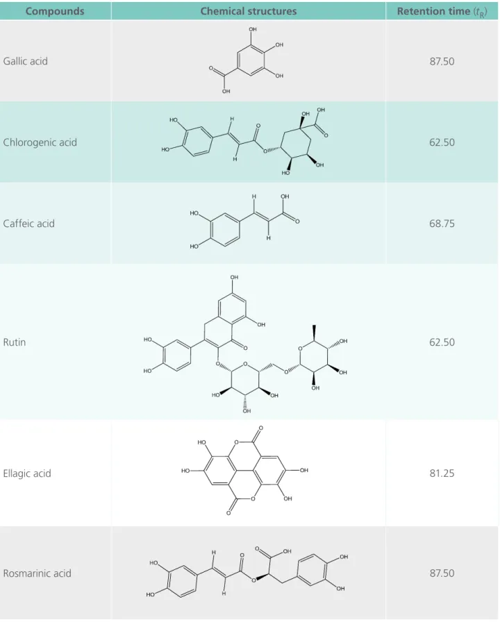
The correlation of chromatographic peaks was achieved by comparing of experimental reten-tion times (tR) with reference standards (Table 1). All chromatographic preliminary phytochemi-cal analysis of AEMC were carried out in tripli-cate at room temperature and revealed phenolic compounds (gallic acid, chlorogenic acid, caffeic acid, ellagic acid and rosmarinic acid) and lavo-noid (rutin) with the following tR: 3,4,5-trihydro-xybenzoic acid(1) tR= 5 min; (1S,3R,4R,5R)-3-[(E)- 3-(3,4-dihydroxyphenyl)prop-2-enoyl]oxy-1,4,5-trihydroxycyclohexane-1 carboxylic acid (2) tR = 14,9 min; (E)-3-(3,4-dihydroxyphenyl)prop-2-enoic acid (3)tR= 15,1 min; 2-(3,4-dihydroxyphenyl)-5,7- dihydroxy-3-[(2S,3R,4S,5S,6R)-3,4,5-trihydroxy-6-[[(2R,3R,4R,5R,6S)-3,4,5 trihydroxy-6-methyloxan-2-yl]oxymethyl]oxan-2-yl]oxychromen-4-one (4)tR
(2R)-Table 1. HPLC analysis of aqueous extract of the fruit of Morinda citrifolia.
Compounds Chemical structures Retention time (tR)
Gallic acid 87.50
Chlorogenic acid 62.50
Caffeic acid 68.75
Rutin 62.50
Ellagic acid 81.25
3-(3,4-dihydroxyphenyl)-2-[(E)-3-(3,4-dihydro-xyphenyl)prop-2-enoyl] oxypropanoic acid (6)tR = 37.5 min.
Cell viability
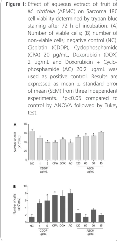
S180 cells viability was evaluated after 72 h following ex vivo exposure to antineoplastics and AEMC. S180 cells treated with CDDP (1 and 5 µg/mL), CPA, DOX, AC and AEMC (30, 60 and 120 µg/mL) showed cell viability reduction (48.7 ± 2.5, 39.2 ± 1.5, 39.5 ± 2.1, 45.5 ± 2.5, 38.5 ± 3.6, 54 ± 2.3, 53.7 ± 2.5 and 50.7 ± 1.3, respectively) when compared with untreated cells (64.5 ± 3.5) (Figure 1, p<0.05).
Apoptosis and necrosis induced by antineoplastics and AEMC in S180 cells
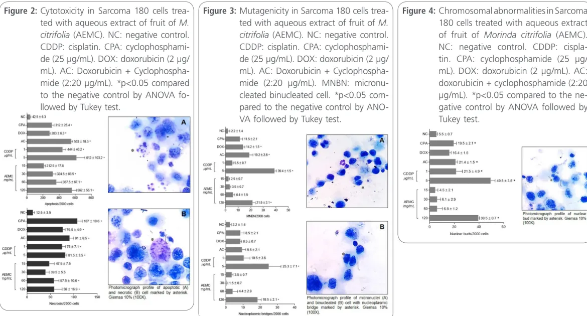
CDDP (1 and 5 µg/mL), CPA, DOX, AC and AEMC (30, 60 and 120 µg/mL) induced apoptosis in S180 cells (444 ± 40.2, 612 ± 103.2, 312 ± 25.4, 283 ± 6.3, 553 ± 18.3, 324.5 ± 66.5, 367.5 ± 67.1 and 562 ± 55.1, respectively) in comparison with negative control (42.5 ± 6.3) and AEMC at 60 and 120 µg/mL produced similar results to the antican-cer agents (Figure 2, p<0.05). Meanwhile, all anti-neoplastics substances also induced the arising of necrosis igures [75 ± 7.1, 81.5 ± 3.5, 107 ± 10.6, 76.5 ± 4.9, 91 ± 8.5, 57.5 ± 10.6, 58 ± 16.9 for CDDP (1 and 5 µg/mL, CPA, DOX, AC and AEMC (60 and 120 µg/mL) respectively (p<0.05)], though these indings were not in lower concentrations of AEMC (p>0.05).
Nuclear abnormalities induced by antineoplasics and AEMC in S180 cells Micronucleus were statistically more found at 5 µg/ mL of CDDP (39.4 ± 1.5), at 120 µg/mL of AEMC (21.5 ± 2.1) and in sarcoma cells treated with DOX (14.2 ± 1.5) and with the association of AC (19.2 ± 2.8) when compared to the negative group (2.2 ± 1.4) (Figure 3). On the other hand, only CDDP (5 µg/mL) and AEMC at concentration of 120 µg/ mL induced signiicant increasing of nucleoplasmic bridges (25.3 ± 7.1 and 18.5 ± 2.1) (p<0.05).
Once again, CDDP (1 and 5 µg/mL), CPA and DOX + CPA (AC) also increased the number of chro-mosomal abnormalities (21.5 ± 4.9, 49.5 ± 3.5, 19.5 ± 2.1 and 21.4 ± 1.5). However, only the highest concentration of AEMC was able to induced the formation of nuclear buds (39.5 ± 0.7) (Figure 4, p<0.05).
Figure 1: Effect of aqueous extract of fruit of
M. citrifolia (AEMC) on Sarcoma 180
Figure 2: Cytotoxicity in Sarcoma 180 cells trea-ted with aqueous extract of fruit of M. citrifolia (AEMC). NC: negative control. CDDP: cisplatin. CPA: cyclophosphami-de (25 µg/mL). DOX: doxorubicin (2 µg/ mL). AC: Doxorubicin + Cyclophospha-mide (2:20 µg/mL). *p<0.05 compared to the negative control by ANOVA fo-llowed by Tukey test.
Figure 4: Chromosomal abnormalities in Sarcoma 180 cells treated with aqueous extract of fruit of Morinda citrifolia (AEMC). NC: negative control. CDDP: cispla-tin. CPA: cyclophosphamide (25 µg/ mL). DOX: doxorubicin (2 µg/mL). AC: doxorubicin + cyclophosphamide (2:20 µg/mL). *p<0.05 compared to the ne-gative control by ANOVA followed by Tukey test.
DNA damage induced by antineoplastics and AEMC in S180 cells
All concentrations of the AEMC increased the ID after 72 h of incubation (90 ± 15.5, 113 ± 27.5,
122 ± 1.4, 147 ± 4.2 and 216 ± 9.1 for 5, 15, 30, 60 and 120 µg/mL, respectively) (Figure 5, p<0,05) when compared to negative control (21.5 ± 5.6). Meanwhile, the positive control CDDP presented ID values of 282 ± 47.5 and 329 ± 43.8 for 1 and 5 µg/mL, respectively. Regarding to FD, CPPD also caused signiicant increasing (66.5 ± 4.9 and 89.5 ± 7.7, respectively) and only the lowest concentration of the AEMC did not induce FD increasing (60.2 ± 5.8, p>0.05) (Figure 5).
Evaluation of oxidative damage induced by antineoplastics and AEMC in S.
cerevisiaestrains
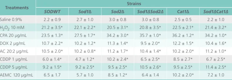
All antineoplastics tested (CDDP, CPA, DOX and AC) induced oxidative damage in S. cerevisiae strains
(SODWT, Sod1∆, Sod2∆, Sod1∆Sod2∆, Cat1∆ and
Cat1∆Sod1∆). On the other, AEMC at 120 µg/mL induced oxidative damage only in the strain with mutation for mitochondrial superoxide dismutase (Sod2∆) and catalase (Cat1∆) (Table 2).
Figure 5: Genotoxicity in Sarcoma 180 cells trea-ted with aqueous extract of fruit of M. citrifolia (AEMC). NC: negative control. CDDP: cisplatin. *p<0.05 compared to the negative control by ANOVA fo-llowed by Tukey test.
Tabl 2. Inhibition growth induced by antineoplastics and aqueous extract of fruit of M. citrifolia (AEMC) in Saccharomyces cerevisiae strains.
Treatments Strains
SODWT Sod1∆ Sod2∆ Sod1∆Sod2∆ Cat1∆ Sod1∆Cat1∆
Saline 0.9% 2.2 ± 0.9 2.7 ± 1.0 3.0 ± 0.8 3.0 ± 0.8 2.5 ± 0.5 2.2 ± 1.0 H2O2 10 mM 21.2 ± 3.5* 22.1 ± 2.2* 20.5 ± 3.1* 20.8 ± 3.5* 22.5 ± 2.1* 21.4 ± 3.2* CPA 20 µg/mL 23.5 ± 1.3* 27.5 ± 1.7* 34.2 ± 3.0* 35.7 ± 1.0* 36.2 ± 1.2* 34.2 ± 1.0* DOX 2 µg/mL 10.7 ± 2.2* 10.2 ± 1.2* 11.3 ± 1.4* 9.5 ± 2.0* 12.2 ± 1.5* 10.4 ± 1.6* AC 20:2 µg/mL 10.5 ± 2.0* 10.2 ± 0.8* 11.2 ± 1.7* 10.4 ± 1.4* 10.2 ± 2.0* 11.2 ± 1.0* CDDP 1 µg/mL 6.0 ± 1.4* 4.7 ± 1.2* 10.2 ± 2.4* 6.5 ± 2.5* 8.5 ± 2.7* 6.7 ± 2.5* CDDP 5 µg/mL 9.2 ± 1.5* 9.2 ± 2.5* 9.5 ± 2.5* 10.5 ± 2.6* 9.5 ± 2.5* 11.4 ± 2.5* AEMC 120 µg/mL 6.5 ± 1.7 5.7 ± 1.0 8.5 ± 1.2* 6.4 ± 1.4 10.2 ± 2.0* 7.2 ± 1.0
Discussion
Cancer is represented by a pool of diseases related to the accumulation of multiple genetic changes that leads to genetic instability (DNA damage) and repair failures and they have been associated with the diagnosis and prognosis of neoplasms. Such instability can be mediated through chromosomal changes, that can be detected by, cytogenetical tec-niques [17]. Assays as CBMN, Single Cell Gel Elec-trophoresis and oxidative analyzes in S. cerevisiae
strainshave been proposed as biomarkers to assess cytogenetic alterations and measure DNA damage [18].
Primary cultures of tissues that preserve the ori-ginal tumor microenvironment injected into mice has been used as models of human sarcomas [19], allowing the understanding of the molecular bio-logy of cancer and cellular changes after exposu-re to antiproliferative agents. Some tools, such as Cytokinesis-Block Micronucleus Assay (CBMN), a multi-endpoint assay that measures chromosomal damage; micronuclei (MN), relecting chromoso-me breaks; nucleoplasmic bridges (NPB), relecting chromosomal rearrangements and nuclear buds (NBUD), relecting gene ampliication, along with other cellular events such as apoptosis and necrosis [14] are often used to evaluate pharmacological and toxicological effects of bioactive natural and synthe-tic compounds in cellular models.
S180 cells provide a paradigm to identify unknown pharmacogenetic variants associated with drug-in-duced cytotoxicity [20]. Herein, all chemotherapics used (CDDP, CPA, DOX, AC) and AEMC induced apoptosis, necrosis and reduced cell viability of sar-coma 180 cells.
CDDP is capable to induce adduct formation effects and peculiar DNA-depentent functions, in-cluding inhibition of replication and transcription, cell cycle arrest, and DNA damage that can lead to the cellular death and apoptosis, thus may followed by mutations [21]. Our results suggest that genetic factors involved in cytotoxicity also contribute to
cisplatin-induced apoptosis. CDDP have been fu-lly ascribed to its ability to generate unrepairable DNA lesions, hence inducing either a permanent proliferative arrest known as cellular senescence or apoptosis induction by mitochondrial pathway [22].
CPA is metabolically activated by the enzyme CYP2B6 and induced cell death via formation of DNA adducts by it’s active metabolite, phosphora-mide mustard. These adducts are the most physio-logically relevant because of their ability to block the replication of DNA and its association with cytotoxicity [23]. These DNA damages presented as nuclear abnormalities, such as micronuclei and formation of nuclear buds, were also found after treatment with CDDP (5 µg/mL), AEMC (120 µg/ mL), DOX and with the association of AC. Howe-ver, only CDDP (5 µg/mL) and AEMC (120 µg/mL) induced signiicant increasing of nucleoplasmic bridges. Moreover, all concentrations of the AEMC increased the ID and FD after 72h of incubation, but only the lowest concentration did not induce FD increasing.
cycle arrest and apoptosis, protein kinases changes and inhibition of COX-2 activity [25] and ellagin acid induces apoptosis in human pancreatic ade-nocarcinoma and hepatocarcinoma (HepG2) cells [26], sugesting mechanism involving mitochondrial membrane depolarization, release of protein C and caspases activation [27], activation of transcription factors and anti-apoptotic proteins dysregulation [28].
In a similar way, the lavonoid rutin also plays an-titumor activity by induction of DNA damage in cells mutant for BRCA gene, cell cycle arrest (G2/M) in neuroblastoma cell lines by apoptosis-inducing me-chanism, as well as regulation of gene expression suppressing tumor [29]. These are major bioactive constituents of AEMC responsible for various phar-macological activities [30].
Necrosis was also identiied in antineoplastics- and AEMC-sarcoma treated cells. In toxic stimulus analyzes, higher doses regularly develops concomi-tant features of apoptosis and necrosis. Under such conditions, severity and not speciicity of the sti-mulus selectively determines how cell death occurs. If the necrosis prevails, early lesions on the plas-ma membrane occur instead of cell shrinking [21]. Therefore, depending on the concentration used, many different processes may be inluenced and/or altered, suggesting that dose-dependent regulation of cellular process relects signalization triggered by bioactive compounds as stated here and by others. For example, previous studies with the diterpenoid casearin X also reported characteristics of apoptosis or secondary necrosis [31].
Micronuclei, biomarkers of genotoxic events and chromosomal instability were induced by AEMC (120 µg/mL), while the other nuclear abnormality, such as NPB, were produced by AECM and CDDP. The MN is frequently originated during anaphase from lagging acentric chromosome or chromatid fragments caused by misrepair of DNA breaks or unrepaired DNA breaks. Mal-segregation of whole chromosomes at anaphase may also lead to MN
formation as a result of hypomethylation of re-peat sequences in centromeric and pericentromeric DNA, defects in kinetochore proteins or assem-bly, dysfunctional spindle and defective anaphase checkpoint genes. NPB originates from the dicen-tric chromosomes, may occur due to misrepair of DNA breaks, telomere end fusions, and could also be observed when defective separation of sister chromatids at anaphase due to failure of deca-tenation. NBUD represents the process of elimi-nation of ampliied DNA, DNA repair complexes and possibly excess chromosomes from aneuploid cells [14].
The yeast S. cerevisiae also provides a power-ful experimental instrument for the study of drugs’ effect on eukaryotic cells [32]. Herein, oxidative da-mage in S. cerevisiae point out that the AEMC (120 µg/mL) induced oxidative damage similar to CDDP, DOX and AC at the concentrations tested to the strains Sod2∆ and Cat1∆. Hung et al. [33] have su-ggested that caffeic acid, presented in AECM, redu-ces mitochondrial membrane potential and induredu-ces apoptosis [34].
indu-ces cardiotoxicity by lipid peroxidation [7] caused by oxidative stress [8].
Despite of these genotoxic effects, M. citrifolia
has been used in traditional therapies against several chronic diseases, including cancer. Damnacanthal, a compound isolated from M. citrifolia, increases anti-tumorigenic activity in human colorectal cancer cells [39]. Futhermore, pre-clinical studies have suggest that Noni may have antitumor proper¬ties due to the immune system stimulating using macrophages, natural killer and T cells dependent ways [40].
It is likely that chemicals presented in AECM, such as gallic acid, chlorogenic acid, caffeic acid, ellagic acid and rosmarinic acid and rutin, are involved in the cytotoxicity on sarcoma and S. cerevisiae mu-tants cells, by direct ou indirect (oxidative) DNA da-mages, leading to apoptosis. In vitro and in vivo
investigations are in progress with sarcoma 180 mo-dels to detail the mechanisms(s) of action.
Conclusion
The aqueous fruit extract of Morinda critrifolia
(AEMC) induced cytogenetic damages with increa-sing in the formation of micronuclei, nuclear buds and nucleoplasmic bridges like the antineoplastics, Cisplatin, Cyclophosphamide and Doxorubicin taken as standards in Sarcoma 180 cells. Additionally, AEMC also exhibited a Cisplatin like oxidative da-mage in mitochondrial superoxide dismutase and cytosolic catalase diicient Saccharomyces cerevisiae
strains. Phytochemicals, present in AEMC fruit such as gallic, caffeic, chlorogenic and ellagic acid and lavonoid rutin may be responsible for the antitumo-ral effects. Furtther researches are recommended to ind out the responsible chemical moity(s) with the appropriate action mechanism(s) of our found activities.
Acknowledgments
The authors thank the Genetic and Toxicological Research Laboratory, Centre for Pharmaceutical Te-chnology (NTF), Federal University of Piaui (UFPI), Teresina, Piaui, Brazil.
Conflict of interest None declared.
References
1. Ferlay J, Soerjomataram I, Dikshit R, Eser S, Mathers C, Rebelo M, Parkin DM, Forman D, Bray F. Cancer incidence and mortality worldwide: sources, methods and major patterns in GLOBOCAN 2012. Int J Cancer 2015; 136: 359-386.
2. Lee C, Raffaghello L, Brandhorst S, Safdie FM, Bianchi G, Martin-Montalvo A, Pistoia V, Wei M, Hwang S, Merlino A, et al. Fasting cycles retard growth of tumors and sensitize a range of cancer cell types to chemotherapy. Sci Transl Med 2012; 4: 124-127.
3. Wang H, Zheng X, Fei F, Wang J, Li X, Liu Y, Zhang F. Towards pathway-centric cancer therapies via pharmacogenomic proiling analysis of ERK signalling pathway. Clin Transl Med 2015; 4: 1-10.
4. Macdermed DM, Miller LL, Peabody TD, Simon MA, Luu HH, Haydon RC, Montag AG, Undevia SD, Connell PP. Primary tumor necrosis predicts distant control in locally advanced soft-tissue sarcomas after preoperative concurrent chemoradiotherapy. Int J Radiat Oncol Biol Phys 2010; 76: 1147-1153.
5. Fuchs-Tarlovsky V. Role of antioxidants in cancer therapy. Nutri 2013; 29: 15-21.
6. Marullo R, Werner E, Degtyareva N, Moore B, Altavilla G, Ramalingam SS, Doetsch PW. Cisplatin induces a mitochondrial-ROS response that contributes to cytotoxicity depending on mitochondrial redox status and bioenergetic functions. PLoS One 2013; 8: e81162.
7. Johnkennedy N, Ifeoma UH. Cardioprotective effect of Gnetum bucholzianum leaf extract against doxorubicin cardiomyopathy in Wistar rats. Novel Sci 2012; 1: 61-64.
8. Chennuru A, Saleem MT. Antioxidant, lipid lowering, and membrane stabilization effect of sesamol against doxorubicin-induced cardiomyopathy in experimental rats. Biomed Res Int 2013; 2013: 934-939.
9. Huang Y, Li L. DNA crosslinking damage and cancer - a tale of friend and foe. Transl Cancer Res 2013; 2: 144-154.
10. Taylor BS, Barretina J, Maki RG, Antonescu CR, Singer S, Ladanyi M. Advances in sarcoma genomics and new therapeutic targets. Nat Rev Cancer 2011; 11: 541-557.
12. Wang MY, West BJ, Jensen CJ, Nowicki D, Su C, Palu AK, Anderson G. Morinda citrifolia (Noni): a literature review and recent advances in Noni research. Acta Pharmacol Sin 2002; 23: 1127-1141.
13. Renzi D, Valtolina M, Foster R. The evaluation of the multi-endpoint cytotoxicity assay system. ATLA 1993; 21: 89-96.
14. Fenech M, Kirsch-Volders M, Natarajan AT, Surralles J, Crott JW, Parry J, Norppa H, Eastmond DA, Tucker JD, Thomas P. Molecular mechanisms of micronucleus, nucleoplasmic bridge and nuclear bud formation in mammalian and human cells. Mutagenesis 2011; 26: 125-132.
15. Speit G, Rothfuss A. The comet assay: a sensitive genotoxicity test for the detection of DNA damage and repair. Methods Mol Biol 2012; 920: 79-90.
16. Oliveira GLS, Oliveira FRAM, Alencar MVOBA, Gomes Junior AL, Souza AA, Melo Cavalcante AAC, Freitas RM. Evaluation of antioxidant capacity of the aqueous extract of Cynara scolymus
L. (Asteraceae) in vitro and in Saccharomyces cerevisiae. Afr J Pharm Pharmacol 2014; 8: 136-147.
17. Vincent K, Pichler M, Lee GW, Ling H. MicroRNAs, genomic instability and cancer. Int J Mol Sci 2014; 15: 14475-14491.
18. Gomes-Junior AL, Paz MFCJ, Silva LIS, Carvalho SCS, Sobral ALP, Machado KC, Ferreira PMP, Satyal P, Freitas RM, Melo Cavalcante AAC. Serum oxidative stress markers and genotoxic proile induced by chemotherapy in patients with breast cancer: a pilot study. Oxid Med Cell Longev 2015; 2015: 1-11.
19. Quesada J, Amato R. The molecular biology of soft-tissue sarcomas and current trends in therapy. Sarcoma 2012; 2012: 849456.
20. Ferreira PMP, Costa PM, Costa AM, Lima DJ, Drumond RR, Silva JN, Moreira DR, De Oliveira Filho GB, Ferreira JM, De Queiroz MG, et al. Cytotoxic and toxicological effects of phthalimide derivatives on tumor and normal murine cells. An Acad Bras Cienc 2015; 87: 313-330.
21. Chaney SG, Campbell SL, Bassett E, Wu Y. Recognition and processing of cisplatin- and oxaliplatin-DNA adducts. Crit Rev Oncol Hematol 2005; 53: 3-11.
22. Galluzzi L, Vitale I, Michels J, Brenner C, Szabadkai G, Harel-Bellan A, Castedo M, Kroemer G. Systems biology of cisplatin resistance: past, present and future. Cell Death Dis 2014; 5: e1257.
23. Johnson LA, Malayappan B, Tretyakova N, Campbell C, MacMillan ML, Wagner JE, Jacobson PA. Formation of cyclophosphamide speciic DNA adducts in hematological diseases. Pediatr Blood Cancer 2012; 58: 708-714.
24. Yan Y, Li J, Han J, Hou N, Song Y, Dong L. Chlorogenic acid enhances the effects of 5-luorouracil in human hepatocellular carcinoma cells through the inhibition of extracellular signal-regulated kinases. Anticancer Drugs 2015; 26: 540-546.
25. Kuo YY, Jim WT, Su LC, Chung CJ, Lin CY, Huo C, Tseng J, Huang SH, Lai CJ, Chen BC, et al. Caffeic Acid phenethyl ester is a potential therapeutic agent for oral cancer. Int J Mol Sci 2015; 16: 10748-10766.
26. Zhang HM, Zhao L, Li H, Xu H, Chen WW, Tao L. Research progress on the anticarcinogenic actions and mechanisms of ellagic acid. Cancer Biol Med 2014; 11: 92-100.
27. Mertens-Talcott SU, Percival SS. Ellagic acid and quercetin interact synergistically with resveratrol in the induction of apoptosis and cause transient cell cycle arrest in human leukemia cells. Cancer Lett 2005; 218: 141-151.
28. Vanella L, Di Giacomo C, Acquaviva R, Barbagallo I, Cardile V, Kim DH, Abraham NG, Sorrenti V. Apoptotic markers in a prostate cancer cell line: effect of ellagic acid. Oncol Rep 2013; 30: 2804-2810.
29. Chen H, Miao Q, Geng M, Liu J, Hu Y, Tian L, Pan J, Yang Y. Anti-tumor effect of rutin on human neuroblastoma cell lines through inducing G2/M cell cycle arrest and promoting apoptosis. SCI WORLD J Journal 2013; 2013: 1-8.
30. Pachauri SD, Tota S, Khandelwal K, Verma PR, Nath C, Hanif K, Shukla R, Saxena JK, Dwivedi AK. Protective effect of fruits of Morinda citrifolia L. on scopolamine induced memory impairment in mice: a behavioral, biochemical and cerebral blood low study. J Ethnopharmacol 2012; 139: 34-41.
31. Ferreira PMP, Militão GCG, Lima DJB, Costa NDJ, Machado KC, Santos AG, Cavalheiro AJ, Bolzani VS, Silva DHS, Pessoa C. Morphological and biochemical alterations activated by antitumor clerodane diterpenes. Chem Biol Interact 2014; 222: 112-125.
32. Rojas M, Casado M, Portugal J, Piña B. Selective inhibition of yeast regulons by daunorubicin: a transcriptome-wide analysis. BMC Genomics 2008; 9: 358.
33. Hung WM, Shiao MS, Tsai LC, Chang GG, Chang TC. Apoptotic effect of caffeic acid phenethyl ester and its ester and amide analogues in human cervical cancer ME180 cells. Anticancer Res 2003; 23: 4773-4780.
34. Jaganathan SK. Growth inhibition by caffeic acid, one of the phenolic constituents of honey, in HCT 15 colon cancer cells. Sci World J 2012; 2012: 372345.
35. Valentovic MA, Ball JG, Brown JM, Terneus MV, McQuade E, Van Meter S, Hedrick HM, Roy AA, Williams T. Resveratrol attenuates cisplatin renal cortical cytotoxicity by modifying oxidative stress. Toxicol In Vitro 2014; 28: 248-257.
International Archives of Medicine is an open access journal publishing articles encompassing all aspects of medical scien-ce and clinical practiscien-ce. IAM is considered a megajournal with independent sections on all areas of medicine. IAM is a really international journal with authors and board members from all around the world. The journal is widely indexed and classified Q1 in category Medicine.
Publish in International Archives of Medicine
37. Sadir S, Deveci S, Korkmaz A, Oter S. Alpha-tocopherol, beta-carotene and melatonin administration protects cyclophosphamide-induced oxidative damage to bladder tissue in rats. Cell Biochem Funct 2007; 25: 521-526.
38. Wolter R, Siede W, Brendel M. Regulation of SNM1, an inducible
Saccharomyces cerevisiae gene required for repair of DNA cross-links. Mol Gen Genet 1996; 250: 162-168.
39. Nualsanit T, Rojanapanthu P, Gritsanapan W, Lee SH, Lawson D, Baek SJ. Damnacanthal, a noni component, exhibits antitumorigenic activity in human colorectal cancer cells. J Nutr Biochem 2012; 23: 915-923.