DOI: 10.5897/JMPR2015.6017 Article Number:CC5FB8856796 ISSN 1996-0875
Copyright © 2016
Author(s) retain the copyright of this article http://www.academicjournals.org/JMPR
Journal of Medicinal Plants Research
Full Length Research Paper
Toxicogenetic profile of rats treated with aqueous
extract from
Morinda citrifolia
fruits
Germano Pinho de Moraes
1, Marcus Vinícius Oliveira Barros de Alencar
2,3*, Md. Torequl
Islam
2,4, Lidiane da Silva Araújo
4, Débora Cássia Vieira Gomes
2,3, Rodrigo Mendes de
Carvalho
3, Dione Corrêa
1, Márcia Fernanda Correia Jardim Paz
1,3, Paulo Michel Pinheiro
Ferreira
2,5,6, Ana Amélia de Carvalho Melo-Cavalcante
2,3,5, Alexandre Ferraz
1Ivana Grivicich
1and Jaqueline Nascimento Picada
11
Post-Graduation Program in Cellular Biology Applied to Health (PPGBioSaúde), Lutheran University of Brazil, Canoas,
Brazil.
2
Postgraduate Program in Biotechnology, Federal University of Piauí, Teresina, Brazil.
3Laboratory of Genetic Toxicology, Federal University of Piauí, Teresina, Brazil.
4
Laboratory of Research in Experimental Neurochemistry, Federal University of Piauí, Teresina, Brazil.
5Postgraduate Program in Pharmaceutical Sciences, Federal University of Piauí, Teresina, Brazil.
6Laboratory of Experimental Cancerology, Department of Biophysics and Physiology, Federal University of Piauí,
Teresina, Brazil.
Received 20 November, 2015; Accepted 8 January, 2016
Morinda citrifolia
((Family: Rubiaceae)) is extensively used in traditional medicine due to its
anti-inflammatory, antimicrobial, antitumoral, and anti-hypertensive activities. However, there is no
substantial data about hepatotoxic and toxicogenetic effects. This study evaluated biochemical
changes and hepatotoxic, genotoxic, and mutagenic effects of aqueous extract of the fruit of
M.
citrifolia
(AEMC) in liver, bone marrow, and peripheral blood cells. Animals (
Rattus novergicus
, 5 males
and 5 females) were divided into negative control, positive control (Cyclophosphamide 25 mg/kg), and
AEMC (2.5, 5, and 10 mg/kg, by gavage). AEMC induced increase of aspartate aminotransferase (AST),
alanine aminotransferase (ALT), and alkaline phosphatase (ALP), especially at 10 mg/kg in female (174.8
± 50.7, 221.4 ± 24.6, and 174.7 ± 14.3 U/L) and male (156.5 ± 21.6, 183.7 ± 21.5, and 147.3 ± 17.8 U/L)
(
p
<0.05). Histological analysis of livers showed inflammatory cell infiltration, nuclear fragmentation,
microvacuolization, cellular swelling, points of inflammatoy necrosis, and discrete microvesicular
steatosis. DNA damage in hepatocytes w
as
found in both genders, mainly at 10 mg/kg (Frequency of
Damage: 78.1 ± 4.5 and 70.4 ± 7.3%; Index of Damage: 107.6 ± 14.2 and 136.0 ± 26.9 for male and female,
respectively). Similar results were observed in bone marrow cells. The AEMC 5 and 10 mg/kg induced
micronucleus formation (4.4 ± 0.8 and 7.8 ± 1.1; 7.4 ± 1.1 and 9.6 ± 1.4 for peripheral blood and bone
marrow cells, respectively) (
p
<0.05). These findings suggest clastogenic and/or aneugenic effects and
genetic instability activated by AEMC, indicating precaution regarding the consumption of formulations
or folk preparations based on this plant.
Key words:
Hepatotoxicity, genotoxicity, mutagenicity,
Morinda citrifolia
, noni.
INTRODUCTION
Popular knowledges associated with geographic and
requirements (Firenzuoli and Gori, 2007; WHO, 2011;
Akram et al., 2014; Tuttolomondo et al., 2014; Araujo et
al., 2015). However,
continuous
exposure to synthetic or
natural chemicals present in these preparations may lead
to structural and functional damage to macromolecules,
due to possible toxic effects, which can be measured by
several genotoxic and mutagenic testing methodologies
(Speit and Rothfuss, 2012; Hussin et al., 2014).
Morinda citrifolia
Linn (Fam.: Rubiaceae) is habituated
to the Southeast Asia and is distributed through several
settlers of the Pacific Islands (Wang et al., 2002;
Samoylenko et al., 2006; Ebeling et al., 2014). Aerial
parts of this plant are commonly used in folk medicine as
antibacterial,
antiviral,
antifungal,
antitumoral,
anthelmintic,
contra-ceptive,
hypotensive,
anti-inflammatory, antioxidant, immunomodulating (Nayak and
Shettigar, 2010), as well as antidopaminergic and
antiadrenergic (Pandy et al., 2014). Moreover, its fruit has
medicinal effects
o
n allergy, arthritis, asthma, bacterial
infections, cancer, diabetes, hypertension, menstrual
disorders, obesity, gastric ulcers, headaches, sexual
inhibition, insomnia, depression, stress, respiratory
problems, AIDS, multiple sclerosis and drug dependency
(Selvam et al., 2009; Gupta and Patel, 2013; Murata et
al., 2014).
About 96 volatile compounds were identified in ripe fruit
of
M. citrifolia
and more than 200 compounds were
isolated from several parts of the plant, of which as
hexanoic acid, octanoic and asperuloside acid, alcohols,
esters, ketones and lactones are few of them.
Nonetheless, the phytochemical composition is not
complete yet (Potterat and Hamburger, 2007; Assi et al.,
2015). Although folk uses of
M. citrifolia
fruits
are
an
earlier report, still there is a lack of consistent data
regarding hepatotoxic, genotoxic, mutagenic, and
cytotoxic effects. Thus, studies addressing DNA damage
in eukaryotic cells are important to undersand the risk of
cellular injury. This study evaluated the possible
biochemical changes and hepatotoxic, genotoxic and
mutagenic effects of aqueous extract of the fruit of
M.
citrifolia
(AEMC) in liver, bone marrow and peripheral
blood cells in Wistar rats.
MATERIALS AND METHODS
Plant and extract preparation
Fruits of M. citrifolia were collected in 2014 in the municipality of Altos, Piauí, Brazil, (05° 02‟20” S–latitude, 42° 27‟39” O– longitude) at 187 m above sea level. The botanical identification was held at the Center for Environmental Sciences of Tropic Ecotonal Northeast, Teresina, Piauí (voucher number: 21644). After collection, the fruits were dried in a forced air oven for 8 days at a maximum temperature of 45°C (±1°C). Then, they followed by
course grinding and were preserved in an amber glass.
HPLC analysis
High performance liquid chromatograph (HPLC) analysis was performed in AEMC to determine the presence of flavonoids and phenolic compounds. Briefly, Waters 2695 liquid chromatograph equipped with autosampler and a variable wavelength UV/VIS detector (Waters 2487 Detector Dual Absorbance, 190 to 700 nm). Columns: Waters Spherisorb ODS2 (5 µm, 4.6×250 mm). The mobile phases consisted of acetonitrile (A) HPLC grade purchased and 0.1% H3PO4 aqueous solution (B, filtered using a Millipore
system). The following were the gradient conditions: 5% (A); 15% (A) for 10 min; 35% (A) for 40 min; 100% (A) for 15 min and 5% (A) for 5 min. Total run time was 70 min at a flow rate of 1.8 ml/min. Injection volumes were 20 µl. The AEMC was solubilized in acetonitrile (30%, HPLC grade and 0.1% H3PO4 aqueous solution,
70%).
Animals and treatment
A total of 50 Wistar adult Rattus norvegicus of both sexes weighing 200 to 250 g were purchased from the Faculty of Health, Life Sciences and Technology of Piauí (NOVAFAPI), Teresina, Piauí, Brazil. The animals were kept in plastic cages (6 animals/cage) with maintenance of diet (Purina, Brazil) and water ad libitum in air-conditioned temperature of 25°C with a photoperiod of 12 h light/dark cycle. The investigational protocols were approved by the local Ethical Committee on Animal Research at NOVAFAPI (Process No. #0039/10) and are in accordance with the national (Colégio Brasileiro de Experimentação Animal – COBEA) and international standard for the care and use of experimental laboratory animals (EEC Directive, 1986).
Animals (10 per group: 5 males and 5 females) were randomly distributed into the following groups: negative control (NC; untreated animals); positive control (Cyclophosphamide 25 mg/kg, i.p.) and three for AEMC (2.5, 5 and 10 mg/kg, by gavage). Animals of the positive control received cyclophosphamide (CPA) for 2 days and were sacrificed on the 3rd day. After 14 days of treatment, the rats were intraperitoneally anaesthetized with a solution composed of ketamine and xylazine at a dose of 40 and 5 mg/kg, respectively. Then, blood (5 ml) from the portal vein was collected to evaluate biochemical parameters. For the micronucleus test, blood was collected from the caudal vein. Liver tissue samples were collected for histopathological analysis and comet assay. In addition, bone marrow samples from both femurs were collected for micronucleus test and comet assay.
Biochemical parameters
Alanine aminotransferase (ALT), aspartate aminotransferase (AST), alkaline phosphatase (ALP) and gamma-glutamyl transferase (GGT) were measured by using commercially available assay kits (Labtest Diagnóstica S.A., Brazil).
Liver histopathological analysis
For histological analysis, the livers of the remaining animals of each group were fixed in 10% formalin for 72 h. To examine
*Corresponding author. E-mail: [email protected].
morphological changes by light microscopy (Olympus, Tokyo, Japan) at a magnification of 200 and 400X, small pieces were processed, embedded in paraffin and 3 to 5 μm thick sections were prepared and stained with Hematoxylin and Eosin (H&E).
Comet assay
The alkaline comet assay was performed by the method earlier described by Speit and Rothfuss (2012). Briefly, 10 µl of the cell suspension (hepatocytes and bone marrow mononuclear cells) was mixed with a thin agarose layer low melting point (0.75%) (90 µl) and placed on pre-covered slides with normal melting point agarose (1.5%). The slides were immersed in lysing solution (2.5 mol/L NaCl, 100 mmol/L EDTA and 10 mmol/L Tris, pH 10 with 1% Triton X-100 and 10% of DMSO) up to 72 h at 4°C. The slides were then incubated in alkaline buffer (300 mmol/L NaOH and 1 mmol/L EDTA; pH >13) for 20 min followed by exposition to an electrical current 300 mA and 25 V (0.90 V/cm) for 15 min (electrophoresis). Finally, the slides were neutralized with Tris buffer 0.4 M, pH 7.5 and stained with silver solution. The results were expressed in index of damage (ID) and frequency of damage (FD). The ID was calculated from the visual assessment of the damage classes (0 to 4), extracting an index that expresses the overall damage suffered by the cells (100 cells/slide in duplicate). However, in injured cells, theDNAmigratesfromthecore to the anode during electrophoresis, showing a similar comet tail. From these images, the cells were sorted between classes 1 (minimal damage) and 4 (maximum damage), 0 denoted for intact nucleus. The FD was calculated based on the equation: FD = 100 – N0, where FD is the frequency
of damage; N0: number of tailless cells.
Micronucleus frequency assay
The micronucleus test was carried out according to Mavournin et al. (1990) and Kasamoto et al. (2013). Briefly, the collected bone marrow was mixed with 0.3 ml of fetal bovine serum on the slide previously coded. Additionally, peripheral blood was collected from the tails of live animals. The smears were performed by extender blade and after 30 min of drying, the slides were fixed in methanol for 10 min. Then, the slides were stained with Giemsa (Merck) in phosphatebuffer0.2mol/L,pH 5.8.The counting of normochromatic erythrocytes (NCE), polychromatic erythrocytes (PCE) and micronuclei in polychromatic erythrocytes (MNPCE) were carried out by photomicrography at 100X. In each animal, 2000 PCE were analyzed. The ratio of PCE/NCE was determined by evaluating the frequency of PCE in 500 erythrocytes per animal.
Statistical analysis
Results were expressed as mean ± standard deviation (SD) (n=10). In order to determine the differences, data were compared by one-way analysis of variance (ANOVA) followed by the Tukey test (p<0.05).
RESULTS
Identification of compounds by HPLC analysis
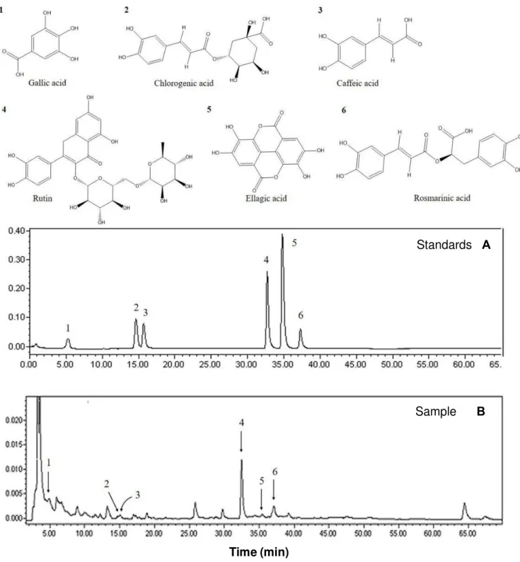
Qualitative HPLC analysis of AEMC is as shown in Figure
1a. The correlation of chromatographic peaks was
achieved by comparing experimental retention times (t
R)
with reference standards (Figure 1b). All chromatographic
operations were carried out in triplicate at ambient
temperature. According to the chromatogram obtained for
AEMC, gallic acid, chlorogenic acid, caffeic acid, ellagic
acid, rosmarinic acid and flavonoid like-rutin with the
following retention time (
t
R) were found:
3,4,5-trihydroxybenzoic acid (1)
t
R: 5.0 min;
(1S,3R,4R,5R)-3-[(E)-3-(3,4-dihydroxyphenyl)prop-2-enoyl]oxy-1,4,5
tri
hydroxycyclohexane-1-carboxylic acid (2)
t
R: 14.9 min;
(E)-3-(3,4-dihydroxyphenyl)prop-2-enoic acid (3)
t
R: 15.1
min;
2-(3,4-dihydroxyphenyl)-5,7-dihydroxy-3-[(2S,3R,
4S,5S,6R)-3,4,5-trihydroxy-6-[[(2R,3R, 4R,5R,
6S)-3,4,5-trihydroxy-6-methyloxan-2-yl]oxymethyl]oxan-2-yl]
oxychromen-4-one (4)
t
R: 32.5 min;
2,3,7,8-Tetrahydroxy-chromeno[5,4,3-cde] chromene-5,10-dione (5)
t
R: 35.0
min;
(2R)-3-(3,4-dihydroxyphenyl)-2-[(E)-3-(3,4-dihydroxyphenyl)prop-2-enoyl]oxypropanoic acid (6)
t
R:
37.5 min.
In vivo
hepatotoxicity
Hepatotoxicity of AEMC in rats was observed by
an
increase (
p
<0.05) of the values of AST, ALT and ALP,
especially at the highest dose (10 mg/kg) in female
(174.8 ± 50.7, 221.4 ± 24.6 and 174.7 ± 14.3 U/L) and
male (156.5 ± 21.6, 183.7 ± 21.5 and 147.3 ± 17.8 U/L)
when compared with the negative control [(female: 32.1 ±
13.6, 41.1 ± 9.8 and 53.5 ± 2.7 U/L) and male (33.2 ±
14.8, 49.9 ± 12.0 and 59.7 ± 4.7 U/L), respectively].
Furthermore, high levels of ALP were found in all
AEMC-treated animals (Table 1) and male animals also showed
GGT increasing at the dose of 10 mg/kg (1.2 ± 0.2 U/L) in
comparison with
the
untreated group (0.9 ± 0.1 U/L)
(
p
<0.05). Similarly, histological analysis of livers also
revelaed signs of injury, corroborating the alteration in
serum enzymes, as morphological changes in both
genders at 10 mg/kg, which showed histoarchitecture
preservation with inflammatory infiltrating cells in the
perivascular space (Figure 2a), nuclear fragmentation of
hepatocytes (suggestive of apoptosis) (Figure 2b),
microvacuolization (Figure 2c), cellular swelling (Figure
2d), points of inflammatoy necrosis (Figure 2e) and
discrete microvesicular steatosis (Figure 2f), all findings
indicative of hepatotoxicity.
DNA damage in liver and bone marrow cells
Standards
A
Sample
B
Time (min)
Figure 1. HPLC chromatogram of a standard mixture (A) and aqueous extract of the fruit of M. citrifolia (AEMC) (A). 3,4,5-trihydroxybenzoic acid (1) tR: 5.0 min; (1S,3R,4R,5R)-3-[(E)-3-(3,4-dihydroxyphenyl)prop-2-enoyl]oxy-1,4,5-trihydroxycyclohexane-1-carboxylic acid (2) tR: 14.9 min; (E)-3-(3,4-dihydroxyphenyl)prop-2-enoic acid (3) tR: 15.1 min; 2-(3,4- dihydroxyphenyl)-5,7-dihydroxy-3-[(2S,3R,4S,5S,6R)-3,4,5-trihydroxy-6-[[(2R,3R,4R,5R,6S)-3,4,5-trihydroxy-6-methyloxan-2-yl]oxymethyl]oxan-2-yl]oxychromen-4-one (4) tR: 32.5 min; 2,3,7,8-Tetrahydroxy-chromeno[5,4,3-cde]chromene-5,10-dione (5) tR: 35.0 min; (2R)-3-(3,4-dihydroxyphenyl)-2-[(E)-3-(3,4-dihydroxyphenyl)prop-2-enoyl]oxypropanoic acid (6) tR: 37.5 min.
damage at 5 mg/kg in bone marrow cells of males only
(44.6 ± 4.4) and at 10 mg/kg it oc
c
urred in both genders
(78.4 ± 5.7 and 68.1 ± 8.6, for male and female,
respectively) (
p
<0.05). On the other hand, CPA, positive
control, caused increased in FD and ID levels in a
n
Table 1. Biochemical markers evaluated in ratstreated with aqueous extract of fruit of Morinda citrifolia (AEMC).
Marker
Treatment
Negative control CPA 25 mg/kg AEMC
2.5 mg/kg 5 mg/kg 10 mg/kg
Male Female Male Female Male Female Male Female Male Female
ALT (U/L) 32.1 ± 13.6 33.2 ± 14.8 263 ± 83.4* 267.1 ± 53.2* 80.8 ± 11.1 81.1 ± 28.2 129 ± 22.3* 107.1 ± 15.4* 156.5 ± 21.6* 174.8 ± 50.7* AST (U/L) 41.1 ± 9.8 49.9 ± 12.0 368 ± 47.5* 316.4 ± 78.5* 112.0 ± 23.0* 90.7 ± 8.93 116.7 ± 44.4* 83.4 ± 14.4 183.7 ± 21.5* 221.4 ± 24.6* GGT (U/L) 0.9 ± 0.1 1.0 ± 0.1 1.3 ± 0.2* 1.3 ± 0.3* 0.9 ± 0.1 0.9 ± 0.1 1.2 ± 0.1 1.0 ± 0.2 1.2 ± 0.2* 1.2 ± 0.9 ALP (U/L) 53.5 ± 2.7 59.7 ± 4.7 163.3 ± 18.8* 155.9 ± 11.1* 120.9 ± 12.4* 116.7 ± 10.5* 113.2 ± 5.6* 123.5 ± 20.8* 147.3 ± 17.8* 174.7 ± 14.3*
Values are mean ± SD (n=10 animals/group). CPA: Cyclophosphamide; ALT: alanine aminotransferase; AST: aspartate aminotransferase; GGT: gamma glutamyltransferase; ALP: alkaline phosphatase. ANOVA followed by Tukey test. *p<0.05 compared to the negative control.
Table 2. Frequency of DNA damage (FD) evaluated by alkaline comet assay in bone marrow cells and hepatocytes of rats treated with aqueous extract of fruit of Morinda citrifolia (AEMC).
Group Bone marrow (%) Liver (%)
Male Female Male Female
Negative control 12.1 ± 5.7 17.6 ± 3.2 19.1 ± 6.9 20.6 ± 9.5 CPA 29.8 ± 8.7* 74.6 ± 9.9* 62.8 ± 7.4* 73.8 ± 5.3* AEMC 2.5 mg/kg 18.4 ± 8.3 12.1 ±7.3 41.6 ± 4.6* 26.1 ± 4.6 AEMC 5 mg/kg 44.6 ± 4.4* 26.8 ± 5.5 37.2 ± 5.8* 61.2 ± 4.1* AEMC 10 mg/kg 78.4 ± 5.7* 68.1 ± 8.6* 78.1 ± 4.5* 70.4 ± 7.3*
Values are mean ± SD (n=10 animals/group). CPA: Cyclophosphamide 25 mg/kg. *p<0.05 compared to the negative control by ANOVA followed by Tukey test.
Induction of micronucleus formation
For complementary evaluation, the mutagenicity
of AEMC was evaluated by the MN assay in bone
marrow polychromatic erythrocytes and peripheral
blood cells (Figure 4). Again, the doses 5 and 10
mg/kg showed a significant increase in MN (4.4 ±
0.8 and 7.8 ± 1.1; 7.4 ± 1.1 and 9.6 ± 1.4 for
peripheral blood and bone marrow cells,
respectively) (p<0.05). In addition to these results
of mutagenicity by clastogenic and/or aneugenic
effects, cytotoxicity was also evident, since the
reduction in the PCE/NCE ratio was found in all
doses tested, mainly at 10 mg/kg, which
showed a
significant decrease in PCE/NCE ratio for males
(0.8 ± 0.1%) and females (1.0 ± 0.1%) (Figure 5).
DISCUSSION
Phytochemical analysis of methanolic extract of
M. citrifolia
indicated the presence of flavonoids,
A
B
C
D
E
F
Figure 2. Liver histopathology analyzes of rats treated with aqueous extract of fruit of Morinda citrifolia (AEMC) at 10 mg/kg. A: Perivascular inflammatory infiltrate (200X). B: Nuclear fragmentation (400X). C: Microvacuolization (400X). D: Cellular swelling (400X). E: Lobular necroinflammatory (200X). F: Microvesicular steatosis (200X). Hematoxylin-Eosin staining.
related to the scavenging of free radicals and as
prooxidant metals (antioxidant), although there are
reports for toxic effects of compounds isolated from the
Figure 3. Genotoxicity in (A) bone marrow and (B) hepatic cells in rats treated with aqueous extract of fruit of Morinda citrifolia (AEMC). NC: negative control; CPA: cyclophosphamide 25 mg/kg. Values are mean ± S.D. (n=10 animals/group). *p<0.05 compared to the negative control by ANOVA followed by Tukey test.
et al., 2006), leaves (West et al., 2006; Lopez-Cepero et
al., 2007) and juice (Sivagnanam et al., 2011; Mrzljak et
al., 2013) of M. citrifolia are hepatotoxic in humans, since
preparations for them are rich inanthraquinones and
coumarins (scopoletin) (Ee et al., 2009), they induce
generation of free radicals derived from oxygen and
trigger oxidative stress. Free radicals cause depletion of
intracellular reduced glutathione and mitochondrial
membrane potential, thus initiating lipid peroxidation and
eventually, cell death (Su et al., 2005; Bussmann et al.,
2013).
Herein, using biochemistry and morphological analysis,
it was shown that the AEMC at 2.5, 5 and 10 mg/kg
induced hepatotoxicity in rats, as confirmed by levels of
AST, ALT, ALP and GGT (this latter only at 10 mg/kg).
Levels of ALT, AST and ALP were found to be
approximately 2 to 6-fold higher in
M. citrifolia
treated
group than those seen in untreated animals. Hepatic
„„leakage‟‟ enzymes are usually cytosolic, which gain
access into circulation by leaking out of the cytoplasm
either by reversible (membrane blebs) or irreversible
(mitochondrial membrane damage) hepatic injury (Gores
et al., 1990; Van Hoof et al., 1997). Following acute injury
resulting in moderate to severe zonal necrosis by a liver
toxicant, there is generally a moderate to marked
increase in the serum ALT and AST activities which
returns to normal within a few days indicative of
resolution of injury. Although signals of tissue impairment
have also been seen, since even necrosis
are
found with
conjunctive tissue preservation, liver always presents
good regeneration capacity, generally achieving a
complete hepatic restoration (Hall and Cash, 2012).
Although our studies have linked
M. citrifolia
consumption
with liver damage, some assessments suggest it may
protect liver from CCl
4-induced damage (Wang et al.,
2008).
Figure 4. Mutagenicity in (A) bone marrow and (B) peripheral blood cells of rats treated with aqueous extract of fruit of Morinda citrifolia (AEMC). NC: Negative control; CPA: cyclophosphamide 25 mg/kg; MN: micronuclei; MNPCE: polychromatic erythrocytes with micronuclei. Values are mean ± S.D. (n=10 animals/group). *p<0.05 compared to the negative control by ANOVA followed by Tukey test.
nucleus (Fairbairn et al., 1995).
It should be emphasized that genotoxic damages can
be repaired (Azqueta et al., 2014). However, extensive
genotoxic injuries probably led to unrepaired genoma.
This probably occurred at higher doses of AEMC, since
DNA injury and micronuclei figures were more usually
found. In such condition, when a damage of superior
intensity leads to unrepaired genomic instability at the
chromosome/molecular
level
and
such
defects
accumulate point mutations in the DNA, it can reflect on
chromosome breakage, chromosome rearrangement,
chromosome loss, non-disjunction, gene amplification,
necrosis and apoptosis, resulting in micronuclei, mainly
from chromosome breaks or whole chromosomes that fail
to engage with the mitotic spindle when the cell divides
(Fenech et al., 2011). Notably, studies confirm that the
high frequency of MNs increases the risk of developing
tumors, because cancer is based on the accumulation of
mutations on genetic material (Cardinale et al., 2012;
Bhatia and Kumar, 2013).
Genotoxicity and mutagenicity findings can be
attributed to the effects of complex mixtures of bioactive
compounds present in
M. citrifolia
fruits. One of its
components, caffeic acid (CA), has been related to
induction of cell cycle arrest and apoptosis, protein
kinases changes and inhibition of cyclooxygenase-2
(COX-2) activity (Kuo et al., 2015) and can generate
hydrogen peroxide and hydroxyl radicals (Ruiz-Laguna
and Pueyo, 1999). Ellagic acid (EA) has anticarcinogenic
Figure 5. Cytotoxicity in bone marrow cells of rats treated with aqueous extract of fruit of Morinda citrifolia (AEMC). NC: negative control. CPA: Cyclophosphamide 25 mg/kg. MN: micronuclei; NCE: normochromatic erythrocyte; PCE: polychromatic erythrocytes; MNPCE: polychromatic erythrocytes with micronuclei. Values are mean ± SD (n=10 animals/group). *p<0.05 compared to the negative control by ANOVA followed by Tukey test.
common manifestation of drug toxicity and accounts for
greater than 50% of acute liver failure cases. Hepatic
damage is the largest obstacle to the development of
drugs and is the major reason for withdrawal of drugs
from the market (Cullen and Miller, 2006).
Conclusion
Outcomes pointed that the AEMC, at 5 and 10 mg/kg,
predominantly, induces hepatotoxicity, genotoxicity and
mutagenicity in liver, bone marrow and peripheral blood
cells of rats. These findings suggest clastogenic and/or
aneugenic effects and genetic instability activated by
M.
citrifolia
, which indicates precaution regarding the
con-sumption of medicinal formulations or folk preparations
based on this plant.
Conflict of Interests
The authors have not declared any conflict of interests.
ACKNOWLEDGEMENTS
The authors wish to thank the Brazilian agencies
Conselho Nacional de Desenvolvimento Científico e
Tecnológico (CNPq) and Fundação de Amparo à
Pesquisa do Estado do Piauí (FAPEPI) for the financial
support.
REFERENCES
Akram M, Hamid A, Khalil A, Ghaffar A, Tayyaba N, Saeed A, Ali M, Naveed A (2014). Review on medicinal uses, pharmacological, phytochemistry and immunomodulatory activity of plants. Int. J. Immunopathol. Pharmacol. 27(3):313-319.
Araujo EJF, Oliveira GAL, Sousa LQ, Bolzani VS, Cavalheiro AJ, Tomé AR, Peron AP, Santos AG, Cito AMGL, Pessoa C, Freitas RM, Ferreira PMP (2015). Counteracting effects on free radicals and histological alterations induced by a fraction with casearins. An. Acad. Bras. Cienc. 87(3):1791-807.
Assi RA, Darwis Y, Abdulbaqi IM, Khan AA, Vuanghao L, Laghari MH (2015). Morinda citrifolia (Noni): A comprehensive review on its industrial uses, pharmacological activities, and clinical trials. Arabian J. Chem. In Press.
Aziz MY, Omar AR, Subramani T, Yeap SK, Ho WY, Ismail NH, Ahmad S, Alitheen NB (2014). Damnacanthal is a potent inducer of apoptosis with anticancer activity by stimulating p53 and p21 genes in MCF-7 breast cancer cells. Oncol. Lett. 7(5):1479-1484.
Azqueta A, Slyskova J, Langie SA, O'Neill IG, Collins A (2014). Comet assay to measure DNA repair: approach and applications. Front.
Genet. 5:288.
http://journal.frontiersin.org/article/10.3389/fgene.2014.00288/full Bhatia A, Kumar Y (2013). Cancer cell micronucleus: an update on
clinical and diagnostic applications. APMIS 121(7):569-581.
Cardinale F, Bruzzi P, Bolognesi C (2012). Role of micronucleus test in predicting breast cancer susceptibility: a systematic review and meta-analysis. Br. J. Cancer. 106(4):780-90.
Chen H, Miao Q, Geng M, Liu J, Hu Y, Tian L (2013). Anti-tumor effect of rutin on human neuroblastoma cell lines through inducing G2/M cell cycle arrest and promoting apoptosis. Sci. World J. 2013:1-8. Cullen JM, Miller RT (2006). The role of pathology in the identification of
drug-induced hepatic toxicity. Exp. Opin. Drug Metab. Toxicol. 2(2):241-247.
Deng S, West BJ, Palu AK, Zhou BN, Jensen CJ (2007). Noni as an anxiolytic and sedative: a mechanism involving its gamma-aminobutyric acidergic effects. Phytomedicine 14(7-8):517-522. Ebeling S, Naumann K, Pollok S, Wardecki T, Vidal YSS, Nascimento
JM, Boerries M, Schmidt G, Brandner JM, Merfort I (2014). From a traditional medicinal plant to a rational drug: understanding the clinically proven wound healing efficacy of birch bark extract. PLoS One 9(1):e86147.
Ee GC, Wen YP, Sukari MA, Go R, Lee HL (2009). A new anthraquinone from Morinda citrifolia roots. Nat. Prod. Res. 23(14):1322-1329.
EEC Directive (1986). Council Directive of 24 November 1986 on the approximation of laws, regulations and administrative provisions of the Member States regarding the protection of animals used for experimental and other scientific purposes (86/609/EEC).
Fairbairn DW, Olive PL, O'Neill KL (1995). The comet assay: a comprehensive review. Mutat. Res. 339(1):37-59.
Fenech M, Kirsch-Volders M, Natarajan AT, Surralles J, Crott JW, Parry J (2011). Molecular mechanisms of micronucleus, nucleoplasmic bridge and nuclear bud formation in mammalian and human cells. Mutagenesis 26(1):125-132.
Ferreira PMP, Militão GCG, Lima DJB, Costa NDJ, Machado KC, Santos AG, Cavalheiro AJ, Bolzani VS, Silva DHS, Pessoa C (2014). Morphological and biochemical alterations activated by antitumor clerodane diterpenes. Chem. Biol. Interact. 222C:112-125.
Firenzuoli F, Gori L (2007). Herbal medicine today: clinical and research issues. Evid. Based Complement. Altern. Med. 4(1):37-40.
Furusawa E, Hirazumi A, Story S, Jensen J (2003). Antitumour potential of a polysaccharide-rich substance from the fruit juice of Morinda citrifolia (Noni) on sarcoma 180 ascites tumour in mice. Phytother. Res. 17(10):1158-1164.
Gores CJ, Herman B, Lemasters JJ (1990). Plasma membrane bleb formation and rupture: A common feature of hepatocellular injury. Hepatology 11(4):690-698.
Gupta RK, Patel AK (2013). Do the health claims made for Morinda citrifolia (Noni) harmonize with current scientific knowledge and evaluation of its biological effects. Asian Pac. J. Cancer Prev. 14(8):4495-4499.
Hall P, Cash J (2012). What is the real function of the liver “function” tests? Ulster Med. J. 81:30-36.
Hussin F, Eshkoor SA, Rahmat A, Othman F, Akim A (2014). The Centella asiatica juice effects on DNA damage, apoptosis and gene expression in hepatocellular carcinoma (HCC). BMC Complement. Altern. Med. 14:32.
Kasamoto S, Masumori S, Hayashi M (2013). In vivo micronucleus assay in mouse bone marrow and peripheral blood. Methods Mol. Biol. 1044:179-189.
Kuo YY, Jim WT, Su LC, Chung CJ, Lin CY, Huo C, Tseng JC, Huang SH, Lai CJ, Chen BC, Wang BJ, Chan TM, Lin HP, Chang WS, Chang CR, Chuu CP (2015). Caffeic Acid phenethyl ester is a potential therapeutic agent for oral câncer. Int J. Mol. Sci. 16(5):10748-10766.
Lopez-Cepero AJM, Castilla LS, Olvera FMD, Vidal AA (2007). Hepatotoxicity caused by a Noni (Morinda citrifolia) preparation. Rev. Esp. Enferm. Dig. 99(3):179-181.
Maeda J, Roybal EJ, Brents CA, Uesaka M, Aizawa Y, Kato TA (2014). Natural and glucosyl flavonoids inhibit poly (ADP-ribose) polymerase activity and induce synthetic lethality in BRCA mutant cells. Oncol. Rep. 31(2):551-556.
Mavournin KH, Blakey DH, Cimino MC, Salamone MF, Heddle JÁ (1990). The in vivo micronucleus assay in mammalian bone marrow and peripheral blood. A report of the U.S. Environmental Protection Agency Gene-Tox Program. Mutat. Res. 239(1):29-80.
Millonig G, Stadlmann S, Vogel W (2005). Herbal hepatotoxicity: Acute hepatitis caused by a Noni preparation (Morinda citrifolia). Eur. J. Gastroenterol. Hepatol. 17(4):445-447.
Mrzljak A, Kosuta I, Skrtic A, Kanizaj TF, Vrhovac R (2013). Drug-induced liver injury associated with Noni (Morinda citrifolia) juice and fenobarbital. Case Rep. Gastroenterol. 7(1):19-24.
Murata K, Abe Y, Shinohara K, Futamura-Masuda M, Uwaya A, Isami F, Matsuda H (2014). Anti-allergic activity of the Morinda citrifolia extract and its constituents. Pharmacogn. Res. 6(3):260-265.
Nayak S, Shettigar R (2010). Morinda citrifolia: A review. J. Pharm. Res. 3(8):1872-1874.
Nualsanit T, Rojanapanthu P, Gritsanapan W, Lee SH, Lawson D, Baek SJ (2012). Damnacanthal, a noni component, exhibits antitumorigenic activity in human colorectal cancer cells. J. Nutr. Biochem. 23(8):915-923.
Pachauri SD, Tota S, Khandelwal K, Verma PR, Nath C, Hanif K, Shukla R, Saxena JK, Dwivedi AK (2012). Protective effect of fruits of Morinda citrifolia L. on scopolamine induced memory impairment in mice: a behavioral, biochemical and cerebral blood flow study. J. Ethnopharmacol. 139(1):34-41.
Pandy V, Narasingam M, Kunasegaran T, Murugan DD, Mohamed Z (2014). Effect of noni (Morinda citrifolia Linn.) fruit and its bioactive principles scopoletin and rutin on rat vas deferens contractility: An ex vivo study. Sci. World J. 2014:909586.
Potterat O, Hamburger M (2007). Morinda citrifolia (Noni) fruit--phytochemistry, pharmacology, safety. Planta Med. 73(3):191-199. Pourrut B, Pinelli E, Celiz Mendiola V, Silvestre J, Douay F (2015).
Recommendations for increasing alkaline comet assay reliability in plants. Mutagenesis 30(1):37-43.
Ramesh S, Radhakrishnan M, Anburaj R, Elangomathavan R, Patharajan S (2012). Physicochemical, phytochemical and antimicrobial studies on Morinda citrifolia L. fruits at different maturity stages. Int J. Pharm. Sci. 4(5):473-476.
Ruiz-Laguna J, Pueyo C (1999). Hydrogen peroxide and coffee induce G:C-->T:A transversions in the lacI gene of catalase-defective Escherichia coli. Mutagenesis 14(1):95-102.
Samoylenko V, Zhao J, Dunbar DC, Khan IA, Rushing JW, Muhammad I (2006). New constituents from noni (Morinda citrifolia) fruit juice. J. Agric. Food Chem. 54(17):6398-6402.
Selvam P, Murugesh N, Witvrouw M, Keyaerts E, Neyts J (2009). Studies of antiviral activity and cytotoxicity of wrightia tinctoria and Morinda citrifolia. Indian J. Pharm. Sci. 71(6):670-672.
Speit G, Rothfuss A (2012). The comet assay: a sensitive genotoxicity test for the detection of DNA damage and repair. Methods Mol. Biol. 314:79-90.
Stadlbauer V, Fickert P, Lackner C, Schmerlaib J, Krisper P, Trauner M, Stauber RE (2005). Hepatotoxicity of Noni juice: Report of two cases. World J. Gastroenterol. 11(30):4758-4760.
Su BN, Pawlus AD, Jung HA, Keller WJ, McLaughlin JL, Kinghorn AD (2005). Chemical constituents of the fruits of Morinda citrifolia (Noni) and their antioxidant activity. J. Nat. Prod. 68(4):592-595.
Tuttolomondo T, Licata M, Leto C, Savo V, Bonsangue G, Letizia Gargano M, Venturella G, La Bella S (2014). Ethnobotanical investigation on wild medicinal plants in the Monti Sicani Regional Park (Sicily, Italy). J. Ethnopharmacol. 153(3):568-586.
Van der Bij GJ, Bogels M, Oosterling SJ, Kroon J, Schuckmann DT, De Vries HE, Meijer S, Beelen RHJ, Van Egmond M (2008). Tumor infiltrating macrophages reduce development of peritoneal colorectal carcinoma metástases. Cancer Lett. 262(1):77-86.
Van Hoof VO, Deng JT, De Broe ME (1997). How do plasma membranes reach the circulation? Clin. Chim. Acta 266(1):23-31. Wang MY, Anderson G, Nowicki D, Jensen J (2008). Hepatic protection
by noni fruit juice against CCl(4)-induced chronic liver damage in female SD rats. Plant Foods Hum. Nutr. 63(3):141-145.
Wang MY, West BJ, Jensen CJ, Nowicki D, Su C, Palu AK, Anderson G (2002). Morinda citrifolia (Noni): a literature review and recent advances in Noni research. Acta Pharmacol. Sin. 23(12):1127-1141. West BJ, Jensen CJ, Westendorf J (2006). Noni juice is not hepatotoxic.
World J. Gastroenterol. 12(22):3616-3619.
Yu EL, Sivagnanam M, Ellis L, Huang JS (2011). Acute hepatotoxicity after ingestion of Morinda citrifolia (Noni Berry) juice in a 14-year-old boy. J. Pediatr. Gastroenterol. Nutr. 52(2):222-224.
Yüce B, Gülberg V, Diebold J, Gerbes AL (2006). Hepatitis induced by Noni juice from Morinda citrifolia: a rare cause of hepatotoxicity or the tip of the iceberg? Digestion 73(2-3):167-70.