Modulation of in
fl
ammatory and oxidative status by exercise attenuates
cardiac morphofunctional remodeling in experimental
Chagas cardiomyopathy
Rômulo D. Novaes
a,b,⁎
, Reggiani V. Gonçalves
c, Arlete R. Penitente
b, Luiz Henrique M. Bozi
d, Clóvis A. Neves
e,
Izabel R.S.C. Maldonado
e, Antônio J. Natali
f, André Talvani
baInstitute of Biomedical Sciences, Department of Structural Biology, Federal University of Alfenas, 37130-000 MG, Brazil bDepartment of Biological Sciences and NUPEB, Federal University of Ouro Preto, 35400-000 MG, Brazil
cDepartment of Animal Biology, Federal University of Viçosa, 36570-000 MG, Brazil dSchool of Physical Education and Sport, University of São Paulo, 05508-030 SP, Brazil eDepartment of General Biology, Federal University of Viçosa, 36570-000 MG, Brazil fDepartment of Physical Education, Federal University of Viçosa, 36570-000 MG, Brazil
a b s t r a c t
a r t i c l e
i n f o
Article history:
Received 2 December 2015
Received in revised form 20 March 2016 Accepted 28 March 2016
Available online 31 March 2016
Aims:The rational basis that explains the benefits of exercise therapy on Chagas cardiomyopathy (ChC) is poorly understood. This study investigated the impact of an exercise program on exercise performance, heart parasitism, immunoinflammatory response,fibrogenesis, oxidative damage, and cardiomyocytes contractility in experimen-tal ChC.
Main methods:Wistar rats were subjected to a 9-week treadmill running training and challenged with
Trypanosoma cruzi.Control animals remained sedentary. Physical and metabolic performance, cardiac morphol-ogy, cytokines, chemokines, nitric oxide, oxidative tissue damage, cardiomyocyte morphology and contractility were analyzed.
Keyfindings:Exercise training was efficient to improve physical performance and anaerobic threshold in trained animals. By increasing cardiac and serum levels of cytokines (TNF-α, IFN-γ, and IL-6), chemokines (MCP-1 and CX3CL1), the myocardial activity catalase and superoxide dismutase, and reducing lipid and protein oxidation in cardiac tissue, exercise training seem to be a beneficial strategy to mitigate the progression and severity of Chagas-associated cardiomyopathy.
Significance:The protective adaptations to the host triggered by exercise training contributed to reduce cardiac parasitism, inflammation,fibrosis and cardiomyocytes atrophy. Although exercise training does not affect nitric oxide levels in cardiac tissue from infected animals, this strategy enhanced the efficiency of endogenous antiox-idant mechanisms, restricting oxidative tissue damage with positive repercussions to cardiomyocytes biome-chanics in rats.
© 2016 Elsevier Inc. All rights reserved. Keywords:
Animal model Cardiovascular pathology Chagas disease Histopathology
Trypanosoma cruzi
1. Introduction
Chagas disease caused by the hemoflagellate protozoan Trypanosoma cruzirepresents a neglected disease worldwide[1,2]. At the moment, Chagas disease has no cure afterT. cruzispreads and para-sitizes multiple organs of the vertebrate host [2,3]. Infectious
cardiomyopathy is the most serious clinical manifestation of Chagas dis-ease and around 14,000 patients/year die due to congestive heart fail-ure, complex dysrhythmias, heart blocks and thromboembolic events [3,4]. As a consequence of population migration from Central and South American endemic countries, Chagas disease has also become a serious health problem in non-endemic areas, especially in the United States of America and European countries[3,5,6].
Although the therapeutic management of Chagas disease is almost exclusively based on antiparasitic chemotherapy (i.e., benznidazole and nifurtimox)[1,3,7], non-pharmacological strategies such as exercise training therapy has emerged as a complementary approach for the treatment of Chagas cardiomyopathy (ChC)[8,9,10]. Even though scien-tific investigations in this area are scarce, there are recent studies indi-cating that chagasic patients exposed to a 12-week aerobic exercise training period presented significant increase in physical performance Abbreviations:CAT, catalase; ChC, Chagas cardiomyopathy; CX3CL1, chemokine
C-X3-C Motif Ligand 1; ELISA, enzyme-linked immunosorbent assay; IFN-γ, interferon gamma; IL-6, interleukin 6; LT, lactate threshold; LV, left ventricle; MDA, malondialdehyde; MCP-1, monocyte chemoattractant protein-1; NO, Nitric oxide; RA, right atrium; ROS, reactive ox-ygen species; RNS, reactive nitrogen species; SOD, superoxide dismutase; TTF, time until fatigue; TNF-α, tumor necrosis factor-alpha.
⁎ Corresponding author at: Department of Structural Biology, Federal University of Alfenas, Rua Gabriel Monteiro da Silva, 700, Alfenas 37130-000, Minas Gerais, Brazil.
E-mail address:[email protected](R.D. Novaes).
http://dx.doi.org/10.1016/j.lfs.2016.03.053
0024-3205/© 2016 Elsevier Inc. All rights reserved.
Contents lists available atScienceDirect
Life Sciences
markers such as maximum oxygen consumption, peak of heart rate, chronotropic response, brain-derived neurotrophic factor serum levels, exercise tolerance, and health-related quality of life[8,9,11]. In mice, an 8-week pre-infection treadmill training program proved to be a resis-tance factor in the development of acute infection caused byT. cruzi. This program was associated with reductions in total parasitemia and weight loss induced by the infection, afinding potentially related to im-munomodulatory mechanisms triggered by exercise[12].
There is evidence that chronic exercise training of moderate inten-sity is associated with the installation of a complex cardioprotective phenotype[13]. Particularly in response to infections, physical training is able to modulate the immune system to a Th1 phenotype[14,15], es-sential to promote cell-mediated immunity and resistance against T. cruzi[12,16]. Furthermore, enhancing the endogenous antioxidant defenses is an important adaptation induced by exercise training that justifies the applicability of exercise therapy in the treatment of infec-tious diseases[13,14,17]. It is recognized that ChC has a worse prognosis than other non-inflammatory cardiomyopathies (i.e. ischemic, hyper-tensive, and idiopathic)[18], and the pivotal difference between these pathologies is an intense inflammatory process coupled with oxidative heart tissue damage. Therefore, it seems plausible that modulating the inflammatory process and oxidative cardiac status by exercise therapy could mitigate the progression and severity of ChC. This study investi-gated the impact of a structured exercise training program on blood and heart parasitism, inflammation and oxidative cardiac damage, as well as the morphological and functional properties of cardiomyocytes in experimental ChC.
2. Materials and methods
2.1. Animals and experimental design
Fifty-six male Wistar rats (16-weeks-old) were randomized into four groups containing 14 animals each. Two of these groups were sub-jected to aerobic treadmill training (trained), and the others were main-tained without exercise (sedentary). After the training period, animals of one trained and one control group (not trained) were infected with T. cruzi.The four groups were: sedentary not infected (SN = 14), seden-tary infected (SI = 14), trained not infected (TN = 14) and trained in-fected (TI = 14). The animals were maintained in a controlled environment (temperature 22 ± 2 °C, humidity 60–70%, and 12/12 h dark/light inverted cycles) with free access to chow and water. All ex-perimental procedures were conducted in accordance with the National Institutes of Health guide for the care and use of Laboratory animals (NIH Publication No. 8023, revised 1978). The study was approved by the Animal Research Ethics Commission of the Federal University of Viçosa, Brazil (protocol 30/2009).
2.2. Pre-infection treadmill running program
All animals in the TN and TI groups were subjected to an incremental running training program on a motor-driven treadmill (Insight Instruments®, Ribeirão Preto, Brazil), 5 days/week, for 9 weeks as previously described[19]. The parameters of velocity, inclination and time applied to running training were, respectively: Week 1 and 2) 17 m/min, 0% grade for 15 min. Exercise duration was increased 5 min/day until 60 min/session by the end of week 2. Weeks 3 and 4) 17 m/min, 10% grade for 60 min. Week 5) 17 m/ min, 10% grade for 8 min at (warm-up), followed by 45 min at 20 m/min, and 5 min at 17 m/min (warm-down). Weeks 6, 7, 8 and 9) 20 m/min, 10% grade for 8 min (warm-up), followed by 45 min at 23 m/min, and 5 min at 18 m/min (warm-down). Exercise training was initiated 24 h after determining the basal indexes of physical performance.
2.3. Analysis of physical performance
To evaluate the effect of exercise training and inactivity on the vol-untary physical performance, all animals were subjected to a treadmill progressive running protocol[20]. This protocol was repeated three times on alternating days before and after the exercise program. For seven consecutive days, the animals were familiarized with the tread-mill by running at 10 m/min, 5% inclination for 5 min/day. After 48 h, the analysis of physical performance was performed on three consecu-tive days at 5% inclination and initial speed at 10 m/min. The running speed was increased by 1 m/min every 2 min until fatigue. The fatigue was established as the moment at which the animals were unable to maintain the running cadence and interrupted the race by over 10 s. Traveled distance (m), time until fatigue (min), and workload (W; kg m) were applied as performance markers. Workload was calculated as W = animal weight (kg) × time until fatigue (min) × running speed (m/min) × sineθ(treadmill inclination)[21]. All indexes were considered as the average values obtained from the three physical per-formance tests.
2.4. Analysis of metabolic adaptation induced by exercise training
Twenty-four hours after voluntary physical performance evaluation, the same treadmill running protocol was used to investigate metabolic adaptation induced by exercise training[20]. However, treadmill veloc-ity was increased by 1 m/min every 3 min. During each speed transition, peripheral blood was collected to measure lactate levels (Accutrend Lactate®, Roche, Basel, Switzerland). The transition point between the aerobic metabolism and anaerobic metabolism was determined by assessing the lactate threshold (LT), which is represented by the infl ec-tion point in which lactate blood levels exhibit exponential growth and lose linearity in accordance with increases in exercise intensity (time and velocity)[22].
2.5. Infection and parasitemia
Infected animals were inoculated with T. cruzi (600,000 trypomastigotes [Y strain]/100 g body weight, i.p.) obtained from the blood of infected mice[23]. The Y strain was selected due to high tro-pism by cardiac tissue and marked virulence[1]. The animals were inoc-ulated 72 h after the last physical performance evaluation [12]. Confirmation of infection and parasitemia curves were determined using peripheral blood according Brener's protocol[24]. Nine weeks after inoculation, the animals were sacrificed under anesthesia with xylazine (2 mg/kg, i.p.) and ketamine (10 mg/kg, i.p.).
2.6. Biometry and histopathology
The atria and ventricles of six animals in each group were dissected and weighed separately. The indexes of cardiac, atrial and ventricular hypertrophy were calculated by the ratios of heart, atrium and ventricle weight to body weight, respectively. Heart fragments werefixed for 48 h (10% w/v formaldehyde in 0.1 M phosphate buffer, pH 7.2)[21]. The fragments of right atrium and left ventricle were embedded in par-affin, sectioned at 4-μm thickness and stained by hematoxylin-eosin. Histological sections were examined using a photomicroscope (Olym-pus BX-60®, Tóquio, Japan). Sixty histologicalfields (×400 magnifi ca-tion) were randomly sampled and the myocardium (total area = 1.41 × 10⁶
μm2) was analyzed[21].
Sections stained with H&E was used to assess the presence and in-tensity of the inflammatory process in the infected animals compared to tissue cellularity observed in hearts from control animals. Tissue cel-lularity was evaluated in a test area of 3.4 × 10³
μm2at a magni fication of ×1000 across 5 random microscopicfields of each animal in a total of 170 × 103
μm2of heart tissue for each group. Nuclei of cardiomyocytes were excluded from this count[21].
2.7. Scanning electron microscopy and spectrophotometry for collagen
Myocardialfibrosis was evaluated by scanning electron microscopy. RA and LV samples not used for brightfield microscopy were subjected to a NaOH maceration method for isolation of the collagen matrix[25]. The samples were immersed in a 10% NaOH solution (6 days), rinsed in distilled water, immersed in 1% tannic (4 h), rinsed in distilled water overnight and post-fixed in 1% osmium tetroxide (2 h). The fragments were dehydrated in ethanol and to critical point (CPD030, Bal-tec, North Rhine-Westphalia, Germany), coated with gold, and observed using a scanning electron microscope (Leo 1430VP, Zeiss, Jena, Thurin-gia, Germany).
The collagen content was additionally quantified by a previously de-scribed micromethod[26]. For each group, twenty sections (8-μm thick-ness) stained with Fast Green and Sirius Red were used to measure total protein and collagen content in cardiac tissue using by spectrophotom-etry (Evolution-300, Thermo Scientific, Waltham, MA, USA). Using this method, the maximal absorbance to the Fast Green (605 nm) and Sirius Red (540 nm) dyes corresponds to the tissue level of non-collagenous proteins and collagen, respectively.
2.8. Cardiomyocyte isolation and morphology
At sacrifice, the hearts (n = 8/group) were rapidly removed and wereflushed with a Hepes-Tyrode solution (10 glucose, 5 Hepes, 10
creatine, 20 taurine, 5.4 KCl, 130 NaCl, 0.4 NaH2PO4, 1.4 MgCl2, and 0.75 CaCl2 [mM, pH 7.4]). Cardiomyocytes were isolated by a collagenase-protease method using a Langendorff perfusion apparatus [27]. Briefly, the heart was perfused and digested with collagenase type II and the right atrium (RA) and left ventricle (LV) were separated. Ventricular and atrial cardiomyocytes were isolated by mechanical dis-persion at 37 °C for 5 min,filtered, centrifuged, and resuspended in Hepes-Tyrode solution. Only quiescent cardiomyocytes showing clear cross striations were studied. Measurements of cell length and width were obtained using a previously described edge detection system [27]. From these variables myocyte volume (V, pl) was calculated: Vol-ume (pl) = length (μm) × width (μm) × (7.59 × 10⁻³pl/μm2).
Sarco-mere size was measured using an optical densitometry analysis of myocyte Z lines in an IonWizard A/Dconverter (Ionoptix, Milton, MA, USA).
2.9. Biomechanics of isolated cardiomyocytes
Isolated cardiomyocytes were placed in an experimental chamber mounted on the stage of an inverted-type phase contrast microscope (Eclipse-TS100, Nikon, Tokyo, Japan). The cells were perfused with Tyrode's solution (10 glucose, 10 HEPES, 5.4 KCl, 1 MgCl2, 140 NaCl, and 1.8 CaCl2[mM, pH 7.4, ~28 °C]). Myocytes were stimulated with platinum electrodes at 20 V, 5 ms pulses, and 3 Hz. Cells were examined in scanning mode on a PC monitor coupled with a video camera
Fig. 1.Effect of exercise training on physical performance in rats beforeT. cruziinfection. SN, sedentary not infected; SI, sedentary infected; TN, trained not infected; TI, trained infected. Data are expressed as mean ± standard deviation.a,b,cDifferent letters in the columns denote statistical difference among the groups (p
b0.05).
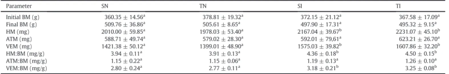
Table 1
Effect of exercise training on biometrical variables in control andT. cruziinfected rats.
Parameter SN TN SI TI
Initial BM (g) 360.35 ± 14.56a 378.81 ± 19.32a 372.15 ± 21.12a 367.58 ± 17.09a
Final BM (g) 509.76 ± 36.86a 505.61 ± 8.65a 497.90 ± 17.31a 495.32 ± 9.15a
HM (mg) 2010.00 ± 59.85a 1978.03 ± 53.40a 2167.04 ± 39.67b 2231.07 ± 45.10b
ATM (mg) 588.71 ± 49.74a 579.02 ± 28.30a 592.01 ± 79,61a 623.21 ± 26.70a
VEM (mg) 1421.38 ± 50.12a 1399.01 ± 48.90a 1575.03 ± 39.82b 1607.86 ± 32.20b
HM:BM (mg/g) 3.94 ± 0.11a 3.91 ± 0.13a 4.36 ± 0.18b 4.50 ± 0.15b
ATM:BM (mg/g) 1.15 ± 0.22a 1.15 ± 0.06a 1.19 ± 0.13a 1.26 ± 0.10a
VEM:BM (mg/g) 2.80 ± 0.24a 2.77 ± 0.11a 3.18 ± 0.21b 3.25 ± 0.08b
Data are expressed as mean ± SD. SN, sedentary no infected; SI, sedentary infected; TN, trained no infected; TI, trained infected. BM, body mass; HM, heart mass; ATM, atrium mass; VEM, ventricle mass.a,b,cDifferent letters in rows indicate statistical difference between the groups (p
(Ionoptix, CCD100V Myo-Cam, MA, USA). Cell shortening was measured using a real-time motion edge detection system (Ionoptix, MA, USA) [27]. From consecutive cardiomyocytes contractions (8 to 16), cell shortening, maximal rate of contraction and relaxation were calculated [28].
2.10. Cytokines immunoassay
The cytokine concentrations in serum and myocardium were mea-sured by ELISA. The measurements in serum were performed for all an-imals 24 h after the last exercise session. Nine weeks afterT. cruzi inoculation, RA and LV fragments were removed from 6 animals per group and used in this immunoassay. The cytokines TNF-α, IFN-γ, IL-6 and the chemokines CCL-2/MCP-1 and CX3CL1 were assayed following the manufacturer's instructions (Promega, Madison, WI, USA). The reac-tion was revealed using peroxidase-conjugated streptavidin (Vector Lab., CA, USA) and the substrate containing 3.3′,5,5″ -tetramethylbenzidine (Promega, WI, USA). Optical densities were read by spectrophotometry at 450 nm, and the levels of cytokines were
calculated by extrapolating the optical densities obtained from a stan-dard curve using recombinant cytokines.
2.11. Oxidative/nitrosative stress markers
The same RA and LV fragments used in the cytokine immunoassays were used for assessing oxidative and nitrosative markers. The status of lipid peroxidation was determined by analyze the tissue levels of malondialdehyde (MDA). In this analysis, 100 mg of frozen heart was homogenized in PBS and incubated with thiobarbituric acid to deter-mine the levels of thiobarbituric acid-reactive substances[29]. Protein oxidation was verified by the quantification of protein carbonyls in car-diac tissue using the 2.4-dinitrophenylhydrazine (DNPH) procedure [30]. Catalase (CAT) and Superoxide dismutase (SOD) were analyzed to estimate the antioxidant status of the cardiac tissue. CAT activity was evaluated by measuring the rate of decomposition of hydrogen per-oxide (H2O2), according to the protocol described by Aebi[31]. SOD ac-tivity was estimated by a method described by Sarban et al.[32], in which xanthine oxidase produce H2O2reducing nitroblue tetrazolium. Nitric oxide (NO) was indirectly determined by the quantification of ni-trite/nitrate levels in serum and cardiac tissue using the Griess method [33]. Total protein levels were measured using the Bradford method [34].
2.12. Statistical analysis
Data were reported as mean and standard deviation (mean ± SD). Data distribution was verified by D'agostino-Pearson test. Parameters of exercise tolerance, biometry, biochemical and morphological data, were compared using analysis of variance ANOVA one-way followed by Tukey'spost-hoctest. Parasitemia, cytokines and cell contractile func-tion were compared using the Kruskal-Wallis test. A probability of pb0.05 was considered statistically significant.
3. Results
The initial andfinal body mass of the animals was similar in all groups (pN0.01). In both groups infected withT. cruzithere was a
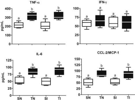
Fig. 3.Effect of exercise training on serum levels of cytokines and chemokines in rats before infection withT. cruzi. SN, sedentary not infected; SI, sedentary infected; TN, trained not infected; TI, trained infected. Box: interquartile interval, horizontal line: median, whiskers: superior and inferior quartiles.a,b,cDifferent letters in the columns denote statistical
difference among the groups (pb0.05).
Fig. 2.Effect of exercise training on metabolic adaptations in rats beforeT. cruziinfection. SN, sedentary not infected; SI, sedentary infected; TN, trained not infected; TI, trained infected. Data are expressed as mean ± standard deviation. The dotted boxes indicate the lactate threshold in both groups investigated.
significant increase in the absolute and relative whole heart and ventric-ular mass compared to that of control animals (pb0.01) (Table 1).
As shown inFig. 1, the initial level of physical performance obtained in the incremental running test was similar in all groups. Nine weeks after the treadmill running program, all variables of physical perfor-mance were significantly higher in both trained groups than those in sedentary animals (pb0.01).
The lactate threshold was reached late in trained animals compared to animals that remained sedentary during the experimental period (Fig. 2). At the beginning of the experiment, the lactate threshold oc-curred at similar time in both groups (data not shown).
Twenty-four hours after the last training session, exercised animals presented high levels of TNF-α, IL-6 and CCL2/MCP-1 (pb0.01), how-ever they had similar values for IFN-γcompared to control animals (pN0.05) (Fig. 3).
After inoculation withT. cruzi, blood parasitism was detected on day five in all infected animals, which coincided with the peak of parasitemia in trained animals (Fig. 4). In the sedentary group, the peak of parasitemia was observed on the sixth day. Parasitemia was
higher in sedentary (mean 14.42 ± 6.10; median 10.13 trypomastigotes/0.1 mL of blood × 103) compared to trained (mean 8.41 ± 4.84; median 6.00 trypomastigotes/0.1 mL of blood × 103) animals (pb0.05).
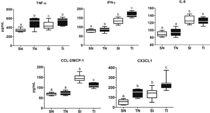
Nine weeks after exercise training, cardiac levels of TNF-αand CX3CL1 were higher in trained compared to sedentary uninfected animals (Fig. 5). All infected animals presented high levels of TNF-α, IFN-γ, IL-6, CCL2/MCP-1, and CX3CL1 compared to those of the control sedentary animals (pb0.01). In trained and infected animals, the cardiac levels of TNF-α, IFN-γand CX3CL1 were higher, CCL2/MCP-1 was lower (pb0.01) and IL-6 was similar compared to sedentary and infected animals (pN0.01).
Infected sedentary animals presented marked inflammatory infiltrate, cardiomyocyte hypotrophy (Fig. 6) and collagen content compared to the other groups (Table 2).
In all non-infected animals, thin collagenfibers were distributed in a well-organized, three-dimensional structure. Scanning electron microscopy revealed diffuse fibrosis with thick collagen fibers distributed in a disorganized pattern in all infected animals (Fig. 7). Tissuefibrosis was confirmed by spectrophotometric quantification of collagen in the myocardium (Table 2). All these variables showed lower values in infected and trained animals.
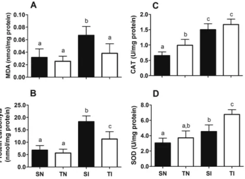
Myocardial levels of malondialdehyde and carbonyl proteins were similarly low in trained and sedentary uninfected control animals (pN0.05) (Fig. 8). Infected animals presented significant protein oxidation compared to the other groups (pb0.05). Lipid and protein oxidation was attenuated by exercise in infected animals (pb0.05). Ex-ercise training was also efficient in stimulating CAT (pb0.05) but not SOD activity (pN0.05) in the myocardium. In addition, trained and in-fected animals presented increased SOD activity (pb0.05), but similar results for CAT (pb0.05) compared to sedentary and infected animals. Both trained groups presented NO2−/NO3−serum levels significantly higher than sedentary animals (pb0.001). In uninfected animals, this adaptation was not maintained 9 weeks after stopping training (pN0.05). In infected animals, NO2−/NO3−levels in serum and myocar-dium were similar and significantly higher in trained and sedentary animals compared to those in both uninfected control groups (pN0.05) (Fig. 9).
Fig. 5.Effect of exercise training on cardiac levels of cytokines and chemokines in control andT. cruziinfected rats. SN, sedentary not infected; SI, sedentary infected; TN, trained not infected; TI, trained infected. Box: interquartile interval, horizontal line: median, whiskers: superior and inferior quartiles.a,b,cDifferent letters denote statistical difference among the
groups (pb0.05).
The morphological properties of isolated atrial and ventricular cardiomyocytes are shown inTable 3. The lower width and cellular vol-ume reinforces the evidence of cardiomyocyte hypotrophy in both in-fected groups. Sedentary, but not trained and inin-fected animals, also presented lower sarcomere length compared to the other groups.
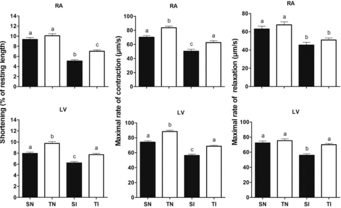
The biomechanical characteristics of isolated atrial and ventricular cardiomyocytes are shown inFig. 10. In uninfected and trained animals, it was observed a greater contraction velocity in atrial and ventricular cardiomyocytes and amplitude of shortening in ventricular cardiomyocytes (pb0.05). Sedentary and infected animals presented reduced cell shortening, contraction and relaxation velocity compared to control animals (pb0.05). In infected and trained animals, all param-eters investigated were higher compared to the animals that remain sedentary (pb0.05), except relaxation velocity in atrial cardiomyocytes (pN0.05).
4. Discussion
In the present study, by modulating the cardiac inflammatory pro-cess and oxidative status, exercise training appears to be a potential ef-fective complementary, non-pharmacological strategy to change the pathological repercussions of ChC. Our results suggested that that exer-cise training may induce beneficial adaptations to the host, which seems to be related to the increased production of Th1 cytokines (i.e. IFN-γand
TNF-α), reduction in parasitemia levels, cardiac parasitism, infl amma-tion,fibrosis and cell atrophy. Concurrently, trained animals presented increased activities of antioxidant enzymes, reduced cardiac oxidative damage and cardiomyocyte contractile dysfunction in infected rats.
As expected, both trained groups presented better physical perfor-mance and metabolic adaptation than sedentary animals, which was ac-companied by increased circulating levels of IL-6 and TNF-αand lower parasitemia. Previous studies have recognized the immunomodulatory effects of physical training by demonstrating that chronic exercise en-hances leukocyte function indexes, including the“oxidative burst”of neutrophils and monocytes, TCD4+/TCD8+ lymphocyte ratio and pro-liferation, antibody synthesis, and cytotoxic activity of NK cells[15,35]. It has been shown that in response to infections physical training is able to stimulate the production of TNF-α, IFN-γ, IL-12, CCL2/MCP-1, CCL3/MIP-1αand CCL4/MIP-1β, molecules involved in the polarization to a Th1 immune response pattern[14,15], which is essential to pro-mote cell-mediated immunity againstT. cruzi[12,16].Thus, the reduced parasitemia observed here can be potentially related to high expression of Th1 cytokines (especially IFN-γand TNF-α), an opposite effect when Th2 cytokines (i.e., IL-10, IL-4 and TGF-β) are predominant, since these molecules increase the susceptibility to infection[16,36]. However, it should be considered that the expression of Th2 response could modu-late the tissue damage caused by the immune response directed against the parasite[16].
Table 2
Effect of preinfection treadmill training on collagen content and myocardial cellularity in control andT. cruziinfected rats.
Variables SN TN SI TI
Right atrium
Collagen (μg/mg protein) 17.52 ± 1.40a 16.77 ± 1.07a 28.60 ± 1.55b 23.71 ± 1.13c
MC (cells/170 × 103μm2) 101.15 ± 15.65a 117.90 ± 18.95a 356.40 ± 14.01b 179.60 ± 16.62c Left ventricle
Collagen (μg/mg protein) 20.59 ± 1.33a 18.86 ± 1.13a 30.93 ± 1.85b 25.29 ± 1.17c
MC (cells/170 × 103μm2) 94.10 ± 19.49a 115.25 ± 17.03a 625.60 ± 84.57b 403.00 ± 71.65c
SN, sedentary no infected, SI, sedentary infected, TN, trained no infected; TI, trained infected. MC, myocardial cellularity. Data are expressed as mean ± standard deviation.a,b,cDifferent
letters in rows indicate statistical differences between the groups for the same cardiac segment (pb0.05).
Fig. 6.Representative photomicrographs of the left ventricle from control (A and B) and infected (C and D) rats (DAPI and Phalloidin staining under epi-fluorescence microscopy). (A) SN, sedentary not infected; (B) TN, trained not infected; (C) SI, sedentary infected; (D) TI, trained infected. Intracellular amastigotes ofT. cruziwere identified in both infected groups (highlighted in panels C and D, H&E staining).
In addition to probably modifying the plasma levels of cytokines, ex-ercise training seems to reduce cardiac parasitism, inflammation andfi -brosis. In Chagas disease, the establishment of a pro-inflammatory response is critical in controlling the intense parasitism of the acute phase, however, this response often leads to the installation of a fibrosing cardiomyopathy in the chronic phase[4,37]. In humans with chronic heart failure, physical training was able to reduce CCL2 serum levels, an event associated with reduced macrophage recruitment, in-flammation and heart failure progression[38]. A similarfinding was ob-served in the present study, in which infected and trained animals presented lower CCL2 levels and tissue cellularity compared to infected
and sedentary animals. In individuals with coronary artery disease, physical training was effective in stimulating an anti-inflammatory cardioprotective phenotype, reducing IL-1, IL-6, and IFN-γand increas-ing IL-10 circulatincreas-ing levels, a potent anti-inflammatory cytokine[39]. In humans with chronic heart failure, physical training also reduced TNF-α and IL-6 plasma levels[38], which are cytokines that directly modulate tissue inflammation and cardiac function, in addition to affecting the production of reactive oxygen (ROS) and nitrogen (RNS)-derived spe-cies that are involved in the pathogenesis of ChC[35,38].
As expected, NO serum levels were increased in trained animals, a mechanism admittedly associated with an increased tissue expression
Fig. 8.Effect of exercise training on the levels of oxidative markers and activity of antioxidant enzymes in the myocardium from control andT. cruziinfected rats. SN, sedentary not infected; SI, sedentary infected; TN, trained not infected; TI, trained infected. Data are expressed as mean ± standard deviation. As there were no statistical differences between the right atrium and left ventricle, the data represent the average for both cardiac segments.a,b,cDifferent letters in the columns denotes statistical difference between the groups (p
b0.001).
of inducible and endothelial nitric oxide synthase (iNOS and eNOS) trig-gered by exercise training[13,35,40]. Although, this adaptation has been transitory, the host ability to produce NO in response toT. cruzi in-fection was not affected by exercise training. In addition, significant re-active tissue damage (i.e., MDA and protein carbonyls) was observed in infected animals, which was attenuated by exercise training. In humans [17]and animals[41,42], the installation of a oxidant status of pro-gressive intensity overT. cruziinfection has been described, in which the endogenous antioxidant reservation is not sufficient to counteract the high ROS and RNS production and limit tissue damage[41–45]. It has been demonstrated that the recruitment of inflammatory cells to car-diac tissue activates a respiratory burst that culminates in the intensive formation of ROS such as superoxide anions (O2–), hydroxyl radicals (OH–) and hydrogen peroxide (H
2O2)[42,43]. These reactive species are also produced by cardiomyocytes, an event that not only represents a host defense mechanism against the parasite, but a mitochondrial dys-function that occurs during cell parasitism. Thus, ROS/RNS production by cardiomyocytes seems to be a susceptibility factor for myocardial damage, cell death, increased inflammation and myocardial dysfunction in ChC[42–45], which can be mitigated by antioxidant strategies[41].
In the present study, the increased activity of antioxidant enzymes seems to constitute a remarkable cardiac adaptation induced by exer-cise training in infected animals. Operating predominantly on skeletal and cardiac muscle, it is recognized that chronic exercise can increase the expression and activity of antioxidant enzymes such as superoxide dismutase, glutathione peroxidase, glutathione-S-transferase and cata-lase, enhancing tissue efficiency in neutralize the excess of ROS/RNS [13,46]. Although in cardiomyopathies with other etiologies there is a compensatory reaction of antioxidant enzymes to neutralize the excess of reactive species, this mechanism is limited in Chagas disease and seems to be insufficient to prevent reactive myocardial damage and
inflammatory cardiomyopathy progression[42,43]. In humans, the ex-tent to which ChC worsens, there is a gradual reduction in GPX, SOD and GST activities in blood elements[47]. Due to a reduction in expres-sion of these enzymes in more advanced stages of heart failure, endog-enous antioxidant insufficiency seems to contribute to the progression of ChC[44,47]. Thus, by enhancing the enzymatic antioxidant defenses, physical training could be potentially beneficial in mitigating oxidative tissue damage and the severity of ChC.
It is known that cytokines and chemokines can change the redox balance in multiple organs[42,48]. There is evidence thatT. cruzi expo-sure increases the circulating levels of TNF-α, IL-1 and IL-6, which par-ticipate in parasite control by stimulating ROS/RNS synthesis[42,43]. Our results corroborated this association between cytokines and ROS/ RNS in infected sedentary animals, which presented high levels of TNF-αand IL-6 together with increased lipid and protein oxidative damage. In cardiomyocytes infected withT. cruzi, the production of re-active species can be increased by TNF-α, IFN-γand IL-1β, which does not occur when non-infected cardiomyocytes are treated with these molecules[42,43]. A key role has been reported for TNF-αand IFN-γ in inducing NO synthesis in animals infected withT. cruzi, a molecule di-rectly involved in host resistance to infection and in the control of parasitemia and cell parasitism[39,47]. In humans, TNF-αand NO levels were inversely correlated with the levels of antioxidant enzymes SOD and GPx, and directly correlated with the severity of ChC[47], a propo-sition apparently applicable to sedentary but not trained animals.
Currently, investigations on the intrinsic contractile properties of cardiac myocytes in Chagas disease are scarce, constituting a neglected issue. In this study,T. cruziinfection markedly impaired cardiomyocyte contractility, which seems to be partially restored by exercise training. Our results support the evidence of a potential negative influence of re-active stress on cardiomyocyte contractility. Studies conducted by our
Table 3
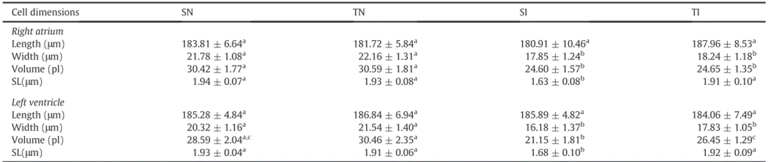
Effect of preinfection treadmill training on morphological parameters of isolated cardiomyocytes from control andT. cruziinfected rats.
Cell dimensions SN TN SI TI
Right atrium
Length (μm) 183.81 ± 6.64a 181.72 ± 5.84a 180.91 ± 10.46a 187.96 ± 8.53a
Width (μm) 21.78 ± 1.08a 22.16 ± 1.31a 17.85 ± 1.24b 18.24 ± 1.18b
Volume (pl) 30.42 ± 1.77a 30.59 ± 1.81a 24.60 ± 1.57b 24.65 ± 1.35b
SL(μm) 1.94 ± 0.07a 1.93 ± 0.08a 1.63 ± 0.08b 1.91 ± 0.10a Left ventricle
Length (μm) 185.28 ± 4.84a 186.84 ± 6.94a 185.89 ± 4.82a 184.06 ± 7.49a
Width (μm) 20.32 ± 1.16a 21.54 ± 1.40a 16.18 ± 1.37b 17.83 ± 1.05b
Volume (pl) 28.59 ± 2.04a,c 30.46 ± 2.35a 21.15 ± 1.81b 26.45 ± 1.29c
SL(μm) 1.93 ± 0.04a 1.91 ± 0.06a 1.68 ± 0.10b 1.92 ± 0.09a
SN, sedentary no infected, SI, sedentary infected, TN, trained no infected; TI, trained infected. SL, sarcomere length. Data are expressed as mean ± standard deviation.a,b,cDifferent letters in
rows indicate statistical difference between the groups for the same cardiac segment (pb0.05).
Fig. 9.Effect of exercise training on nitrite/nitrate (NO2−/NO3−) levels in serum and cardiac tissue from control andT. cruziinfected rats. SN, sedentary not infected; SI, sedentary infected;
TN, trained not infected; TI, trained infected. Data are expressed as mean ± standard deviation. As there were no statistical differences between the right atrium and left ventricle, the data represent the average for both cardiac segments.a,b,cDifferent letters in the columns denotes statistical difference between the groups for the same cardiac segment (p
b0.001).
research group revealed for thefirst time thatT. cruziinfection changes the intrinsic mechanical properties of cardiomyocytes, reducing the am-plitude and cell contraction velocity[21,23,28]. Apparently, these changes are unstable and are influenced by the profile of cytokine ex-pression, which varies with infection[28]. Thus, high IFN-γ, TNF-α and MCP-1/CCL2 serum levels were correlated with poor contractile re-sponses in cardiomyocytes fromT. cruziinfected mice, indicating an im-portant role of these molecules in modulating cell biomechanics in experimental Chagas disease[28]. It has been proposed that contractil-ity dysfunctions in ChC may also be associated with pro-oxidant events that generate cumulative damage to myocardium biomolecules, which can determine morphofunctional changes in cardiomyocytes and even-tually cell death [23]. In fact, the morphological properties of cardiomyocytes (i.e., width, volume and sarcomere length) were im-paired in infected and sedentary animals, which seems to be partially at-tenuated by physical training. As cell morphology is directly associated with cardiomyocyte biomechanics, it is possible to assume that by restricting cell atrophy and geometric disorders in sarcomeric unities, exercise training can improve cardiomyocyte contractility, counteracting the typical cardiac catabolic pattern observed inT. cruzi infection. Our research group also found that reactive tissue damage can be associated with profound cardiac pathological remodeling, espe-cially stromal expansion and microvascular damage, events potentially associated with cardiomyocyte biomechanical dysfunction inT. cruzi in-fection[23]. Thus, by enhancing the antioxidant defenses and limiting oxidative tissue damage, exercise training could attenuate the installa-tion of cardiac morphological abnormalities, especially the remarkable myocardiumfibrosis, cell atrophy, and cardiomyocytolysis, which to-gether with severe inflammation represent peculiar pathological as-pects in ChC[5,37].
5. Conclusion
Taken together, the results suggested that pre-infection exercise training could be a relevant complementary non-pharmacological
strategy to change the pathological repercussions of ChC. By modulating the immune response and enhancing the efficiency of endogenous anti-oxidant mechanisms, exercise training seems to improve the host resis-tance to infection. This is probably associated with multiple factors such as reduction of parasitemia, parasitism, cardiac inflammation,fibrosis, and reactive tissue damage, with positive repercussions to cardiomyo-cyte contractile function in rats.
Conflict of interest statements
There are no conflicts of interest. All authors contributed to data col-lection and analysis, article preparation and have approved thefinal manuscript.
Acknowledgments
This work was supported by the Brazilian funding agencies Conselho Nacional de Desenvolvimento Científico e Tecnológico (CNPq) (454901/ 2014-3) and Fundação de Amparo à Pesquisa do Estado de Minas Gerais (FAPEMIG) (APQ-02309-14). The authors André Talvani and Antônio J. Natali have research fellowships from Conselho Nacional de Desenvolvimento Científico e Tecnológico (CNP-q), Brazil. The authors thank the“Núcleo de Microscopia and Microanálise - NMM”of the Federal University of Viçosa by assistance in scanning electron microscopy.
References
[1] M.T. Bahia, I.M. de Andrade, T.A. Martins, et al., Fexinidazole: a potential new drug candidate for Chagas disease, PLoS Negl. Trop. Dis. 6 (2012), e1870.
[2] M.C. Nunes, W. Dones, C.A. Morillo, J.J. Encina, A.L. Ribeiro, Council on Chagas disease of the Interamerican Society of Cardiology, Chagas disease: an overview of clinical and epidemiological aspects. J. Am. Coll. Cardiol. 62 (2013) 767–776.
[3] WHO, World Health Organization, Chagas disease American trypanosomiasis, Fact sheet 340 (2015).
[4] A. Rassi-Jr, A. Rassi, J.A. Marin-Neto, Chagas disease, Lancet 375 (2010) 1388–1402.
Fig. 10.Effect of exercise training on cardiomyocytes contractility in control andT. cruziinfected rats. SN, sedentary not infected; TN, trained not infected; SI, sedentary infected; TI, trained infected; RA, right atrium; LV, left ventricle. Data are expressed as mean ± standard error. Were evaluated eight animals per group.a,b,cDifferent letters in the columns denotes statistical
[5] R.A. Guerri-Guttemberg, D.R. Grana, G. Ambrosio, J. Milei, Chagas cardiomyopathy: Europe is not spared! Eur. Heart J. 29 (2008) 2587–2591.
[6] P.J. Hotez, E. Dumonteil, L. Woc-Colburn, et al., Chagas disease:“the new HIV/AIDS of the Americas”, PLoS Negl. Trop. Dis. 6 (2012), e1498.
[7] J.R. Coura, Present situation and new strategies for Chagas disease chemotherapy: a proposal, Mem. Inst. Oswaldo Cruz 104 (2009) 549–554.
[8] M.M. Lima, M.O. Rocha, M.C. Nunes, et al., A randomized trial of the effects of exer-cise training in Chagas cardiomyopathy, Eur. J. Heart Fail. 12 (2010) 866–873.
[9] M.M. Lima, M.C. Nunes, B. Nascimento, et al., Improvement of the functional capac-ity is associated with BDNF and autonomic modulation in Chagas disease, Int. J. Cardiol. 167 (2013) 2363–2366.
[10] B.R. Nascimento, M.M. Lima, Mdo.C. Nunes, et al., Effects of exercise training on heart rate variability in Chagas heart disease, Arq. Bras. Cardiol. 103 (2014) 201–208.
[11] M.M. Lima, M.C. Nunes, M.O. Rocha, F.R. Beloti, M.C. Alencar, A.L. Ribeiro, Left ven-tricular diastolic function and exercise capacity in patients with Chagas cardiomyop-athy, Echocardiography 27 (2010) 519–524.
[12] C. Schebeleski-Soares, R.C. Occhi-Soares, S.M. Franzói-de-Moraes, et al., Preinfection aerobic treadmill training improves resistance againstTrypanosoma cruziinfection in mice, Appl. Physiol. Nutr. Metab. 34 (2009) 659–665.
[13] S.K. Powers, S.L. Lennon, J. Quindry, J.L. Mehta, Exercise and cardioprotection, Curr. Opin. Cardiol. 17 (2002) 495–502.
[14] T. Lowder, D.A. Padgett, J.A. Woods, Moderate exercise protects mice from death due to influenza virus, Brain Behav. Immun. 19 (2005) 377–380.
[15] C. Malm, Exercise immunology: the current state of man and mouse, Sports Med. 34 (2004) 555–566.
[16] M.M. Teixeira, R.T. Gazzinelli, J.S. Silva, Chemokines, inflammation andTrypanosoma cruziinfection, Trends Parasitol. 18 (2002) 262–265.
[17] L.B. Maçao, F.D. Wilhelm, R.C. Pedrosa, et al., Antioxidant therapy attenuates oxida-tive stress in chronic cardiopathy associated with Chagas' disease, Int. J. Cardiol. 123 (2007) 43–49.
[18] C.P. Silva, C.H. Del Carlo, M.T. Oliveira Junior, Why do patients with chagasic cardio-myopathy have worse outcomes than those with non-chagasic cardiocardio-myopathy? Arq. Bras. Cardiol. 91 (2008) 358–362.
[19] W. Lunz, M.C.G. Peluzio, C.M.G.C. Dias, A.P.B. Moreira, A.J. Natali, Long-term aerobic swimming training impairs the development of colon cancer in rats treated with 1.2-dimethyl-hydrazine, Braz. J. Med. Biol. Res. 41 (2008) 1000–1004.
[20] L.G. Koch, S.L. Britton, Artificial selection for intrinsic aerobic endurance running ca-pacity in rats, Physiol. Genomics 5 (2001) 45–52.
[21] R.D. Novaes, A.R. Penitente, R.V. Gonçalves, Effects ofTrypanosoma cruziinfection on myocardial morphology, single cardiomyocyte contractile function and exercise tol-erance in rats, Int. J. Exp. Pathol. 92 (2011) 299–307.
[22] APS, American Physiological Society, Resource Book for the Design of Animal Exer-cise Protocols, 2006.
[23]R.D. Novaes, A.R. Penitente, R.V. Gonçalves, et al.,Trypanosoma cruziinfection in-duces morphological reorganization of the myocardium parenchyma and stroma, and modifies the mechanical properties of atrial and ventricular cardiomyocytes in rats, Cardiovasc. Pathol. 22 (2013) 270–279.
[24] Z. Brener, Therapeutic activity and criterion of cure on mice experimentally infected withTrypanosoma cruzi, Rev. Inst. Med. Trop. São Paulo 4 (1962) 389–396.
[25] M.A. Rossi, M.A. Abreu, L.B. Santoro, Images in cardiovascular medicine. Connective tissue skeleton of the human heart: a demonstration by cell-maceration scanning electron microscope method, Circulation 97 (1998) 934–935.
[26] A. López-De León, M. Rojkind, A simple micromethod for collagen and total protein determination in formalin-fixed paraffin-embedded sections, Histochem. Cytochem. 33 (1985) 737–743.
[27] A.J. Natali, L.A. Wilson, M. Peckham, D.L. Turner, S.M. Harrison, E. White, Different re-gional effects of voluntary exercise on the mechanical and electrical properties of rat ventricular myocytes, J. Physiol. 541 (2002) 863–875.
[28] D. Roman-Campos, H.L.L. Duarte, P.A. Sales-Jr, et al., Changes in cellular contractility and cytokines profile duringTrypanosoma cruziinfection in mice, Basic Res. Cardiol. 104 (2009) 238–246.
[29]J.A. Buege, S.D. Aust, Microsomal lipid peroxidation, Methods Enzymol. 52 (1978) 302–310.
[30] R.L. Levine, D. Garland, C.N. Oliver, et al., Determination of carbonyl content in oxi-datively modified proteins, Methods Enzymol. 186 (1990) 464–478.
[31] H. Aebi, Catalasein vitro, Methods Enzymol. 105 (1984) 121–126.
[32]S. Sarban, A. Kocyigit, M. Yazar, U.E. Isikan, Plasma total antioxidant capacity, lipid peroxidation, and erythrocyte antioxidant enzyme activities in patients with rheu-matoid arthritis and osteoarthritis, Clin. Biochem. 38 (2005) 981–986.
[33] D. Ricart-Jané, M. Llobera, M.D. López-Tejero, Anticoagulants and other preanalytical factors interfere in plasma nitrate/nitrite quantification by the Griess method, Nitric Oxide 6 (2002) 178–185.
[34] M.M. Bradford, A rapid and sensitive method for the quantitation of microgram quantities of protein utilizing the principle of protein dye-binding, Anal. Biochem. 7 (1976) 248–254.
[35] M. Gleeson, Immune function in sport and exercise, J. Appl. Physiol. 103 (2007) 693–699.
[36]W. Savino, D.M. Villa-Verde, D.A. Mendes-da-Cruz, et al., Cytokines and cell adhe-sion receptors in the regulation of immunity toTrypanosoma cruzi, Cytokine Growth Factor Rev. 18 (2007) 107–124.
[37] M.B. Soares, R.S. de Lima, L.L. Rocha, Gene expression changes associated with myo-carditis andfibrosis in hearts of mice with chronic chagasic cardiomyopathy, J. In-fect. Dis. 202 (2010) 416–426.
[38] S. Adamopoulos, J. Parissis, D. Karatzas, et al., Physical training modulates proinfl am-matory cytokines and the soluble Fas/soluble Fas ligand system in patients with chronic heart failure, J. Am. Coll. Cardiol. 39 (2002) 653–663.
[39] R.T. Gazzinelli, I.P. Oswald, S. Hieny, S.L. James, A. Sher, The microbicidal activity of interferon-gamma-treated macrophages againstTrypanosoma cruziinvolves an L-arginine-dependent, nitrogen oxide-mediated mechanism inhibitable by interleukin-10 and transforming growth factor-beta, Eur. J. Immunol. 22 (1992) 2501–2506.
[40] B.A. Kingwell, Nitric oxide-mediated metabolic regulation during exercise: effects of training in health and cardiovascular disease, FASEB J. 14 (2000) 1685–1696.
[41] J.J. Wen, N.J. Garg, Mitochondrial generation of reactive oxygen species is enhanced at the Qo site of the complex III in the myocardium ofTrypanosoma cruzi-infected mice: beneficial effects of an antioxidant, J. Bioenerg. Biomembr. 40 (2008) 587–598.
[42] S. Gupta, V. Bhatia, J.J. Wen, Y. Wu, M.H. Huang, N.J. Garg,Trypanosoma cruzi infec-tion disturbs mitochondrial membrane potential and ROS producinfec-tion rate in cardiomyocytes, Free Radic. Biol. Med. 47 (2009) 1414–1421.
[43] S. Gupta, J.-J. Wen, N.J. Garg, Oxidative stress in Chagas disease, Inter. Perspect. In-fect. Dis ID190354 (2009) 1–8.
[44] J.J. Wen, G. Vyatkina, N.J. Garg, Oxidative damage during chagasic cardiomyopathy development: role of mitochondrial oxidant release and inefficient antioxidant de-fense, Free Radic. Biol. Med. 37 (2004) 1821–1833.
[45] J.J. Wen, P.C. Yachelini, A. Sembaj, R.E. Manzur, N.J. Garg, Increased oxidative stress is correlated with mitochondrial dysfunction in chagasic patients, Free Radic. Biol. Med. 41 (2006) 270–276.
[46]A.M. Niess, H.H. Dickhuth, H. Northoff, E. Fehrenbach, Free radicals and oxidative stress in exercise–immunological aspects, Exerc. Immunol. Rev. 5 (1999) 22–56.
[47] R. Pérez-Fuentes, J.F. Guégan, C. Barnabé, et al., Severity of chronic Chagas disease is associated with cytokine/antioxidant imbalance in chronically infected individuals, Int. J. Parasitol. 33 (2003) 293–299.
[48] B. Halliwell, J.M.C. Gutteridge, Free Radical in Biology and Medicine, third ed. Oxford University Press, Oxford, 1999.