HEPATITIS A: IN VITRO ISOLATION OF AN AGENT WITH UNUSUAL GROWTH REQUIREMENTS FROM A CLINICAL CASE OCCURRING IN COSTA RICA’ ’ 2
W. Pelon,Ph.D.;3 J.H. Miller, Ph.D.;‘+ and J.E. Deas, M.S.5
An entevovirus-like agent with atypical properties has been isolated in vitro from a hepatitis case in Costa Rica. Many of
its pvopevties ave consistent with hepatitis agents isolated in marmosets and observed in human feces.
Introduction
This communication describes a viral agent isolated in cell cultures that had been maintain- ed under special conditions following inocula- tion with a fecal specimen from a case of clinical hepatitis in Costa Rica. Investigations have indicated that this agent possesses several characteristics which distinguish it from known enterovirus types.
Picornaviruses are small (15-30 nm) ribo- nucleic acid-containing viruses which are fur- ther subdivided into rhinoviruses and entero- viruses (I). The former are differentiated from the latter on the basis of significant reductions in infectious virus titer following exposure to pH 3.0 for 1-3 hours (2). Despite one uncon- firmed report of rhinovirus isolation from feces, attempts at isolation from fecal samples col- lected from infected patients and volunteers
‘Supported in part by the United States Army Medical Research and Development Command (Con- tract DADA 17-68-C-8023) and in part by the Edward G. Schlieder Educational Foundation and Public Health Service Research Grant AI-02347. The findings of this renort are not to be construed as an official position of any department of the- United States Government unless so designated by authorized docu- ments.
2Also appearing in Spanish in Boletin de la Oficina Sanitaria Panamericana.
3Associate Professor, Department of Microbiology and Deoartment of Tronical Medicine and Medical Parasitology, Louisiana Siate University Medical Cen- ter, 1542 Tulane Avenue, New Orleans, Louisiana.
4Professor,, Department of Tropical Medicine and Medical Parasrtology, Louisiana State University Medi- cal Center.
5Medical Research Specialist, Department of Tro- pical Medicine and Medical Parasitology, Louisiana State University Medical Center.
have not been successful (3). In addition, all rhinovirus types tested have been found to be acid-labile (2).
To date, despite persistent efforts, all at- tempts at in vitro isolation of the HeA virus or viruses responsible for hepatitis A have been unsuccessful. However, an agent having picorna- virus-like characteristics has been transmitted to marmosets from HeA cases (4, 5). Also, a spherical virus-like antigen (diameter 27 nm) was reported recently in the feces of a human volunteer with HeA (6).
Current enterovirus isolation methods (7) have not succeeded in detecting the HeA agent. Since these employ culture media containing 0.11 to 0.22 per cent NaHCO, the use of a more stable acid environment for HeA virus isolation was explored. The rest of this article is devoted to reporting the course and outcome of that work.
Methods and Results
Fecal Samples Tested
Fecal samples were collected for a double- blind study by the staff of the Louisiana State University International Center for Medical Research and Training (LSU-ICMRT) located in San Jose, Costa Rica. Specimens were obtained from 35 hepatitis A cases occurring in a geographic area found to be endemic for the disease (8, 9). Fifty-five control specimens were also collected from individuals living outside the endemic area. All specimens were prepared as 10 per cent extracts.
Pelon, et al. HEPATITIS A-ISOLATION OF AN AGENT 213
Virus Isolation Studies
All fecal samples were inoculated into prima- ry cultures of Rhesus monkey renal cells and into cultures of HEp-2 cells for enterovirus isolation (7). Because of our interest in viruses that could not be isolated in this way, no further attention was given to specimens from which virus isolations were obtained. The re- maining specimens were tested in cultures of human embryonic diploid lung cell WI-38.
Medium I. Confluent cultures of cells were maintained in Eagle’s basal medium (EBM), which was prepared in Earle’s balanced salt solutitin (Earle’s BSS) with 10 per cent fetal calf serum (FCS) and 0.11 per cent NaHCO, (final concentrations). The cultures were then inoculated, incubated at 36OC, and observed for cytopathic effects (CPE). The pH of the medium ranged from 7.4 at the time of inoculation to 6.6 following three days of incubation.
Medium II. In a parallel study, cell cultures were maintained in a serum-free EBM, which was prepared in a glucose-free Hanks’ BSS, gassed with CO, and inoculated. Following incubation at 36”C, the acidity of this medium stabilized at approximately pH 6.8 and re- mained unchanged during the period of incuba- tion. Fecal extracts frequently were toxic to cells maintained in this serum-free acidic envi- ronment, necessitating the passage of fluids from degenerated cultures into fresh cell cul- tures being maintained under similar condi- tions.
Findings. Most of the fecal extracts were found to produce CPE only in the conventional medium (I), or in both I and II. In one instance, however, CPE were observed only among cul- tures maintained in the stable glucose-free acid environment of Medium II. This result was obtained in cultures inoculated with a fecal extract designated CR69(076) (10). It was later determined that the specimen in question had been collected from a seven-year-old male, whose case had been clinically diagnosed as hepatitis A. The agent responsible for the observed CPE in Medium II was successfully
reisolated from this same fecal specimen, utiliz- ing the procedures described below.
Replication
of
CR69(076) VirusDifficulties in propagating the CR69(076) agent in medium I or II caused us to adopt the following methodology, which permitted rapid and consistent replication: Confluent mono- layer cultures of WI-38 cells were maintained in an EBM prepared from 100X concentrates of vitamin and amino acid mixtures and from a 200 millimolar solution of glutamine in modi- fied Hanks’ BSS containing an equivalent quan- tity of galactose in lieu of glucose (II, 12). This medium (Medium III), which was gassed with CO,, also included 0.035 per cent NaHCO,, 0.6 per cent FCS, sodium pyruvate (55 mg/ liter) (II), alanine (45 mg/liter) (II), and 5-bromodeoxy-uridine (BrDUR) (20 pg/ml). Two ml of the medium were added to each culture, and the cultures were allowed to incubate at 36V for 24 hours prior to inocula- tion. Following gassing, the pH of the medium was 6.0. Following incubation, the medium reached an equilibrium at pH 6.7-6.8.
With this method, infectious virus titers of CR69(076) were found to range from 106m9 to 107+4 tissue culture infectious doses (TCID, c) per ml. Maximum titers were usually reached by the fourth or fifth day of incubation following inoculation.
In brief, few or no CPE were observed in media containing the usual proportions of NaHCO, (0.11 or 0.22 per cent) or in media which had become alkaline owing to improper- ly sealed culture tubes. It was also observed that virus proliferation would occur, but at a lesser rate, if a medium containing a high concentration of NaHCO, was frst saturated with CO, and then added to cell cultures and inoculated.
214 PAHO BULLETIN * Vol. VIII, No. 3, 1974
inoculated cultures ranged from pH 7.4 at the time of inoculation to pH 6.6 following incuba- tion. To clarify this apparent inconsistency, parallel infectious virus titrations were con- ducted with cell cultures maintained in Medium I and with others maintained in Medium III, as described above. Because of the excessive acid pH that developed in the course of incubation, Medium I was changed on the third and sixth days of incubation.
The results of these titrations are presented in Table 1. It may be noted that the maximum infectious virus titer was almost attained after the third day of incubation in Medium III, while the amount of virus detected in the Medium I cultures was substantially less. After this latter medium was changed, the virus titer determined on the sixth day indicated that the amount of infectious virus had doubled; but only a slight increase was noted in the cultures containing Medium III during the same period. A second change of Medium I on the sixth day resulted in additional increase of the infectious virus titer, though to a lesser degree, by day nine. It should be mentioned here that if medium changes were not made, the CPE would regress; infected cells would become detached from the glass and enter the medium, leaving behind a cell sheet of normal appear- ance.
TABLE I-Infuence of different maintenance me-
dia upon the infectious virus titer of CR69 (076). Days of
incubation Medium Ia Medium IIIb
3 lo2.Sc lo6.27
6 104.95 106.7
9 105.35 106.87
aEBM in Earle’s BSS, 10% FCS, 0.11% NaHC03.
Medium changed in inoculated cultures on days 3 and 6 of incubation.
bEBM and Hank’s BSS (galactose), 0.6% FCS, 0.035% NaHCOJ, BrDUR (20 &ml), Na pyruvate (55 mg/l), alanine (45 mg/l), and gassed with CO2.
Medium not chanced duke incubation.
‘To determine more precisely the 50% tissue culture dose endpoint, each virus dilution was inocu- lated into 10 WI-38 tube cultures (0.2ml/tube), and the TClD50 endpoint calculated by the method of Reed and Muench.
These findirigs suggest that the CR69(076) agent not only requires an acid environment for its replication but replicates best inside a narrow acid pH range. In Medium I, cellular metabolism results in a rapid shift of the hydrogen ion concentration from the alkaline side of neutrality to the relatively acid side, quickly passing through that range optimal for viral replication and permitting only limited infection by CR69(076) to occur.
The replication of rhinovirus types requires an acid environment; but it also proceeds better at an incubation temperature of 33OC than at one of 36OC. Therefore, studies were con- ducted to determine whether a similar situation existed with CR69(076). Simultaneous dupli- cate titrations were carried out with CR69(076) virus stock. One titration was incubated at 33OC, the other at 36OC. Similar titrations were also carried out with ECHO 25 and these were incubated in the same manner. In both in- stances, higher infectious virus titers and more rapid virus proliferation were observed at 36oC than at 33OC.
Efforts to detect viral hemagglutinins with human “0” erythrocytes at various tempera-
tures (4OC, 20°C, and 37OC) utilizing normal saline and phosphate buffers (pH 5.7, 6.1, 6.5, 7.1, 7.5, and 8.0) as diluents were unsuccessful. Cytopathic Effects of CR69(076)
The CPE produced by CR69(076) in cul-
tures of WI-38 cells were similar to those
produced by enteroviruses. Infected cells- stained with Giemsa, and with hematoxylin and eosin-often showed swollen nuclei which later became pyknotic. Although neither cytoplas- mic nor nuclear inclusions were apparent, the cytoplasm of some cells contained eosinophilic masses like those seen in enterovirus-infected cells.
Pelon, et al. HEPATITIS A-ISOLATION OF AN AGENT 215
found to be optimal for CR69(076). Similar negative results were obtained with cultures of VERO, HEp-2, HeLa, and Chang liver cells. However, CPE were noted in cultures of a diploid human foreskin fibroblast cell line (HuFs-6j6, utilizing the methodology described for the replication of CR69(076).
Physical and Chemical Characteristics
of
CR69(076)
Unlike the enterovirus types, samples of CR69(076) showed a significant loss of infec- tious virus titer following storage at -7OOC.
Infected culture fluid, clarified by centrifu- gation, was distributed in screw-capped vials in 1 ml aliqudts and stored in a mechanical deep freezer at -7OOC. The vials were then removed periodically and their contents was titrated. As shown in Table 2, a substantial loss of infec- tious virus titer was observed following storage for three months. Subsequent lowering of the storage temperature to -90°C reduced the rate of loss.
The stability of infectious CR69(076) virus following exposure to 60°C for one hour was also investigated. Dilutions of CR69(076) and ECHO 25 were made with sterile deionized water. These were then incubated in a water bath at 60°C for one hour, placed in an ice bath, and inoculated into cultures of WI-38 cells, utilizing the medium most suitable for each virus type. No evidence of CPE was observed in cultures inoculated with dilutions of ECHO 25 exposed to 60°C, though CPE were observed with similar dilutions maintained at 20°C. In contrast, CPE were observed (and confirmed by passage) in one of four cultures of WI-38 cells inoculated with a 10m2 dilution of CR69(076), following its exposure to 60°C for 1 hour. CPE were not observed with other dilutions. A similar lack of complete inactiva- tion following exposure to 60°C for 1 hour has been reported for the marmoset HeA Agent (5).
CR69(076) samples showed no loss of infec- tivity titer in comparison to untreated controls
‘A product of North American Biologicals, Inc.
‘FABLE Z-Stability of CR69 (076) stock stored at -7ooc.
Months of storage
0 107.15
1 105*7
3 101.99
a10 WI-38 tube cultures/dilution.
following overnight exposure to 20 per cent diethyl ether at 4OC or following three-hour exposure to pH 3.0 at 20°C. Proliferation in cells previously exposed to BrDUR (20 ,ug/ml) confirmed the ribonucleic acid composition of this agent.
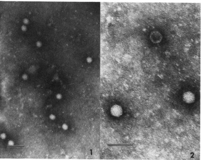
Electron micrographs of concentrated cell culture fluids infected with CR69(076) revealed icosahedral particles having diameters of ap- proximately 29 nm (see Plates 1 and 2). This is within the picomavirus size range and is con- sistent with the description of the virus-like antigen previously reported (6).
Relationship
of
CR69(076) to Known Entero- virusesWhen tested against type-specific enterovirus antisera by neutralization, CR69(076) pro- duced no inhibition with antisera for ECHO virus types l-24, 26, 27, 29-32; Coxsackie group A, types l-3,9; Coxsackie group B, types 1-6; and poliovirus, types l-3. However, rabbit antiserum for ECHO virus type 25 was found to neutralize 100 TCID,, of CR69(076) at a dilution of 1:400. By comparison, it was determined that this same antiserum would neutralize 1,000 TCID s,, of the homologous ECHO vims (type 2.5, strain JV-4) at a 1: 10,000 dilution.
216
PAHO BULLETIN
lVol. VIII, No. 3, I974
PLATES 1 and 2-Representative
samples of virus particles obtained from frozen/thawed
cell cultures centrifuged to settle cellular debris. The supematent was then centrifuged at
150,000 revolutions per hour for three hours, and the resulting pellet was suspended in 10 per
cent formalin and negatively stained with 2 per cent phosphotungstic acid. The bar in Plate 1
represents 100 nm and that in Plate 2 represents 60 nm.
to a minor degree, poliovirus types 1 and 2 (7).
Cross-neutralization studies of CR69(076) are
in progress.
Relationship
ofCR69(076) to HeA
Paired sera collected from disease cases that
occurred during epidemics of hepatitis A
(1967) and hepatitis B (1964) in Costa Rica (8,
9) were examined by neutralization and com-
plement-fixation procedures using CR69(076)
as the antigen (see Table 3). Of 20 paired sera
collected during the 1964 (hepatitis B) epi-
demic, only one pair showed a significant rise in
the antibody level of the convalescent serum. In
contrast, when paired sera from the 1967
(hepatitis A) epidemic were investigated, eight
out of 24 serum pairs (33 per cent) showed
significant increases in the antibody levels of
the convalescent sera. It is of interest to note
TABLE 3-Antibody
responses of paired sera from
44 Costa Rican hepatitis cases to CR69 (076).
Year
Hepatitis type of each
epidemic
No. of No. of paired sera with paired sera
tested
significant antibody resDonsesa
1964 B 20 l( 5%)
1967 A 24 8 (33%)
aAn
antibody level in the convalescent serum representing at least a fourfold rise.that subsequent investigations of sera for Aus-
tralia antigen revealed the nature of the epi-
demics (9).
Discussion
The enterovirus-like nature of the HeA agent
isolated in marmosets (.5), the demonstration
Moon, et al. HEPATITIS A-ISOLATION OF AN AGENT 217
by electron microscopy of an enterovirus-like antigen in feces of a human volunteer with hepatitis A (6), and the absence of any in vitro isolation of the etiologic agent, indicate that current enterovirus isolation procedures have limitations despite the success achieved in isolating many enterovirus types. The need for new approaches to virus isolation has also been brought out by electron microscopic demon- stration of orbivirus-like particles in fecal sam- ples from infants with diarrhea and the failure to isolate such agents from positive-appearing samples by conventional techniques (14, 15).
This report presents data regarding an agent whose presence would have remained un- detected if only conventional isolation methods were employed. Regardless of the role of CR69(076) in disease, these observations are felt to be significant and should be given further consideration in those instances where efforts to isolate a causative disease agent have been unsuccessful.
The CR69(076) agent’s need of a limited hydrogen ion concentration in order to achieve optimum replication is a unique characteristic, unlike any of the features attributed to known enterovirus types. Conceivably, it may apply in principle to many of the Coxsackie A virus types which infect man, but which to date have only been isolated in newborn mice, as well as to newly found virus-like agents such as the orbiviruses.
From our investigations, it appears that extreme acidity does not affect adsorption of the virus on the cell surface, interfering instead with virus penetration. Although the way this penetration is accomplished remains uncertain, enzymatic activity has been considered a pos- sible mechanism. This activity would be influ- enced by the existing hydrogen ion concentra- tion. Once penetration is accomplished, virus replication does not appear to be affected by the existing extracellular environment. It is presumed that a similar situation exists inside the cells when the extracellular environment is within the alkaline range of neutrality as when it is in the acid range.
Our investigations indicate that CR69(076)
is an enteric virus. This conclusion is based on its size, particle morphology, nucleic acid com- position, ether stability, optimal replication at 36OC, resistance to acid pH, and CPE produced. However, CR69(076) also possesses characteris- tics unlike those of known enteric virus types. This has been shown by its limited spectrum of host cell types, its limited replication in a narrow pH range on the acid side of neutrality, its failure to replicate under alkaline or ex- tremely acid conditions, and its relative insta- bility at -7OW under the storage conditions described. On the basis of these observations, it is felt that CR69(076) may be the prototype strain of a new group.
CR69(076) is markedly similar in its physi- cal-chemical properties to those described for the HeA virus isolated in marmosets (5). Filtra- tion studies of the latter show it to have a particle size less than 50 nm but greater than 2.5 nm. Further support for the enterovirus-like nature of the HeA virus was obtained by immune electron microscopy studies of fecal samples from a volunteer with HeA (6). These detected a virus-like particle having an icosa- hedral morphology and an average size of 27 nm. Electron micrographs of CR69(076) (Plates 1 and 2) reveal a particle with an icosahedral morphology and an average diameter of 29 nm, two features showing similarity to other entero- viruses and indistinguishable from those of the reported virus-like antigen (5).
Consideration of these data, together with the source of CR69(076) and this virus-like antigen, indicate that further study of the relationship between these agents is needed even without prior demonstration of antigenic similarity.
It is not possible to claim an etiologic relationship between CR69(076) and HeA on the basis of a single virus isolation from a case of HeA and nine immunologic responses from cases of the disease. There is thus an obvious need for additional investigation along the lines indicated here.
218 PAHO BULLETIN * Vol. VIII, No. 3,19 74
the various reported HeA isolates. One need only recall the search for the virus of the common cold, its initial isolation by the senior author and colleagues (16) and the existence today of at least 100 distinct serologic types. Conceivably, CR69(076) may not have been the prevailing virus type involved in the 1967 epidemic; and the serologic responses obtained in 33 per cent of the paired sera from cases of clinical hepatitis may represent infections caused by a second virus type occurring at a low level within a geographic area known to be endemic for the disease (9).
Consideration should also be given to the disease itself, for the very nature of hepatitis may make serologic diagnosis difficult, particu- larly if the infected individual experienced a prolonged acute or preicteric phase7 of the syndrome. Under such conditions, it is reason- able to assume that the immune response already has been elicited by the time jaundice becomes apparent, medical attention is sought, and appropriate serum samples are collected for study. Under such conditions it is possible that maximum levels of antibody may already have been attained; or, if antibody increases were detected, these may not be within a range that
7The phase of the disease preceding the appearance of jaundice.
should be considered significant. It is obvious, though perhaps not possible in practice, that initial or acute serum samples should be col- lected early in the preicteric stages of the disease in order to permit a true serologic evaluation.
The desirability of isolating additional strains of the CR69(076) virus should also be considered. It is possible that a loss of infec- tious virus titer, as the result of prolonged storage of specimen material, may have occur- red during our investigations. Such a develop- ment could have a most critical effect, particu- larly if the virus were excreted at a low level at the time the specimen was obtained and if the specimen were collected in the later stages of the disease. It is therefore felt that investigation of fresh fecal samples, collected as close to the onset of jaundice as possible, would be more productive.
Volunteer studies have indicated that virus excretion ceases about one week after the onset of jaundice (17). It has been our experience that patients often have jaundice for several days before seeking medical attention. Because of its clinical course, the collection of a proper specimen appears to be an even more critical problem in eitologic investigations of HeA than in the investigation of other enterovirus-asso- ciated illness. Obviously further investigations in this area are urgently required.
SUMMARY
An enterovirus-like agent, designated CR69(076) and possessing properties unlike the known enteroviruses as well as an unusual growth requirement, was isolated from a fecal sample collected from a seven-year-old male Costa Rican experiencing clinical hepatitis. The agent, isolated in cultures of WI-38 cells, replicates best within a stable and relatively narrow pH range provided by the maintenance medium. Little or no replication has been observed when the medium has a pH above 7.0 or an excessively acid pH.
The agent is not a rhinovirus, being acid- stable and replicating best at a temperature of
about 36OC. Moreover, it failed to infect a spectrum of cells usually susceptible to infec- tion by enterovirus types, and proved relatively unstable when stored at a temperature of -70°C under conditions described in the text.
CR69(076) has a particle diameter of ap- proximately 29 nm and an icosahedral mor- phology. It is ether-resistant and possesses a ribonucleic acid core. Although some antigenic crossing was noted when it was tested with antiserum to ECHO virus type 25, no inhibition was noted when it was tested with antisera to other enteroviruses.
Pelon, et al. HEPATITIS A-ISOLATION OF AN AGENT 219
L
,
tis A, the agent prompted an increased antr- agent isolated in marmosets and previously body response in paired sera from nine cases of observed in the feces of a volunteer with hepatitis A in Costa Rica. Furthermore, many hepatitis A by investigators at the United States of the properties of CR69(076) were found to National Institutes of Health.
be similar to those described for the hepatitis A
ADDENDUM
I)
t
c
*
1
4
In an effort to obtain additional virus isolates, 160 specimens of feces, urine, and liver biopsies were collected from cases of clinical hepatitis occurring in Costa Rica and the United States from 1963 to the present. These were inoculated into WI-38 cell cultures, uti- lizing the methodologies previously described.
Although studies are still in progress, virus isolations have already been obtained from one
liver biopsy specimen, one urine specimen, and two fecal specimens. Past efforts to isolate agents from these same specimens were unsuc- cessful when they were inoculated into cultures of WI-38 and Rhesus monkey cells maintained under the usual culture conditions employed for enteric virus isolation. The relationship of these recent isolates to CR69(076) has not yet been determined.
REFERENCES (I) Green, M. Major groups of animal viruses. In:
Frank L. Horsfall and Igor Tamm (eds.). Viral and Rickettsial Diseasei of Man. Philadelphia, Lippincott, 1965,~~. 11-18.
(2) Kapikian, A.Z. Rhinoviruses. In: Edwin H. Len- ette and Nathalie J. Schmidt (eds.). Diagnostic Procedures for Viral and Rickettsial Infec-
tions. New York, American Public Health Association, 1969, pp. 603640.
(3) TyrreIl, D.A.J. Rhinoviruses. In: S. Gard, C. Hallauer and K.F. Myer (eds.). Virology Mon- ographs (Volume 2). New York, Springer- Verlag, 1965, pp. 67-124.
(4) Mascoli, C.C., O.L. Ittensohn, V.M. Villarejos, J.A. Arguedas, P.L. Provost, and M.R. Hil- leman. Recovery of hepatitis agents in the marmoset from human cases occurring in Costa Rica. Froc Sot Exp Biol Med 142: 276-282, 1973.
(5) Provost, P.J., O.L. Ittensohn, V.M. Viiarejos, J.A. Arguedas, and M.R. Hilleman. Etiologic relationship of marmoset-propagated CR326 hepatitis A virus to hepatitis in man. Proc Sot Exp BiolMed 142: 1257-1267,1973.
(6) Feinstone, SM., A.Z. Kapikian, and R.H. Pur- cell. Hepatitis A: Detection by immune elec- tron microscopy of a viruslike antigen asso- ciated with acute illness. Science 182: 1026-1028, 1973.
(7) Melnick, J.L., and H.A. Wenner. Enteroviruses. In: Edwin H. Lenette and Nathalie Schmidt (eds.). Diagnostic Procedures for Viral and
Rickettsial infections. New York, American Public Health Association, 1969, pp. 529-602. (8) Villarejos, V.M., W. Pelon, B. Picado, J.G. Ortiz, R. Jimenez, and H. Navas. Epidemiologic investigation of an outbreak of infectious hepatitis in Costa Rica. Am JEpidemiol 84: 457466,1966.
(9) Villarejos, V.M., A. Gutierrez, and W. Pelon. Identification of a type B hepatitis epidemic in Costa Rica. Am JEpidemiol 96: 372-378, 1972.
(IO) Pelon, W. Studies on the etiology of infectious hepatitis. In: Department of Tropical Medi- cine and Medical Parasitology, LSU Medical Center, Annual THEMS Progress Report for I972. Washington, D.C., US Army Medical Research and Development Command, 1972, pp. 29-36. (DADA 17-68-C-8023.)
(II) Leibovitz, A. The growth and maintenance of tissue-cell cultures in free gas exchange with the atmosphere. Am J Hyg 78: 173-180, 1963.
(12) Cristofalo, V.J., and Kritchevsky. Growth and glycolysis in the human diploid cell strain WI-38. Proc Sot Exp Biol Med 118: 1109-1113, 1965.
(13) Rosen, L., .I. Kern, and J.A. Bell. Observations on an outbreak of infection with a newly recognized enterovirus (JV4). Am J Hyg 79: 1-6, 1964.
220 PAHO BULLETIN * Vol. VIII, No. 3, 19 74
duodenal mucosa from children with acute non-bacterial gastroenteritis. Lancer 2: 1281-1283, 1973.
(15) Bishop, R.F., G.P. Davidson, I.H. Holmes, and B.J. Buck. Detection of a new virus by electron microscopy of faecal extracts from children with acute gastroenteritis.” Lancet 1: 149-151,1974.
(16) Pelon, W., W.J. Mogabgab, IA. Phillips, and W.E. Pierce. Cytopathogenic agent isolated from
recruits with mild respiratory illnesses. Eacte- riological Proceedings, 1956, pg. 67. (Proceed-
ings of the 56th General Meeting of the i American Society for Microbiology, Houston,
Texas, 29 April-3 May 1956.)
(17) Krugman, S., and R. Ward. Hepatitis viruses. In: R. Deb& and J. Celers (eds.). Clinical Virolo-
gy. Philadelphia, W.B. Saunders, 1970, pp. ? 285-299.
*
HEPATITIS A OUTBREAK IN MINNEAPOLIS, U.S.A.
Twelve cases of hepatitis A were reported to the Minneapolis Health Department in the week ending 22 April 1974. Investigation revealed that seven of the individuals were employees of a large retail store. Since the outbreak appeared to have a common source centered in the store, practicing physicians, hospital admission directors, medical records librarians, and the general public in the Minneapolis-St. Paul area were informed of the outbreak and asked to report all cases of hepatitis A. Between 22 April and 11 May an additional 136 cases were reported, bringing the total to 148. Twenty-eight of the 148 patients were employees of the department store and 77 were other persons who had eaten in the store’s restaurants.
Results of food history questionnaires from 66 store-associated cases and 482 controls implicated lunch served in the store’s basement restaurant as the vehicle of infection; cold sandwiches with lettuce and tossed salads carried the greatest risk. Of 12 patients who ate in the restaurant only once, 10 ate there on 15,16, or 18 March. One of the employees who regularly prepared sandwiches and placed salad mix in bowls by hand became ill on 18 March.
All persons who had eaten at the store since 15 February were advised to get injections of immune serum globulin; the Minneapolis Health Department gave almost 10,000 injections and private physicians gave many more. Health department officials discussed their findings with store managers and offered a series of recommendations which stressed more frequent hand washing for food handlers and less handling of food and ingredients where possible. (U.S. Center for Disease Control, Morbidity and
Mortality, Weekly Report, Volume 23, No. 19, 1974.)
c
1
4.