Vol.50, n. 4 : pp. 587-595 July 2007
ISSN 1516-8913 Printed in Brazil BRAZILIAN ARCHIVES OF
BIOLOGY AND TECHNOLOGY
A N I N T E R N A T I O N A L J O U R N A L
Isolation and Mycelial Growth of
Diehliomyces microsporus
:
Effect of Culture Medium and Incubation Temperature
José Soares do Nascimento1*and Augusto Ferreira da Eira2
1
Universidade Federal de Pelotas; Instituto de Biologia; Departamento de Microbiologia e Parasitologia; C.P. 354; jose@ufpel.tche.br; 96010-900; Pelotas - RS - Brasil. 2Universidade Estadual Paulista; FCA; Departamento de Produção Vegetal (Módulo de Cogumelos); C.P. 237; 18603-970; Botucatu - SP -Brasil
ABSTRACT
The false truffle is one of the main problems in the production of the Agaricus brasiliensis in Brazil and the control of this fungal competitor has been rather difficult due to difficulties in the isolation and cultivation of this pathogen. This experiment was conducted in three stages, the first consisting of the isolation of Diehliomyces microsporus starting from portions of the fruiting body and through the ascospores suspension; second, D. microsporus cultivated in vitro at 15, 20, 25, 30 and 35oC in six different culture media (CSDA, OCDA, PCDA, ODA, PDA, CDA); third, D. microsporus was inoculated on sterilized compost for formation of the fruiting body. The colony formation from tissue of D. microsporus starting from portions of fruiting body was more efficient than germination of the ascospores. Compost medium (CDA) allowed a larger diameter of the D. microsporus colony, followed by the medium made up of compost and potato mixture, favoring a denser composition. The largest mycelial growth speed of D. microsporus occurred when the culture was incubated at 28 and 30oC. Incubation temperatures lower than 15oC or above 35oC inhibited the mycelial growth of D. microsporus completely. The fruiting bodies were obtained easily in sterilized compost and later inoculated along with mycelial competitor.
Key words:Agaricus brasiliensis, false truffle, culinary-medicinal mushrooms, culture media, mushroom disease,
competitor
* Author for correspondence
INTRODUCTION
Brazil is one of the main producers of the mushroom Agaricus brasiliensis (Murrill) S.
Wasser, popularly denominated “Royal-sun-Agaricus” in Brazil and Himematsutake in Japan. The growth of this mushroom started in Brazil in the 1990s, raising great interest in the Asian and North American markets due to its medicinal properties, especially its anti-oncogenic activity.
A. brasiliensis is grown in the hot months,
especially during the spring, summer and autumn. Because it is a mushroom of hot climate and low technology systems, those conditions favor the
emergence of pest, such flies, nematodes and colembolas. The bacteria and mainly the fungi act as competitors or causes of diseases, resulting in low yields (Nascimento and Eira, 2003b; Nascimento, 2003).
The problems that affect the yields of A. brasiliensis are countless because it is a recent
culture. Lately, the appearance of false truffle has been suggested (Diehliomyces microsporus Diehl
In 2000, farmers from the Brazilian states of São Paulo and Paraná suggested the presence of structures similar to deformed origins, commonly called “popcorns”, during the fruiting period. When these structures appear, A. brasiliensis does
not produce and more severe recurrences take place in subsequent cultivations. After
identification studies, the contamination was found to be caused by D. microsporus (Nascimento and
Eira, 2001). This fungus is considered as of the most critical competitor in the cultivation of A. brasiliensis (Nascimento and Eira, 2003a).
The isolation and the culture conditions for cultivation of D.microsporus have been described
by many authors. From its first isolation, accomplished by Diehl and Lambert (1930), it was deduced that oats medium and soil extract favored the mycelial growth. Zaayen and Pol-Luiten (1977 and 1978) obtained the best mycelial growth in malt and potato medium; however, this growth demonstrated an instability in the formation of the colony. There have been several studies in relation to incubation temperature. According to Bisset et al. (1982), D. microsporus needs high
temperatures (22-30oC) for germination of
ascospores; however, the mycelium also grows at low temperatures (16oC), but the ideal temperature for the mycelial growth is 26oC. Sharma (1998) submitting the ascospores to 45oC, followed by incubation at 33oC, reduced the germination of ascospores significantly (4%) when compared in relation to incubation at 27oC. The lowest
temperature presented 70% germination after a 72 h incubation period. Thus, this research had as objectives: establish a methodology for the isolation of D. microsporus; to study the
cultivation conditions for D.microsporus (medium
and temperatures) and to obtain ascostroms of D. microsporus on sterilized composed.
MATERIAL AND METHODS
Isolation of D. microsporus
The ascostroms of the false truffle (competitor fungus) were collected from A. brasiliensis
cultivations. In aseptic conditions, the fungus was
isolated on PDA and CDA media. For obtaining
medium was sterilized at 121oC for 20 minutes.
The same procedure was followed with CDA medium, in which 20g of dry and ground compost prepared for the cultivation of A. brasiliensis was
used. The culture media were poured in to Petri dishes (90mm x 15mm) (15mL/plate).
The treatments consisted of two different ways of
D. microsporus isolationwith 10 replications. For
both, ascostroms were used within 24h after collection. For the first isolation form, the ascostroms were opened manually in aseptic conditions to remove the inner mass with a nickel-chrome needle in the shape of an “L” and transferred to a central surface medium. The other isolation form consisted of softening the inner mass of several fruiting bodies, which was thereupon diluted in sterile water. The suspension was filtered throng sterilized cotton wool to remove asci and mycelial fragments, thus obtaining the ascospores. The suspension of spores previously prepared was on the surface of the medium, at a concentration of 4.8 x 10³ ascospores
mL-1. Incubation was performed in Petri dishes at
25oC in the absence of light for 12 days. The
evaluation consisted of estimating the number of colonies formed by D.microsporus and the index
of contamination for other microorganisms.
In vitro cultivation condition for D. microsporus
A culture of D. microsporus (DMI 00/01) was
multiplied in other culture media (Table 1) on Petri dishes (90mm x 15mm).These culture media (Table 1) were prepared from the extracts. After distribution the media in Petri dishes, the culture was transferred and the plates were incubated at 25oC. After mycelial growth, 0.5cm diameter
culture discs were transferred to the center of other Petri dishes which contained the same culture medium and incubated according to the treatments. The treatments consisted of the interaction of the temperature (15, 20, 25, 30 and 35oC) and the
culture medium (Table 1) in a completely randomized factorial design with seven replications. The evaluation consisted of daily measurements of mycelium growth (colony diameter) of D.microsporus.
fruiting bodies production in 10 replications. The top of the flasks was sealed with cotton to enable gaseous changes with the environment and incubated at 25oC in the absence of light for 12
days. After the mycelial growth, the fruiting
bodies of D. microsporus were observed in an
optical microscope (in sections stained with lactofenol and Amann’s blue).
Table 1 - Composition of medium used on Diehliomyces microsporus cultivation in vitro.
Medium Composition
CSDA 10g compost (after phase II) and 10g soil were added to 1L distilled water, boiled for 15min and, to the resulting liquid extract, 10g dextrose and 15g agar were added. After that, the volume was made 1L was completed with distilled water. The medium was sterilized at 121oC for 30 minutes.
OCDA 10g oat and 10g of compost were added to 1L distilled water, boiled for 15min and 10g dextrose and 15g agar were added. The volume was then made 1L with distilled water. The medium was sterilized at 121oC for 30min.
ODA 20g of oat was added in 1L distilled water, boiled for 15min and 10g dextrose and 15g agar were added to the liquid extract. Subsequently, the volume of 1L was made with distilled water. The medium was sterilized at 121oC for 30min.
PDA 150g chopped potato was added to a liter of distilled water, boiled for 15min and 10g dextrose and 15g agar were added to the liquid extract. Then the volume for 1L was made with distilled water. The medium was sterilized at 121oC for 30min.
PCDA 75g chopped potato and 10g compost were added to 1L distilled water, boiled for 15min and 10g dextrose and 15g agar were added to the liquid extract. After, the volume for 1L was made with distilled water. The medium was sterilized at 121oC for 30min.
CDA 20g compost was added to 1L distilled water, boiled for 15min and 10g dextrose and 15g agar were added to liquid extract. The volume for 1L was made with distilled water. The medium was sterilized at 121oC for 30min.
RESULTS AND DISCUSSION
Isolation of D. microsporus
False truffle contaminations became evident during the cultivation of A. brasiliensis by the
formation of ascostroma of D. microsporus.
Usually signs of the false truffle were observed in the final phase of mycelial growth or more commonly during the cropping of the mushrooms (Fig. 1). The young ascostroma had firmer texture and when they ripened they were fragile and desintegrate. At this point, spores were disseminated through air.
When the ascospores were separated from the ascostroma, they had a low percentage of germination and when the isolation was made starting from portions of the recently picked young ascostrom, there was a largest colony formation (Table 2). In view of these results, it was believed that the colony formed the D. microsporus fruiting
bodies were due to vegetative propagation
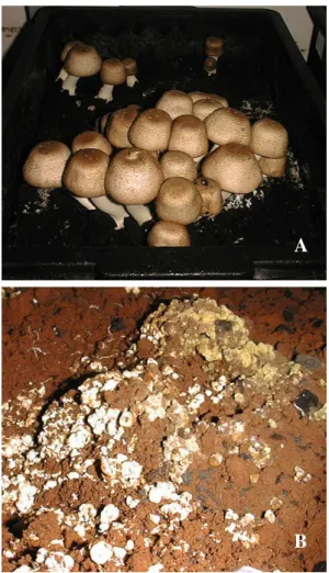
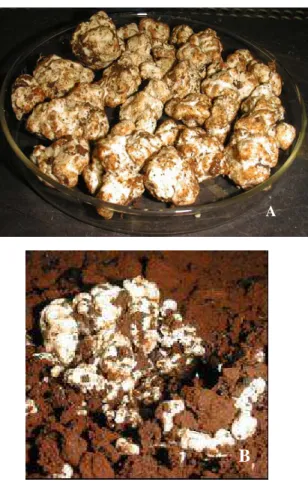
Figure 1 - Fruiting bodies of the Agaricus brasiliensis (A) and Diehliomyces microsporus (B) in Brazil.
Table 2 - Formation of colonies and contaminations starting from Diehliomyces microsporus (DMI) isolation in vitro
Treatments Colony formed (DMI) Contamination No growth
(%)
Ascostroma portion 60 10 30
Ascospores suspension 20 30 50
In vitro cultivation conditions of the D.
microsporus
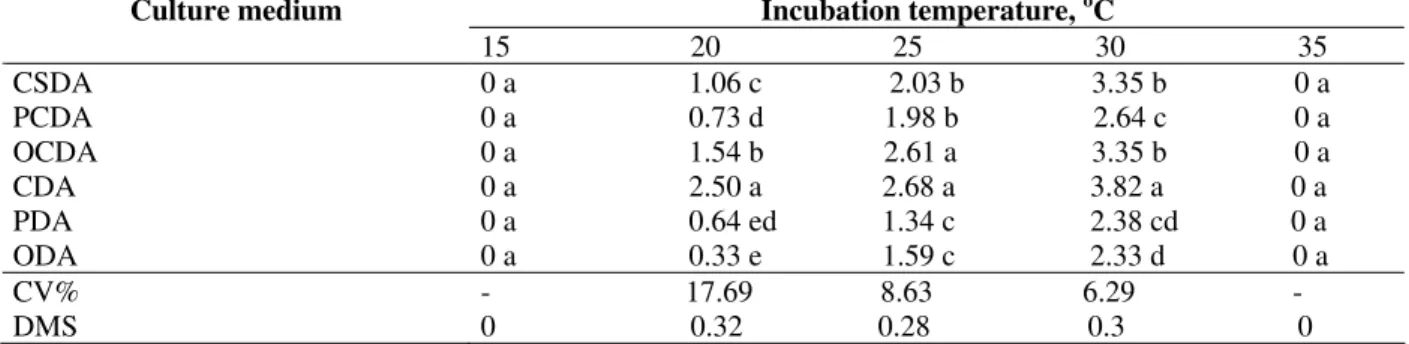
The average mycelial growth data of in vitro
cultivation the D. microsporus in different culture
media and temperatures are shown in Table 3. At 15 and 35oC, there was no colony formation in any
to the starch based ones (ODA and PDA), in which the growth had its peak at 30oC. The mixed
media, made up of the mixture of compost with other such materials as oats, soil or potato favored the mycelial growth of D. microsporus. On the
other hand, at 20, 25 and 30oC, the culture media A
largest density (visualized, but not quantified data) and a colony with a smaller diameter.
Diehl and Lambert (1930) tested several combinations of media from wheat, oat, rice, corn, soil extract to manure extract in the cultivation of
D. microsporus, obtaining better growth for oat
and soil extract based media. That was partly different from the results obtained in this experiment, because the oat based media presented a significantly smaller mycelial growth. However, when testing several culture media for mycelial growth of D. microsporus, Zaayen and Pol-Luiten
(1977) obtained better regular growth in the malt-agar and PDA media. Nevertheless, in most cases, there was formation of sector and fluffy mycelium, what made the measurements difficult. In the mixed medium made up of potato and compost for
A.brasiliensis, this did not happen and there was a
typical radial growth.
A critical point in the cultivation of A.brasiliensis,
in relation to D. microsporus was that both
presented faster growth in the same temperature range, differently from the A. bisporus, in which
the temperature could drop below 18oC during the
body fruiting phase, limiting the progression of the false truffle in this phase of the cultivation. The diameter of the colony of D. microsporus in
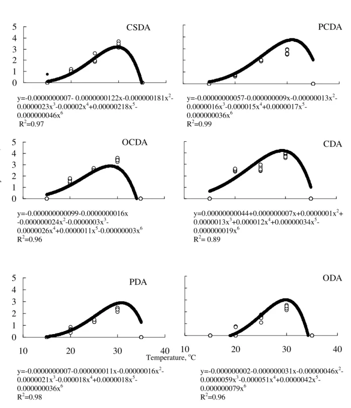
the several culture media and incubation temperature can be expressed through a sixth degree polynomial equation, with a peak at around of 30oC (Fig. 2).
The CDA media yielded a largest peak of mycelial growth of the D.microsporus, as showed in Table
3. In all the media, the null diameter at 15oC
increased gradually according to a temperature increase from 20 to 25oC. It reached its maximum
peak at around 30oC, after which it decreased
quickly when incubated at 35oC, when the colony
diameter was null again. Different results were presented by Zaayen and Pol-Luiten (1977), who obtained maximum growth of D. microsporus at
26oC. The estimated temperatures by the
regression equation would result in the maximum peaks of colony diameter in the respective media are: 29.7oC (CSDA), 31.0oC (PCDA), 28.5oC
(OCDA), 29.3oC (CDA), 30.8oC (PDA) and
29.8oC (ODA).
Table 3 - Colony diameter (cm) of Diehliomyces microsporus cultivated in different culture media in vitro and
incubated at temperatures of 15, 20, 25, 30, 35oC for 7 days.
Incubation temperature, oC Culture medium
15 20 25 30 35 CSDA 0 a 1.06 c 2.03 b 3.35 b 0 a PCDA 0 a 0.73 d 1.98 b 2.64 c 0 a OCDA 0 a 1.54 b 2.61 a 3.35 b 0 a CDA 0 a 2.50 a 2.68 a 3.82 a 0 a PDA 0 a 0.64 ed 1.34 c 2.38 cd 0 a ODA 0 a 0.33 e 1.59 c 2.33 d 0 a CV% - 17.69 8.63 6.29 - DMS 0 0.32 0.28 0.3 0
Tukey’s test: means followed by different letters in the column are significantly different at 5%.
Figure 2 - Colony diameter of Diehliomyces microsporus in different in vitro culture media
incubated at temperatures of 15, 20, 25, 30, 35oC for 7 days.
CSDA: compost-soil-dextrose-agar; PCDA: potato-compost-dextrose-agar; OCDA: oat-compost-dextrose-agar; CDA: compost-dextrose-agar; PDA: potato-dextrose-agar; ODA: oat-dextrose-agar.
ODA
10
20
30
40
y=-0.000000002-0.000000031x-0.00000046x2
-0.0000059x3-0.000051x4+0.0000042x5
-0.000000079x6
R2=0.96
PDA
0
1
2
3
4
5
10
20
30
40
CDA
OCDA
0
1
2
3
4
5
PCDA
CSDA
0
1
2
3
4
5
y=-0.0000000007- 0.0000000122x-0.000000181x2-
0.0000023x3-0.00002x4+0.00000218x5
-0.000000046x6
R2=0.97
y=-0.00000000057-0.000000009x-0.00000013x2
-0.0000016x3-0.000015x4+0.0000017x5
-0.000000036x6
R2=0.99
y=-0.000000000099-0.0000000016x -0.000000024x2-0.0000003x3
-0.0000026x4+0.0000011x5-0.00000003x6
R2=0.96
y=0.00000000044+0.000000007x+0.0000001x2+
0.0000013x3+0.000012x4+0.00000034x5
-0.000000019x6
R2= 0.89
y=-0.0000000007-0.000000011x-0.00000016x2-
0.0000021x3-0.000018x4+0.0000018x5-
0.000000036x6
R2=0.98
Temperature, oC
C
ol
on
y
di
am
et
er
, c
Cultivation of D. microsporus in sterilized compost
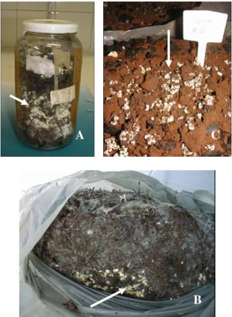
The fruiting bodies of D. microsporus were
checked approximately 30 days after the inoculation of the prepared compost for the cultivation of A. brasiliensis. This medium was
sterilized and inoculated with the competitor only (Fig. 3A). The fruiting bodies of the false truffle were shown to be similar to a great amount of deformed pin or “popcorn-like” structures, as it was called by the growers.
Biotic and abiotic factors such as temperature, humidity, presence of the host or other microorganisms, are still not certain. They can favor the fruiting bodies of the false truffle in the cultivation of mushrooms or even the ones that they can contribute in the performance of the fungus as a parasite in the system.
When the D. microsporus was inoculated in the
sterilized compost, the fruiting bodies of D.
microsporus occurred independently of the
presence of the A. brasiliensis (Fig. 3A). Parallel
to this, during the experiment, the farmers of A. brasiliensis observed ascostroma of the false
truffle before placement of the casing layer (Fig. 3B), and the most significant incidence of the ascostroma occurred during, or after the initial phase of fruiting bodies of A. brasiliensis. When
grown in transparent plastic bags, a regular procedure in mushroom farms, the largest ascostroma concentration was observed at the edge of the plastic. Initially, in the surface of the casing layer, there appear cracks where a lot of the ascostroma are concentrated in the interface compost/casing, usually with a decrease or absence of production of A. brasiliensis (Fig. 3C).
These observations also apply to the cultivation of
A.bisporus and A. bitorquis, according to Kligman
(1944), Zaayen and Pol-Luiten (1978), Sharma (1998).
Figure 3 - Fruiting bodies of Diehliomyces microsporus (indicated by the arrows) with material
incubated at the laboratory and supplied by the farmers of Agaricus brasiliensis
(bags). (A) ascostroma in sterilized compost incubated at 28oC for 35 days; (B)
fruiting bodies in the compost before the casing with approximately 30 days; (C) superficial blooming of the ascostroma in the casing soil.
A C
The ascostroma of D. microsporus presented
variations in size from 5 to 30mm in diameter, had cephalic forms, initially from a yellowish-white coloration to brown in the maturation phase (Fig. 4A). When they grew very closely some ascostroma joined, forming larger and deformed structures (Fig. 4B). These characteristics
confirmed the observations made by Diehl and Lambert (1930), Kligman (1944), Zaayen and Pol-Luiten (1977), Wood and Fletcher (1991), Yadav et al. (2000), during the cultivations of A.bisporus
and A. bitorquis.
Figure 4 - Ascostroma of Diehlioyces microsporus: (A): ascostroma collected in Petri dish; (B)
ascostroma union on casing soil.
CONCLUSIONS
The isolation of D. microsporus from fragments of
recently collected ascostroma was more efficient than the germination of the ascospores. The culture media made from a compost base (CDA) provided largest colony diameter of the D. microsporus, followed by the media made up of
the compost and potato mixture, favoring a denser mycelia. The maximum growth speed of D.
The fruiting bodies of D. microsporus occurred
before the placement of the casing layer, and during the phase of fruiting bodies of A. brasiliensis.
ACKNOWLEDGMENTS
FAPESP (Fundação de Amparo à Pesquisa do Estado de São Paulo) and CAPES (Coordenação
A
RESUMO
A falsa trufa está sendo um dos principais problemas na produção do Agaricus brasiliensis
cultivado no Brasil e o controle deste fungo competidor tem sido difícil, devido às dificuldades encontradas no isolamento e cultivo do patógeno. Este experimento foi conduzido em três etapas, sendo a primeira constituída pelo isolamento de
Diehliomyces microsporus a partir de porções do
ascostroma e através da suspensão de ascósporos; a segunda, o cultivo in vitro de D. microsporus nas
temperaturas de 15, 20, 25, 30 e 35oC e em seis
meios de cultura (CTDA, ACDA, BCDA, ADA, BDA e CDA) e a terceira pela inoculação de D. microsporus no composto (pasteurizado, composto
esterilizado e composto esterilizado com camada de cobertura) para formação dos ascostromas. O isolamento de D. microsporus a partir de
fragmentos do ascostroma recém coletado foi mais eficiente do que a germinação dos ascósporos; o meio de cultura à base de composto (CDA) proporcionou maior diâmetro da colônia de D. microsporus, seguido pelo meio constituído da
mistura de composto e batata, favorecendo um micélio mais denso; a maior velocidade de crescimento de D. microsporus ocorreu quando a
cultura foi incubada entre 28 e 30oC; temperaturas
de incubação menor que15oC ou a acima de 35oC
inibiu completamente o crescimento micelial de D. microsporus; a obtenção de frutificação de D. microsporus foi facilmente obtida em composto
esterilizado e posteriormente inoculado com o competidor.
REFERENCES
Bisset, P.G., Colhoun, J., Gandy, D.G. 1982. Germination of Diehliomyces microsporus ascospores
and determination of their thermal death point.
Transation of the British Mycological Society, 78, 3,
540-542.
Diehl, W.W., Lambert, E.B., 1930. A new truffle in beds of cultivated mushrooms. Mycologia, 22,
223-226.
Kligman, A.M. 1944. Conthol of the truffle in beds of the cultivated mushroom. Phytopathology, 34,
376-384.
Nascimento, J.S. 2003. Etiologia, controle e demanda de energia na prevenção da falsa trufa (Diehliomyces microsporus) em cultivos de Agaricus blazei
Botucatu, 115p. Tese (Doutorado em
Agronomia/Energia na Agricultura) - Faculdade de Ciências Agronômicas, Universidade Estadual Paulista.
Nascimento, J.S., Eira, A.F. 2001. Ocorrência e prejuízos da falsa trufa (Diehliomyces microsporus)
em cultivo do Agaricus blazei Murrill. Congresso
Brasileiro de Micologia, 3, Águas de Lindóia.
Resumos... São Paulo: Sociedade Brasileira de
Micologia, 2001. p.37.
Nascimento, J.S., Eira, A.F. 2003a. Occurrence of false truffle (Diehliomyces microsporus Gilkey) and
damage on the Himematsutake medicinal mushroom (Agaricus brasiliensis S. Wasser et al.). Int. J. Med. Mushr., 5, 1, 87-94.
Nascimento, J.S., Eira, A.F. 2003b. Doenças e competidores do Agaricus blazei. In: Eira, A.F. cultivo do “cogumelo-do-sol” (Agaricus blazei (Murrill) ss. Heinemann). Viçosa: Aprenda Fácil,
p.221-249.
Sharma, V.P. 1998. Biology and management of false truffle (Diehliomyces microsporus) during cultivation
of Agaricus spp. Mushr. Res., 7, 1, 1-12.
Zaayen, A. Van, Pol-Luiten, B. Van Der. 1977. Heat resistance, biology and prevention of Diehliomyces microsporus in crops of Agaricus species. J. Pl. Path., 83, 221-240.
Zaayen, A. Van, Pol-Luiten, B. Van Der. 1978. Heat resistance, some biological aspects and prevention of false truffle (Diehliomyces microsporus). In: Maher,
M.J. (Ed.) Science and cultivation of edible fungi.
Rotterdam: Balkema, 2, 319-336.
Wood, M.W., Fletcher, J.T. 1991. The occurrence of ascocarps of Diehliomyces microsporus, the cause of
false truffle disease. Mushrooms Science XII, 1,
379-384.
Yadav, M.C., Dhar, B.L., Verma, R. N. 2000. Breeding studies on development of high yielding and quality hybrids of Agaricus bitorquis. Mushrooms Science XV, 1, 299-304.