Infection of Human Retinal Pigment
Epithelium with
Chlamydia trachomatis
Ernest Boiko1,2, Dmitrii Maltsev1*, Alevtina Savicheva3, Kira Shalepo3, Tatyana Khusnutdinova3, Alexei Pozniak1, Igor Kvetnoi4, Viktoria Polyakova4, Alexei Suetov1
1Department of Ophthalmology, Military Medical Academy, St Petersburg, Russia,2St. Petersburg Branch of the Academician S. Fyodorov IRTC“Eye Microsurgery”, St. Petersburg, Russia,3Laboratory of
Microbiology, D.O. Ott Research Institute of Obstetrics and Gynaecology, St. Petersburg, Russia,
4Pathology Department, D.O. Ott Research Institute of Obstetrics and Gynaecology, St. Petersburg, Russia
Abstract
Purpose
Little is known about the susceptibility of posterior segment tissues, particularly the human retinal pigment epithelium (hRPE), toChlamydia trachomatis. The purpose of the study was to investigate the possibility of infecting the hRPE withChlamydia trachomatis, and to exam-ine the infectivity of differentChlamydia trachomatisclinical isolates for hRPE cells and the hRPE cell response to the infection.
Methods
Cultured hRPE and McCoy cells were inoculated with eightChlamydia trachomatis(serovar E) clinical isolates at multiplicity of infection (MOI) of 2.0 or 0.3. To detectChlamydia tracho-matis, samples were stained immunohistochemically with anti-major outer membrane pro-tein antibodies at 24h, 48h, and 72h postinoculation (PI). The changes in the expression of signaling molecules and proteins of cytoskeleton and extracellular matrix in hRPE cells were examined immunohistochemically.
Results
All eight clinical isolates demonstrated ability to infect hRPE cells. At equal MOI of 0.3, the infectivity ofChlamydia trachomatisclinical isolates for RPE culture was found to be at least as high as that for McCoy cell culture. At 24h PI, the percentage of inclusion-containing cells varied from 1.5±0.52 to 14.6±3.3% in hRPE cell culture infected at MOI of 2.0 against 0.37±0.34 to 8.9±0.2% in McCoy cell culture infected at MOI of 0.3. Collagen type I, colla-gen type IV, basic fibroblast growth factor, transforming growth factor-beta and interleukin– 8 expression at 48h PI were maximally increased, by 2.1-, 1.3-, 1.5-, 1.5- and 1.6-fold, respectively, in theChlamydia trachomatis-infected compared with control hRPE cell culture specimens (P<0.05).
OPEN ACCESS
Citation:Boiko E, Maltsev D, Savicheva A, Shalepo K, Khusnutdinova T, Pozniak A, et al. (2015) Infection of Human Retinal Pigment Epithelium withChlamydia trachomatis. PLoS ONE 10(11): e0141754. doi:10.1371/journal.pone.0141754
Editor:Eliseo A Eugenin, Rutgers University, UNITED STATES
Received:June 30, 2015
Accepted:October 13, 2015
Published:November 4, 2015
Copyright:© 2015 Boiko et al. This is an open access article distributed under the terms of the Creative Commons Attribution License, which permits unrestricted use, distribution, and reproduction in any medium, provided the original author and source are credited.
Data Availability Statement:All relevant data are within the paper and its Supporting Information files.
Funding:The authors have no support or funding to report.
Conclusions
This study, for the first time, proved the possibility of infecting hRPE cultured cells with Chla-mydia trachomatis, which leads to proproliferative and proinflammatory changes in the expression of signaling molecules and extracellular matrix components.
Introduction
Retinal pigment epithelium (RPE) is critically important for maintaining the structural and functional integrity of the retina and choriocapillaris. Changes in the RPE play a key role in the pathogenesis of various posterior segment disorders, including age-related macular degenera-tion and proliferative vitreoretinopathy.
Numerous factors (including oxidative stress, inflammation, genetic specificity, etc.) condi-tion the modulacondi-tion of biological characteristics of RPE cells, leading to changes in their func-tional activity. Two of the most significant factors influencing the RPE are proinflammatory stimuli and signaling molecules participating in inflammation [1,2]. The marked influence of these molecules on the biological characteristics of the human RPE (hRPE) has been demon-stratedin vitro. This involves the increase in complement expression by RPE cells stimulated by activated macrophages [1] and an enhanced RPE cell migration and proliferative activity stimulated by epidermal (EGF), platelet-derived (PDGF) and basic fibroblast growth factors (bFGF).[2,3]
Infection is one of the main causes of inflammation that develops either due to direct cell infection, or through immune mechanisms. Stimulation of hRPE withC.pneumoniaeantigen [4] and the proinflammatory effect of a combination of virulence factors ofS.aureuson the expression of cytokines in RPE cells [5] have been demonstratedin vitro.In vivo, direct infec-tion of RPE with Varizella Zoster and Cytomegalovirus has been demonstrated in human autopsy specimens [6,7] and in experimental animal studies [8]. Several works have shown the possibility ofin vitrodamage to cultured hRPE cells by neurotropic viruses that can cause ocu-lar involvement (particuocu-larly, retinitis and chorioretinitis) in humans [9–11]. Most of the works on infectious RPE damage, however, are related to viral infections, whereas the effect of bacterial infections has been poorly investigated.
In our previous work, we have demonstrated infection of the RPE with the obligate intracel-lular human bacterial pathogen,Chlamydia trachomatis, following ocular inoculation in rab-bits [12]. Yet, the possibility of infecting the hRPE withChlamydia trachomatisand the pathogenetic significance of such an infectious process remain unknown. Our current aim, therefore, was to investigate the possibility of infecting the hRPE withChlamydia trachomatis, and to examine both the infectivity of different clinical isolates of the pathogen for hRPE cells and the response of hRPE cells to the infection.
Materials and Methods
Ethics statement
i)C.trachomatisclinical isolates: samples ofC.trachomatisclinical isolates (serovar E) were obtained from patients of the Ott Research Institute of Obstetrics and Gynaecology during treatment and diagnostic manipulations. Written consent for sampling of clinical isolates for research purpose was obtained as approved by the Ethics Committees of the Ott Research Insti-tute of Obstetrics and Gynaecology and Military Medical Academy.
ii) globes from deceased donors: prior to harvesting the organ for donation, the legal next of kin of each deceased donor gave a written informed consent for use of the globe for transplan-tation and research (as part of the overall protocol and consent for organ donation).
Cell Cultures
RPE. hRPE cultures were obtained from 6 cadaveric eyes within 36 hours after death, and after the cornea had been removed for transplantation surgery, using a technique described earlier [10]. Briefly, after external tissues were removed, the globe was rinsed with 70% ethanol and twice washed with sterile Hanks’balanced salt solution (HBSS) (GIBCO-BRL, Grand Island, NY). The anterior segment, vitreous and retina were removed. The eyecup was incu-bated with 0.2 mg/ml collagenase type 1 (GIBCO-BRL) for 40 minutes at 37°C and 5% CO2.
One point five to 1.8 × 10⁶cells per donor eye were collected by gentle pipetting, and the result-ing suspension was centrifuged at 300g for 4 min. After removal of supernatant, cells were resuspended in DMEM/F12 medium (Biolot, St.Petersburg, Russia) supplemented with strep-tomycin, penicillin and amphotericin (GIBCO-BRL) and 10% bovine serum albumine (BSA) (GIBCO-BRL), and seeded into culture flasks. Cells were passaged at a 1:3 ratio when 70–80% confluent; usually, each passage was accompanied by a three- to four-fold increase in cell num-bers. The hRPE used in all experiments were from passage 4 to 5. The purity of the cell line was confirmed by detection of cytokeratin 8/18 expression (Table 1).
To perform the experiments, cells were transferred to 96-well flat-bottom plates (5×10³cells per well by the time of infection) five days preinoculation.
McCoy. McCoy cell culture was provided by the Cell Culture Bank of the Ott Institute of Obstetrics and Gynaecology (St. Petersburg, Russia), and grown to confluence in DMEM/F12 containing gentamicin, amphotericin, and 10% BSA. Twenty-four hours preinoculation, after
Table 1. Primary Antibodies Used to DetectC.trachomatisAntigens, Signaling, Cytoskeleton and Extracellular Matrix Molecules.MOMP, major outer membrane protein;VEGF, vascular endothelial growth factor;TNF–α, tumor necrosis factors–α;α-SMA, smooth muscle actin;MMP–9, matrix metallo-proteinase 9;bFGF, basic fibroblast growth factor;TGF–β, transforming growth factorβ;RFU, ready for use
Antigens Host Type Number Dilution Manufacturer
MOMPC.trachomatis Mouse Polyclonal AM088-5M RFU BioGenex, Mountainview, CA MOMPC.trachomatis(fluorescein-isothiocyanate-conjugated) Chicken Polyclonal 039487 RFU LabDiagnostic, Moscow, Russia
VEGF Mouse Monoclonal M7273 1:100 DAKO, Santa Barbara, CA
Collagen type IV Mouse Monoclonal M0785 1:50 DAKO
Interleukin-8 Rabbit Polyclonal ab7747 1:100 ABCAM, Cambridge, MA
TNF–α Rabbit Polyclonal ab6671 1:50 ABCAM
α-SMA Mouse Monoclonal LS-C95386 RFU LSBio, Seattle, WA
MMP-9 Mouse Monoclonal NCL-MMP9-439 1:40 Novocastra, Newcastle, UK
Collagen type I Rabbit Polyclonal ab34710 1:50 ABCAM
bFGF Mouse Monoclonal 610073 1:100 BD Transduction, San Jose, CA
TGF–β Mouse Monoclonal ab64715 1:50 ABCAM
E-cadherin Mouse Monoclonal CM 170 C 1:100 BioCare, Concord, CA
CD105 Rabbit Monoclonal NCL-CD105 1:100 Novocastra
Cytokeratin 8/18 Mouse Monoclonal MS-743-S 1:50 Thermo Scientific, Rockford, IL
being trypsinized, cells were seeded into 96-well plates (3×10⁴cells per well by the time of infection).
Pathogen and Inoculation
EightChlamydia trachomatisclinical isolates (serovar E) of patients from the Ott Institute of Obstetrics and Gynaecology were selected for inoculation of cell cultures. Viability of the path-ogen, its infectivity for cell cultures, andChlamydia trachomatisdoses were tested with McCoy cell culture. Infected McCoy cell monolayers were detached by scraping and disrupted by ster-ile glass beads to release elementary bodies (EBs). Cell debris was removed by centrifugation (500g, 15 min). Each non-control well of a 96-well plate received 100μL of cell lysate contain-ing a fixed number of inclusion formcontain-ing units of aChlamydia trachomatisclinical isolate (Table 2). Each control well of the same plate received 100μL of uninfected cell lysate as a mock infection control. The plates were then centrifuged at 2400 rpm for 60 min at 37°C, and the medium was completely changed with the same medium (DMEM/F12, 10% BSA) to which 1 mg/L cycloheximide (Acros Organics, Geel, Belgium) was added only in the experiments for the assessment of the infectivity of the pathogen. To verify the completion of C. trachomatis life cycle, clinical isolate No. 24032 was repassaged from individual RPE cell culture samples to RPE and McCoy cell cultures using the technique described above 72 hpi. Intracellular inclu-sions were visualized by direct immune fluorescence 48 hours after repassage. For immunohis-tochemistry and direct immunofluorescent staining, the culture medium was removed from plate wells, and the cells were washed twice with HBSS and fixed at 24h, 48h, and 72h postinoculation.
Immunohistochemistry and Immunofluorescence Detection, and
Assessment of the Infectivity of the Pathogen
Immunohistochemistry. For immunohistochemical staining, cells were fixed in 4% para-formaldehyde for 15 minutes at 20°C, permeabilized with 0.01% Triton X-100 for 15 minutes and rinsed 3 times with PBS for 5 minutes each time. They were then blocked for endogenous peroxidase using 3% H2O2in PBS for 15 minutes and again rinsed 3 times with PBS for 5
min-utes each time. This was followed by incubation for 15 minmin-utes in 1% BSA. The cells were then exposed overnight to a primary mouse anti-major outer membrane protein antibody (Table 1). The primary antibodies were detected by incubating the cells with a goat anti-mouse/anti-rab-bit horseradish peroxidase-conjugated secondary antibody (DAKO, Santa Barbara, CA) for 30 min at 20°C. Diaminobenzidine (DAKO) and hematoxylin were used as the chromogen and counterstain, respectively.
Direct immunofluorescent staining. For direct immunofluorescent staining, cells were fixed in 4% paraformaldehyde for 15 minutes at 20°C and rinsed 3 times with PBS for 5 min-utes each time. The cells were then incubated with fluorescein-isothiocyanate-conjugated anti-body toChlamydia trachomatismajor outer membrane protein (Table 1) for 30 min at 37°C in
Table 2. Clinical Isolates ofC.trachomatis, the Doses Used to Infect Cell Cultures and Multiplicities of Infection Corresponding to the Doses and to the Isolates. IFU, inclusion forming units;MOI, multiplicity of infection
Clinical isolate number 1609 3438 24032 27692 5017 27644 685 2556
IFU for RPE 104 104 104 104 104 1.7×10³ 1.7×10³ 1.7×10³
IFU for McCoy 104 104 104 104 104 104 104 104
MOI for RPE 2.0 2.0 2.0 2.0 2.0 0.3 0.3 0.3
MOI for McCoy 0.3 0.3 0.3 0.3 0.3 0.3 0.3 0.3
a dark, humidified chamber; Evans blue was used as a counterstain. After being washed twice in PBS, dried, and coverslipped with PBS/glycerol, the slides were examined using an Axioplan 2 fluorescence microscope (Carl Zeiss, Jena, Germany) (excitation wavelength, 490 nm).
Assessment of the Infectivity of the Pathogen. At each examination time-point, the per-centage of inclusion-containing cells (representing the infectivity of aChlamydia trachomatis
clinical isolate) and the morphology of intracellular inclusions were assessed both in immuno-histochemistry and in direct immunofluorescent staining. The percentage was averaged over 10 fields of view per slide based on the results of two repeated experiments (with hRPE culture from one donor). For each clinical isolate, the percentage of inclusion-containing cells was determined as an average of two repeated experiments.
Immunohisctochemistry of Signaling Molecules and Proteins of
Cytoskeleton and Extracellular Matrix
To examine the post-inoculation changes in the expression of signaling molecules, cytoskeleton and extracellular matrix (ECM) components in RPE cells, cells were plated in 96-well plates (3×10³cells per well) in DMEM:F12 medium supplemented with 10% BSA and without cyclo-heximide. After 72 hours, the medium was changed to DMEM:F12 supplemented with 1% BSA and without cycloheximide. Additionally, non-control wells were infected as described above with clinical isolates Nos.24032 or 27692 (5×10³inclusion forming units per well) that had exhibited the highest infectivity. After 48 hours, the slides were fixed in 4% paraformalde-hyde and stained with antibodies (Table 1) as described above, but without hematoxylin counterstaining.
Optical density of staining was analyzed using Image J (NIH, Bethesda, MD) as follows: (1) Digital color images were processed using the“Subtract Background”function and (2) con-verted to 32-bit greyscale images; (3) The“Measure”function was used to determine mean background pixel values for each of 5 circular cell-free areas (with each circle having a diameter of up to 10 pixels) within a well; (4) Mean pixel values were determined for each of 5 spaced circular areas (with each circle having a diameter of 200 pixels and involving no cell-free subar-eas) showing a homogeneous cell distribution; (5) The data were transferred to Microsoft Excel 2010 (Microsoft Corp., Redmond, WA). Then the mean background pixel value (for all 5 circu-lar cell-free areas) and the mean pixel value (for all 5 spaced circucircu-lar areas showing a homoge-neous cell distribution) were calculated. Thereafter, the difference of these mean values was calculated, with the results normalized to those of control.
Such an approach enabled us to avoid incorporation of areas showing a heterogeneous cell distribution or cell-free areas into the analysis, and to compensate for the different background colors.
Chlamydia trachomatisinfection of cultured cells was confirmed and the infection level was quantified by means of immunohistochemical staining as described above. The experiment was repeated twice with cells at the same passage from different donor eyes. The optical density value was determined as an average of two repeated experiments.
Statistical Analysis
Results
Cell Culture Susceptibility and Dose-dependent Susceptibility to
Chlamydia trachomatis Infection
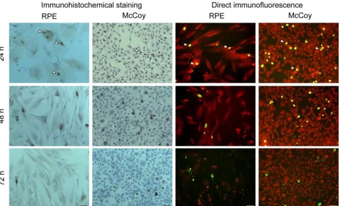
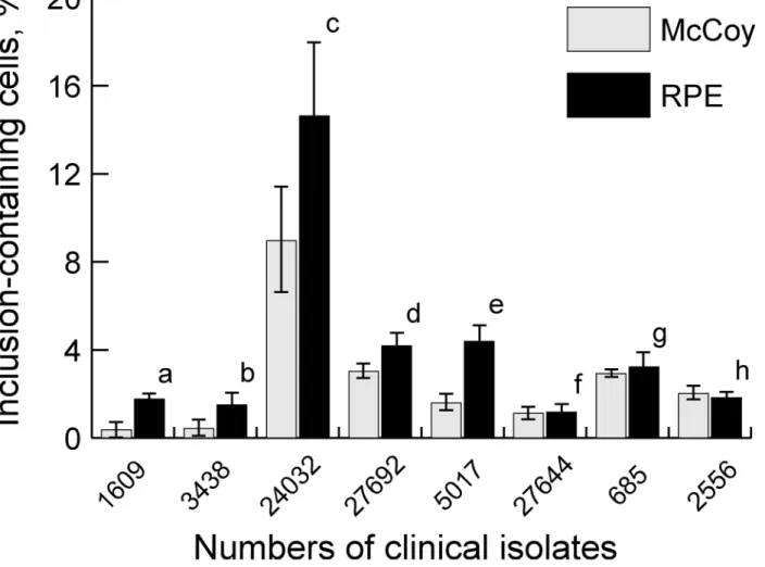
In all non-control samples of RPE and McCoy cell cultures, the pathogen was detected both by immunohistochemistry and by direct immunofluorescent staining (Fig 1). Twenty-four hours postinoculation (hpi), clinical isolates (at equal multiplicity of infection (MOI) of 2.0) demon-strated a wide variation in infectivity, with the percentage of RPE culture inclusion-containing cells ranging from 1.5±0.52% (isolate No.3438) to 14.6±3.3% (isolate No.24032), whereas that of McCoy culture cells infected at equal MOI of 2.0 was 0.37±0.34% (isolate No.1609) to 8.9 ±2.4% (isolate No.24032) (Fig 2). Twenty-four hpi, the variation in the percentage of RPE cul-ture inclusion-containing cells among five clinical isolates used for infection at MOI 2.0 was almost 10-fold (1.5–14.6%). All the five clinical isolates used at a higher MOI for infection of RPE cells than for infection of McCoy cells showed a significantly higher (P<0.05) percentage of inclusion-containing cells for hRPE cell culture than for McCoy cell culture. At equal MOI of 0.3, no statistically significant difference in the percentage of inclusion-containing cells was found between McCoy cells and RPR cells.
Chlamydia trachomatis Life Cycle in Vitro
In the hRPE culture, like in the control culture, the maximal number of intracellular inclusions (mostly small and medium size, perinuclear inclusions) ofChlamydia trachomatiswas observed 24 hours hpi (Fig 1). A few isolated elementary bodies (EBs) (most likely, from the inoculating material) were found on the surface of some cells.
Fig 1. Immunohistochemistry and direct immunofluorescent staining of hRPE and McCoy cell cultures at different time-points postinoculation withChlamydia trachomatis(clinical isolate No.24032).In both types of cultures, both techniques reveal highly immunoreactive inclusions (arrowheads). Twenty-four to 72 h postinoculation, a reduction in the number of intracellular inclusions is observed due to the release of a new generation of EBs to the extracellular environment. Scale bar: 50μm.
Forty-eight hpi, the number of inclusions decreased (Fig 1) due to the release of a new gen-eration of EBs from the largest inclusions. The appearance of cells with a broken cell membrane and release of EBs to the extracellular environment was observed as theChlamydia trachomatis
life cycle was approaching its completion. The released EBs were seen attached to adjacent cells. Seventy-two hpi, a massive release of the EBs from intracellular inclusions was seen in culture specimens; this corresponded to the detection of numerous inclusions exhibiting weak immune responsiveness (Fig 3A) by immunohistochemistry. Numerous EBs were seen in the microscopy projection image of cells without inclusions (likely, on cell membranes) (Fig 3B), which was not observed at 48 hpi. The progression in the reduction in the percentage of inclu-sion-containing cells in hRPE culture was observed in a way similar to that in McCoy culture at 72 hpi (Fig 4). Repassage of the pathogen from RPE cell culture to RPE and McCoy cell cul-tures at 72 hpi was followed by the formation of multipleChlamydia trachomatisinclusions in both RPE and McCoy cell cultures.
Changes in the Expression of Signaling Molecules and Proteins of
Cytoskeleton and Extracellular Matrix in hRPE Cells Grown in Culture
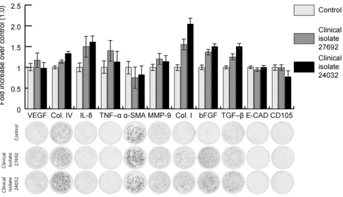
Statistically significant differences in the expression of collagen type IV, IL-8, collagen type I, bFGF and transforming growth factor-β(TGF–β) molecules were revealed between the
Fig 2. Percentage of inclusion-containing cells in hRPE and McCoy cell cultures following inoculation with differentChlamydia trachomatis
clinical isolates.a,P= 0.001; b,P= 0.0002; c,P= 0.01; d,P= 0.039; e,P= 0.018; f,P= 0.76; g,P= 0.18; h, p = 0.22
Chlamydia trachomatis-infected and control specimens as well as between those infected with the two clinical isolates (Fig 5). Additionally matrix metalloproteinase (MMP)-9 were
increased, although not statistically significantly, in the culture specimen infected with clinical isolate No.27692 compared to the control. A similarity in the expression of vascular endothelial growth factor (VEGF), tumor necrosis factors–α(TNF–α),α-smooth muscle actin (α-SMA), MMP-9, E-cadherin and CD105 was observed between theChlamydia trachomatis-infected and control specimens. The number ofChlamydia trachomatisinclusions in the culture speci-men infected with clinical isolate No.24032 was 3.28 times higher than in that infected with clinical isolate No.27692; this value was not statistically significantly different from that obtained during the assessment of infectivity of clinical isolates.
Discussion
This study demonstrates for the first time the possibility of infecting hRPE cells with Chla-mydia trachomatis. Infectivity ofChlamydia trachomatisclinical isolates for RPE culture was found to be at least as high as that for the culture commonly used for the survival and propaga-tion of rather small numbers ofChlamydia trachomatisEBs. Therefore, we found, also for the first time, that the RPE can be considered as highly susceptible toChlamydia trachomatis infec-tion. It is this finding that is of primary importance in this study, since there is no data on natu-ral resistance of specific cell cultures to infectionin vitrowithChlamydia trachomatis. Because a wide variation in the percentage of inclusion-containing cells (1.5% - 14.6%) was observed Fig 3. Immunohistochemistry micrographs of hRPE cell culture at 72 hours postinoculation withChlamydia trachomatisclinical isolate No.24032. (A) Intracellular inclusions (arrowhead) exhibiting weak immune responsiveness (due to release of EBs to extracellular environment). (B) Numerous EBs (arrowheads) in microscopy projection of cells without inclusions. Scale bar: 10μm.
between the hRPE culture specimens inoculated with different clinical isolates, this percentage is determined both by the susceptibility of the cells to infection and by the infectivity of a clini-cal isolate. Interestingly that isolates demonstrating higher levels of infectivity for McCoy cells showed also higher levels of infectivity for RPE cells than those with lower levels of infectivity for McCoy cells, thus evidencing for the absence of specific resistance of RPE cells toC.
trachomatis.
In hRPE cultured cells, the pathogen goes through a complete life cycle, which is completed with the release of numerous new infectious EBs that are capable of maintaining aChlamydia trachomatispopulation. This is first confirmed by the results of further repassages of the patho-gen in RPE culture. Moreover, this fact is supported by (1) the similarity in the dynamics of the percentage of inclusion-containing cells in hRPE and McCoy cultured cells and (2) images of the release of EBs from intracellular inclusions, and images of multiple EBs on cell membranes of RPE cells at 72 hpi.
Pro-fibrogenic and proinflammatory changes in the expression of signaling molecules, cyto-skeleton and ECM components in RPE cells following the infection withChlamydia trachoma-tiswere demonstrated by the selective increase in the expression of collagen type IV, collagen type I, IL-8, bFGF and TGF–β.
Increased expression of collagens (type I and IV) has been observed in patients with tracho-matous conjunctivitis, demonstrating the capacity ofChlamydia trachomatisto modulate the collagen expression [13]. This agrees with the results of this study. Infection of male genital Fig 4. Changes in the percentage of inclusion-containing cells in hRPE and McCoy cell cultures following inoculation withChlamydia trachomatis
(clinical isolate No.24032).
tract (in vivo and in vitro) [14], cervical [15], colonic [15] and some other [13,16] epithelial cell cultures withChlamydia trachomatisis known to upregulate secretion of IL-8. Interleukin-8 is considered to play an important role in the induction of inflammation following infection withChlamydia trachomatis[17].Chlamydia trachomatisinfectionin vitrohas been shown to upregulate TGF-β[18], which promotes scar tissue deposition. Induction of expression of bFGF, which is crucial for remodeling connective tissue, by human smooth muscle cells has been observed followingChlamydia trachomatisinfection [19]. In our study, upregulation of IL-8, TGF-βand bFGF was also found in RPE cells. TGF-βcan increase MMP expression. In our study, the MMP-9 level inChlamydia trachomatis-infected RPE culture was increased, but the value was not statistically significantly different from that of the control. This warrants fur-ther investigation of MMP expression in RPEChlamydia trachomatisinfection, since upregula-tion of MMP-9 (including that in the absence of changes in the expression of TGF-β) was demonstratedin vitroin porcine retinal cell line VIDO R1 [20].
We did not find significant changes in the expression ofα-SMA, VEGF, E-cadherin and CD105. This might be expected, sinceChlamydia trachomatis-induced changes in the expres-sion ofα-SMA, E-cadherin and CD105 have never been reported. In a number of works, upre-gulation of TNF–αexpression inChlamydia trachomatis-infected cell cultures has been reported [18,21,22], sometimes in association with arrest of the pathogen life cycle [21]. RPE culture cells inoculated withChlamydia trachomatisshowed no significant change in expres-sion of TNF–α. This corresponds to a weak response, and partly explains their high susceptibil-ity toChlamydia trachomatisinfection.
One could hypothesize that the increase in expression of collagen type IV, collagen type I, IL-8, TGF–βand bFGF may be partly explained by stimulation of RPE cells withChlamydia
Fig 5. Changes in the expression of signaling molecules and proteins of cytoskeleton and extracellular matrix of hRPE cells grown in culture following inoculation withChlamydia trachomatis(clinical isolates Nos. 24032 and 27692).
trachomatisantigen. However, theChlamydia trachomatisdoses (and, correspondingly, the amounts of introduced antigen) used to compare the effect of two clinical isolates were identi-cal, the difference being only the amount of intracellular inclusions formed. Because the expression was higher in specimens with increased number of inclusions, we believe that infec-tion of the cells substantially contributes to the changes in expression of the meninfec-tioned molecules.
In vitro damage to the hRPE by West Nile virus [9], influenza A viruses [10], cytomegalovi-rus [23], HSV-1 [22], and HIV [24] has been described in literature. Damage to the posterior segment can occur following infection with these viruses[25–27]. The results of cultural studies are therefore in general agreement with infection prevalence in ocular tissues. Because Chla-mydia trachomatiscan spread not only by cell-to-cell contact, but also hematogenously [28], the development of intraocular infection appeared possible and further studies ofChlamydia trachomatisinfectionin vivoof RPE cells are required.
Given the proinflammatory and proproliferative effect ofChlamydia trachomatison RPE cells, which was demonstrated by the upregulation of collagen type IV, collagen type I, IL-8, TGF and FGF, the results of the study should be extrapolated primarily to humans with prolif-erative vitreoretinopathy, because in this disease, production of ECM (including collagen type I) [29], TGF [30,31], FGF [31], and IL-8 [32] is of particular importance.
The present study has several limitations. Particularly, although RPE cultures of different donors may vary in susceptibility to infection due to genetic or other individual features, the assessment of infectivity of the pathogen was performed using the source material of a single donor. Nevertheless, this was necessary to compare the infectivity of different clinical isolates. Addtionally, we assessed the expression of signaling molecules and proteins of cytoskeleton and extracellular matrix using a semi-quantitative method.
In conclusion, this study, for the first time, proved the possibility of infecting hRPE cultured cells withChlamydia trachomatis, which leads to proproliferative and proinflammatory changes in the expression of signaling molecules and ECM components. Furthermore, we dem-onstrated (1) increased susceptibility of RPE cells toChlamydia trachomatisinfection, and (2) variability in infectivity and in the influence on the expression of collagen type IV, collagen type I, IL-8, TGF-βand bFGF among different clinicalChlamydia trachomatisisolates. These results may be of importance in studying the vitreoretinal pathology.
Supporting Information
S1 File. These are raw data from calculation of percentage of inclusion-containing cells in hRPE and McCoy cell cultures following inoculation with different Chlamydia trachomatis clinical isolates and optical density measurement of expression of signaling molecules and proteins of cytoskeleton and extracellular matrix of hRPE cells grown in culture following inoculation with Chlamydia trachomatis.
(XLSX)
Acknowledgments
The authors thank O.V.Oleksiienko for his assistance in translating the article.
Author Contributions
Contributed reagents/materials/analysis tools: A. Savicheva KS IK VP. Wrote the paper: EB DM A. Savicheva KS IK VP.
References
1. Luo C, Zhao J, Madden A, Chen M, Xu H. Complement expression in retinal pigment epithelial cells is modulated by activated macrophages. Exp Eye Res. 2013; 112:93–101. doi:10.1016/j.exer.2013.04. 016PMID:23644095
2. Steindl-Kuscher K, Boulton ME, Haas P, Dossenbach-Glaninger A, Feichtinger H, Binder S. Epidermal growth factor: the driving force in initiation of RPE cell proliferation. Graefes Arch Clin Exp Ophthalmol. 2011; 249:1195–2200. doi:10.1007/s00417-011-1673-1PMID:21494877
3. Kaven CW, Spraul CW, Zavazava NK, Lang GK, Lang GE. Growth factor combinations modulate human retinal pigment epithelial cell proliferation. Curr Eye Res. 2000; 20:480–487. PMID:10980660
4. Fujimoto T, Sonoda KH, Hijioka K, Sato K, Takeda A, Hasegawa E, et al. Choroidal neovascularization enhanced by Chlamydia pneumoniae via Toll-like receptor 2 in the retinal pigment epithelium. Invest Ophthalmol Vis Sci. 2010; 51:4694–4702. doi:10.1167/iovs.09-4464PMID:20393111
5. Lou BS, Yuan ZH, Luo YW, Lin XF. [Effects of Staphylococcus aureus supernatant on expression of various cytokines by neutrophils and retinal pigment epithelium cells]. Zhonghua Yan Ke Za Zhi. 2011; 47:1117–1122. Chinese PMID:22336123
6. Kuppermann BD, Quiceno JI, Wiley C, Hesselink J, Hamilton R, Keefe K, et al. Clinical and histopatho-logic study of varicella zoster virus retinitis in patients with the acquired immunodeficiency syndrome. Am J Ophthalmol. 1994; 118:589–600. PMID:7977572
7. Friedman AH. The retinal lesions of the acquired immune deficiency syndrome. Trans Am Ophthalmol Soc. 1984; 82:447–491. PMID:6100147
8. Zhang M, Xin H, Roon P, Atherton SS. Infection of retinal neurons during murine cytomegalovirus retini-tis. Invest Ophthalmol Vis Sci. 2005; 46:2047–2055. PMID:15914622
9. Munoz-Erazo L, Natoli R, Provis JM, Madigan MC, King NJ. Microarray analysis of gene expression in West Nile virus-infected human retinal pigment epithelium. Mol Vis. 2012; 18:730–743. PMID:
22509103
10. Michaelis M, Geiler J, Klassert D, Doerr HW, Cinatl J Jr. Infection of human retinal pigment epithelial cells with influenza A viruses. Invest Ophthalmol Vis Sci.2009; 50:5419–5425. doi: 10.1167/iovs.09-3752PMID:19553611
11. Zheng M, Atherton SS. Cytokine profiles and inflammatory cells during HSV-1-induced acute retinal necrosis. Invest Ophthalmol Vis Sci. 2005; 46:1356–1363. PMID:15790902
12. Boiko EV, Pozniak AL, Maltsev DS, Suetov AA, Nuralova IV. Chronic ocular Chlamydia trachomatis infection in rabbits; clinical and histopathological findings in the posterior segment. Invest Ophthalmol Vis Sci. 2014; 55:1176–1183. doi:10.1167/iovs.13-13416PMID:24474273
13. Abu el-Asrar AM, Geboes K, Missotten L. Immunology of trachomatous conjunctivitis. Bull Soc Belge Ophtalmol. 2001; 280:73–96. PMID:11486468
14. Al-Mously N, Eley A. Interaction of Chlamydia trachomatis serovar E with male genital tract epithelium results in secretion of proinflammatory cytokines. J Med Microbiol. 2007; 56:1025–1032. PMID:
17644708
15. Mazzoli S, Cai T, Rupealta V, Gavazzi A, Castricchi Pagliai R, Mondaini N, et al. Interleukin 8 and anti-chlamydia trachomatis mucosal IgA as urogenital immunologic markers in patients withC.trachomatis
prostatic infection. Eur Urol. 2007; 51:1385–1393. PMID:17107749
16. Sellami H, Said-Sadier N, Znazen A, Gdoura R, Ojcius DM, Hammami A. Chlamydia trachomatis infec-tion increases the expression of inflammatory tumorigenic cytokines and chemokines as well as com-ponents of the Toll-like receptor and NF-κB pathways in human prostate epithelial cells. Mol Cell Probes. 2014; 28:147–154. doi:10.1016/j.mcp.2014.01.006PMID:24613856
17. Buchholz KR, Stephens RS. Activation of the host cell proinflammatory interleukin-8 response by Chla-mydia trachomatis. Cell Microbiol. 2006; 8:1768–1779. PMID:16803583
18. Rodel J, Straube E, Lungershausen W, Hartmann M, Groh A. Secretion of cytokines by human synovio-cytes during in vitro infection with Chlamydia trachomatis. J. Rheumatol. 1998; 25:2161–2168. PMID:
9818659
20. Käser T, Cnudde T, Hamonic G, Rieder M, Pasternak JA, Lai K, Tikoo SK, Wilson HL, Meurens F. Por-cine retinal cell line VIDO R1 and Chlamydia suis to modelize ocular chlamydiosis. Veterinary Immunol-ogy and ImmunopatholImmunol-ogy. 2015; 166:95–107. doi:10.1016/j.vetimm.2015.06.003PMID:26103808
21. Marangoni A, Bergamini C, Fato R, Cavallini C, Donati M, Nardini P, et al. Infection of human mono-cytes by Chlamydia pneumoniae and Chlamydia trachomatis; an in vitro comparative study. BMC Res Notes. 2014; 7:230. doi:10.1186/1756-0500-7-230PMID:24721461
22. Nie Y, Deng J, Liu S, Huang Q, Wu K. (2007) [Effects of HSV-1 on the growth of human retinal pigment epithelial cells]. Yan Ke Xue Bao 23: 212–218. PMID:18303669
23. Momma Y, Nagineni CN, Chin MS, Srinivasan K, Detrick B, Hooks JJ. Differential expression of chemo-kines by human retinal pigment epithelial cells infected with cytomegalovirus. Invest Ophthalmol Vis Sci. 2003; 44:2026–2033. PMID:12714640
24. Dutt K, York D, Kaplan HJ, Semple E, Verly G, Srinivasan A. Replication of HIV in human fetal retinal cultures and established pigment epithelial cell lines. Invest Ophthalmol Vis Sci. 1989; 30:1535–1541. PMID:2473046
25. Khairallah M, Kahloun R. Ocular manifestations of emerging infectious diseases. Curr Opin Ophthal-mol. 2013; 24:574–580. doi:10.1097/ICU.0b013e3283654e09PMID:24030241
26. Pepose JS, Kreiger AE, Tomiyasu U, Cancilla PA, Foos RY. Immunocytologic localization of herpes simplex type 1 viral antigens in herpetic retinitis and encephalitis in an adult. Ophthalmology. 1985; 92:160–166. PMID:3883280
27. Pepose JS, Hilborne LH, Cancilla PA, Foos RY. Concurrent herpes simplex and cytomegalovirus retini-tis and encephaliretini-tis in the acquired immune deficiency syndrome (AIDS). Ophthalmology. 1984; 91:1669–1677. PMID:6097855
28. Zigangirova NA, Rumyantseva YP, Morgunova EY, Kapotina LN, Didenko LV, Kost EA, et al. Detection of C. trachomatis in the serum of the patients with urogenital chlamydiosis. Biomed Res Int.
2013;2013489489.
29. Feist RM Jr, King JL, Morris R, Witherspoon CD, Guidry C. Myofibroblast and extracellular matrix ori-gins in proliferative vitreoretinopathy. Graefes Arch Clin Exp Ophthalmol. 2014; 252:347–357. doi:10. 1007/s00417-013-2531-0PMID:24276562
30. Hoerster R, Muether PS, Vierkotten S, Hermann MM, Kirchhof B, Fauser S. Upregulation of TGF-ß1 in experimental proliferative vitreoretinopathy is accompanied by epithelial to mesenchymal transition. Graefes Arch Clin Exp Ophthalmol. 2014; 252:11–16. doi:10.1007/s00417-013-2377-5PMID:
23680864
31. Baudouin C, Fredj-Reygrobellet D, Brignole F, Nègre F, Lapalus P, Gastaud P. Growth factors in vitre-ous and subretinal fluid cells from patients with proliferative vitreoretinopathy. Ophthalmic Res. 1993; 25:52–59. PMID:8446368