RevBrasAnestesiol.2016;66(2):208---211
REVISTA
BRASILEIRA
DE
ANESTESIOLOGIA
OfficialPublicationoftheBrazilianSocietyofAnesthesiology www.sba.com.brCLINICAL
INFORMATION
Accidental
catheterization
of
epidural
venous
plexus:
tomographic
analysis
夽
Mariano
Paiva
Souza
a,∗,
Edno
Magalhães
b,c,
Elialba
de
Farias
Cascudo
a,
Marco
Antonio
Dias
Jogaib
a,
Marcelo
Carneiro
da
Silva
aaHospitalRegionaldoGama(HRG),Brasília,DF,Brazil bUniversidadedeBrasília(UnB),Brasília,DF,Brazil
cScientificDepartment,SociedadeBrasileiradeAnestesiologia,Brasília,DF,Brazil
Received12June2012;accepted20March2013 Availableonline31March2014
KEYWORDS
Complications: venous
catheterization; Anesthesia:epidural anesthesia;
Epiduralcatheter; Tomography:venous plexus;
Intervertebralvein; Azygosvein
Abstract
Backgroundandobjectives: Inadvertentvenouscatheterizationsoccurinapproximately9%of lumbarepiduralanestheticprocedureswithcatheterplacementand,if notpromptly recog-nized,canresultinfatalconsequences.Theobjectiveofthisreportistodescribeacaseof accidentalcatheterizationofepiduralvenousplexusanditsrecordingbycomputedtomography withcontrastinjectionthroughthecatheter.
Casereport: Afemalepatientinhersixties,physicalstatusII(ASA),underwentconventional cholecystectomyunderbalancedgeneralanesthesiaandanepiduralwithcatheterfor postop-erativeanalgesia.Duringsurgery,therewasclinicalsuspicionofaccidentalcatheterizationof epiduralvenousplexusbecauseofbloodbackflowthroughthecatheter,confirmedbythe admin-istrationofatestdosethroughthecatheter.Afterthesurgery,aCTscanwasobtainedafter contrastinjectionthroughthecatheter.Contrastwasobservedallthewayfromtheskintothe azygosvein,passingthroughanteriorandposteriorepiduralvenousplexusesandintervertebral vein.
Conclusion:Itispossibletoidentify theactualplacement oftheepiduralcatheter,aswell asto registeran accidentalcatheterization of theepidural venousplexus, usingcomputed tomographywithcontrastinjectionthroughtheepiduralcatheter.
©2014SociedadeBrasileiradeAnestesiologia.PublishedbyElsevier EditoraLtda.Allrights reserved.
夽 StudyconductedattheHospitalRegionaldoGama(HRG),Brasília,DF,Brazil.
∗Correspondingauthor.
E-mails:[email protected],[email protected](M.P.Souza).
Accidentalcatheterizationofepiduralvenousplexus:tomographicanalysis 209 PALAVRAS-CHAVE Complicac¸ões: cateterizac¸ãovenosa; Anestesia:peridural; Cateterperidural; Tomografia:plexo venoso; Veiaintervertebral; Veiaázigo
Cateterizac¸ãoacidentaldoplexovenosoperidural:análisetomográfica
Resumo
Justificativaeobjetivos: Acateterizac¸ãovenosainadvertidaocorreemaproximadamente9% dasanestesias periduraislombarescomintroduc¸ãodecateterecasonão seja prontamente reconhecidapodetrazerconsequênciasfatais.Oobjetivodesterelatoédescreverumcasode cateterizac¸ãoacidentaldoplexovenosoperiduraleoseuregistroportomografia computa-dorizadacominjec¸ãodecontrastepelocateter.
Relatodecaso: Pacientefeminina,sexagenária, estadofísico II(ASA), submetida à colecis-tectomiaconvencionalsobanestesiageralbalanceadaeperiduralcomcateterparaanalgesia pós-operatória.Durantecirurgiahouvesuspeic¸ãoclínicadecateterizac¸ãoacidentaldoplexo venosoperidural,porrefluxodesanguepelocateter,fatoconfirmadopela administrac¸ãode dose-teste pelo cateter. Feita tomografia computadorizada com injec¸ão de contraste pelo cateter,apósoterminodacirurgia.Observadotodootrajetodocontrastedesdeapeleatéa veiaázigo,passandopeloplexovenosoperiduralanterior,posterioreveiaintervertebral.
Conclusão:Épossívelaidentificac¸ãodorealposicionamentodocateterperidural,bemcomo oregistrodacateterizac¸ãoacidentaldoplexovenosoperidural,pormeiodetomografia com-putadorizadacominjec¸ãodecontrastepelocateterperidural.
©2014SociedadeBrasileira deAnestesiologia.PublicadoporElsevierEditoraLtda.Todosos direitosreservados.
Introduction
Inadvertentvenouscatheterizationoccursinapproximately 9%oflumbarepiduralanestheticprocedureswithcatheter placement1 and ifnot promptly recognized,can resultin fatalconsequences,suchasconvulsions,cardiotoxicityand cardiovascularcollapse.1,2
The objective of this report is to describe a case of accidentalcatheterizationoftheposteriorepiduralvenous plexusanditsdocumentationbycomputedtomographywith injectionofiodinatednon-ioniccontrastthroughthe epidu-ralcatheter.
Case
report
A female patient, 63 years old, physical status II (ASA); underwent conventional cholecystectomy under balanced generalanesthesiaandanepidural.Initiallyconscious seda-tionwasapplied,withthepatientproperlymonitoredwith pulseoximetry,continuousECGandnoninvasiveblood pres-sure.Intheleftlateraldecubitusposition,punctureofthe epidural space between T11 and T12 was taken, with a positiveloss ofresistancetest andnegativeaspirationfor CSFor blood,usinga16GTuohyneedlewithitsbevelina cephaladorientation.Afterthepuncture,3mLof2% lido-caine with epinephrine was administered (test dose). No change in heart rate or electrocardiographic tracing was observed, so 20mL of 0.5% ropivacaine was applied, and amulti-fenestrated 16Gepiduralcatheterwasintroduced forthepurposeofpostoperativeanalgesia.Afterthis pro-cedure, suction was done, when reflux of fluid with a small amount of blood was observed. After securing the catheterto the skin and withthe patientin the horizon-taldorsaldecubitumposition,balancedgeneralanesthesia withpropofol,fentanylandsevofluranewasperformed.The
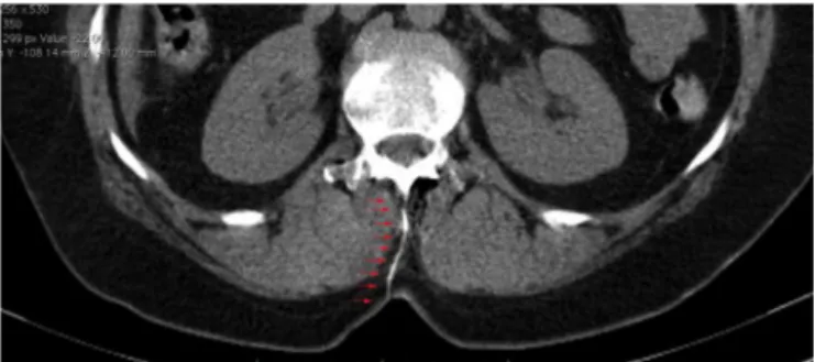
surgicalprocedurewasuneventful.Attheendofthesurgery anewaspirationthroughthecatheterwasperformed,when blood reflux wasagain observed. Then, 3mL of lidocaine withadrenalinewasadministeredthroughthecatheterand subsequentlya40%increaseinheartratewasnoted.After the surgery, the patient, already extubated and in spon-taneous breathing, lucid and oriented was taken to the radiologydepartment.Ahelicalcomputedtomographyscan wasperformedwithinjectionof4mLofiodinatednon-ionic contrastthroughtheepiduralcatheter.The imageanalysis revealed the catheterpath from the skin to theepidural space(Fig.1). This procedure allowed theobservation of theposteriorandanteriorinternalepiduralvenousplexuses (Fig. 2). The intervertebral vein was also identified from its origin in the intervertebral foramen to its confluence withtheazygosvein(Fig.3).Inimagesintheaxial,sagittal andcoronalplanes,itispossibletoidentifytheazygosvein throughoutitsabdominalandthoracicportion(Figs.4---6).
Thecatheterwasremoveduneventfully.Thepatienthad a good clinical course and was discharged on the second postoperativeday,withnocomplaints.
210 M.P.Souzaetal.
Figure2 Anteriorandposteriorepiduralvenousplexuswith contrast.
Figure3 Intervertebralvein.
Discussion
Thecatheterizationoftheinternalepiduralvenousplexus isapossiblecomplication,evenwhentheoperatorfollows thepropertechnique.Severalstudieshavetriedtocorrelate strategiesassociatedwithalowerincidenceofinadvertent catheterizationoftheepiduralvenousplexus.3Mhyreetal., inameta-analysisinvolving30clinicaltrialsandmorethan 12,000patients, concludedthat therisk of venous plexus catheterizationcanbereduced,ifthefollowingstrategies are applied: positioning the patient in lateral decubitus,
Figure4 Azygosveinintheaxialplane.
Figure5 Azygosveininthesagittalplane.
fluidpre-distension,useofsingle-holecathetersandlimiting thedepthofcatheterinsertionto6cmorless.1
Given the relative frequency of accidental epidural venous plexus catheterizationsandthe direconsequences thatanaccidentalintravascularinjectionoflocalanesthetic cancause,itisimperativefortheimmediaterecognitionof thiscomplication bythe anesthesiologist.The administra-tionofatestdose throughthecathetermustbearoutine maneuver, evenwhen the test dose by needle was nega-tive.Whileitispossibletodocumenttheactualplacement of the epidural catheter,aswell as the occurrenceof an
Accidentalcatheterizationofepiduralvenousplexus:tomographicanalysis 211
accidental venous catheterization using computed tomo-graphy with contrast injection through the catheter, its occurrencemust be clinicallyrecognized,sincethe imag-ingtestisnotaccessibletotheanesthesiologistinhis/her dailypractice,becauseof itshigh costorthedifficultyof conductingthispatienttotheradiologydepartmentpre-or perioperatively.
Conflicts
of
interest
Theauthorsdeclarenoconflictsofinterest.
References
1.MhyreJM,GreenfieldMLVH,TsenLC,PollyL.Asystematicreview ofrandomizedcontrolledtrialsthatevaluatestrategiestoavoid epidural vein cannulation during obstretric epidural catheter placement.ObstetrAnesthesiol.2009;108:1232---42.