Sonographic morphometry of the liver and biliary tract
in porcine models submitted to experimental biliary obstruction
*
Estudo ultrassonográfico morfométrico do fígado e trato biliar de suínos submetidos a obstrução biliar experimental
Aline Gomes de Campos1, Edmilson Rodrigo Daneze2, Júverson Alves Terra Júnior3, Aldo Benjamim Rodrigues Barbosa4, Gianne Regina dos Santos Sliuzas2, Alex Augusto da Silva5, Sílvia Azevedo Terra6
Objective: To compare, by means of ultrasonography, pre- and postoperative anatomical changes arising from experi-mentally induced obstructive jaundice in porcine models. Materials and Methods: Six 36-day-old Landrace pigs under-went laparoscopically induced complete biliary obstruction by common bile duct ligation. Results: No difficulty was faced during the procedures and the surgical recovery was uneventful. After seven days, the animals showed jaundice, bilirubinuria and acholic stools. Comparative ultrasonography allowed visualization of hepatomegaly, cholecystomegaly and increased caliber of the common bile duct in all the animals, as well as changes resulting from cholestasis. The morphometric analysis revealed a significant increase in diameter of the gallbladders and left lateral liver lobes. Con-clusion: Pigs represent appropriate experimental models for investigation of obstructive jaundice, and ultrasonography has shown to be sensitive, playing a relevant role in the diagnosis of extrahepatic biliary obstruction in such animals. Keywords: Biometry; Obstructive jaundice; Extrahepatic cholestasis.
Objetivo: Comparar as alterações anatômicas decorrentes de um quadro de icterícia obstrutiva experimental induzida em suínos nos períodos pré e pós-operatório por meio de exame ultrassonográfico. Materiais e Métodos: Seis suí-nos da raça Landrace, com 36 dias de idade, foram submetidos a obstrução biliar completa mediante ligadura do ducto colédoco por cirurgia videolaparoscópica. Resultados: Não ocorreram dificuldades na execução dos procedi-mentos obstrutivos e a recuperação cirúrgica foi eficiente. Decorridos sete dias, os animais apresentaram icterícia, bilirrubinúria e acolia fecal. O exame ultrassonográfico comparativo permitiu visualizar hepatomegalia, colecistomega-lia e aumento no calibre do ducto colédoco em todos os animais, assim como alterações decorrentes da colestase. A avaliação morfométrica revelou aumento significativo nos diâmetros da vesícula biliar e do lobo hepático lateral es-querdo. Conclusão: Os suínos representam um modelo experimental adequado de icterícia obstrutiva, e o exame ultrassonográfico demonstrou-se sensível e relevante no diagnóstico das alterações decorrentes de obstrução biliar extra-hepática nesses animais.
Unitermos: Biometria; Icterícia obstrutiva; Colestase extra-hepática. Abstract
Resumo
* Study developed at Faculdade Dr. Francisco Maeda (FAFRAM/ FE), Ituverava, SP, Brazil.
1. Master, Teacher, Course of Veterinary Medicine, Faculdade Dr. Francisco Maeda (FAFRAM/FE), Ituverava, SP, Brazil.
2. Students, Program of Advanced Studies of Veterinary Prac-tice and Surgery, Faculdade Dr. Francisco Maeda (FAFRAM/FE), Ituverava, SP, Brazil.
3. Master, Associate Professor, Faculdade de Medicina da Universidade Federal do Triângulo Mineiro (UFTM), Uberaba, MG, Brazil.
4. MD, Radiologist, Radiology Department of Santa Casa de Misericórdia de Ituverava, Ituverava, SP, Brazil.
5. PhD, Associate Professor, Faculdade de Medicina da Uni-versidade Federal do Triângulo Mineiro (UFTM), Uberaba, MG, Brazil.
6. PhD, Teacher, Course of Veterinary Medicine at Faculdade Dr. Francisco Maeda (FAFRAM/FE), Ituverava, SP, Brazil. (In memoriam).
Mailing Address: Edmilson Rodrigo Daneze. Avenida Primeiro de Maio, 930, Vila Virgínia. Ribeirão Preto, SP, Brazil, 4030-390,. E-mail: [email protected].
Received October 20, 2012. Accepted after revision January 18, 2013.
Campos AG, Daneze ER, Terra Júnior JA, Barbosa ABR, Sliuzas GRS, Silva AA, Terra SA. Sonographic morphometry of the liver and biliary tract in porcine models submitted to experimental biliary obstruction. Radiol Bras. 2013 Mar/Abr;46(2):89–95.
Therefore, the sonographic analysis of the hepatic and biliary morphometric param-eters in the setting of biliary obstruction is very relevant, helping in the identification and evaluation of the obstruction site, con-tributing in the treatment of the patients.
The imaging evaluation, particularly by ultrasonography (US) of the liver and bil-iary tract has been the object of a number of recent articles in the Brazilian radiologi-cal literature(12–18). US represents the most
utilized imaging method in the primary evaluation of patients with suspected biliary obstruction(4,8,9). Other imaging methods
may also be utilized in the evaluation of the biliary tract, namely, radioisotope chole-scintigraphy, computed tomography (CT),
INTRODUCTION
In the clinical practice, biliary obstruc-tion is a frequent and severe situaobstruc-tion, which can lead to generalized disease(1–3).
Several conditions can trigger extrahepatic biliary obstruction with consequential cholestasis, both in humans and animals(4– 6). Choledocholithiasis, neoplasia and
stenosis present higher incidence(7).
The knowledge of normal anatomy and pathological imaging findings is funda-mental for the detection of conditions in the hepatobiliary system(5,8,9). In the case of
magnetic resonance imaging (MRI), trans-hepatic cholangiography, endoscopic retro-grade cholangiopancreatography, or even radiography, oral cholecystography, and hepatic scintigraphy in some cases(4,7,9).
Bile duct injuries usually affect the up-per part of the duct located close to the he-patic hilum, making it difficult to perform appropriate choledochoduodenal recon-struction(19). Additionally, injuries requiring
biliary repair are commonly associated with long term complications, and the level of the injury and timing of repair are associated with the risk for post-surgical stenosis(20).
With the purpose of developing a more appropriate anatomical and physiological reconstruction for extensive biliary tract injuries, a study was proposed for recon-structing a bile duct by interposition of a “tube” constructed with a small bowel seg-ment, similar to what Monti et al. have pro-posed for continent neobladder drain-age(21). However, such a study requires an
experimental model where one can induce jaundice with an obstructive pattern by li-gating the extrahepatic bile duct, allowing for the analysis, evaluation and comparison of such changes in order to better under-stand the extent of the injury and the con-sequential compromising, as well as pro-viding a previous knowledge on the surgi-cal site, indicating specific techniques for, as a second step proposing the reconstruc-tion of the extrahepatic bile duct.
The present study was aimed at induc-ing obstructive jaundice in pigs, by perform-ing common bile duct ligation through videolaparoscopic surgery. The hepatic morphology histopathologically analyzed and the morphometry of the liver and the biliary tract evaluated at ultrasonography were compared before and seven days after the biliary obstruction. The changes ob-served in the animals were described, thus confirming the success of the procedure.
MATERIALS AND METHODS
The present experimental study was ap-proved by the Committee for Ethics in Research of Faculdade Dr. Francisco Maeda (FAFRAM/FE) under No. 19/2008.
Experiment outline
For the present study, six 36-day-old pigs (Sus scrofa domestica) of the Landrace
race with mean weight of 9.17 (± 1.69) kg, originated from a single offspring, were uti-lized. Such animals were followed-up on and clinically evaluated on a daily basis since birth.
Pre- and post-obstruction sonographic evaluation
All the animals underwent ultrasonog-raphy scans on the day before the obstruc-tive procedure, and post-obstruction scans were performed seven days after the pro-cedure.
The animals under food and water fast-ing for six hours were sedated with acepromazine (0.2 mg/kg/intramuscular) and placed on a 50 cm surgical positioner. US was performed with the animal in dor-sal decubitus, in the B-mode (SIUI CTS-310B apparatus, with a 5.0 MHz convex transducer), utilizing gel as acoustic cou-pling medium. For the purpose of compara-tive analysis, the following measurements were performed: major and minor longitu-dinal diameters of the left lateral hepatic lobe; major and minor cross-sectional and longitudinal diameters of the gallbladder; and the cross-sectional diameter of the cho-ledochal duct on each animal. Additionally, the hepatic parenchyma and the abdominal cavity were analyzed in the search for any possible alterations. The images were printed and recorded on video and the ob-tained morphometric values were recorded on a table attached to the clinical records regarding each animal. Such adopted pro-cedures were standardized, both for the pre-obstruction and the post-pre-obstruction ultra-sonography scans.
Anesthetic and surgical procedures
The preoperative procedures were com-mon to all animals, which were submitted to food and water fasting for six hours prior to the procedure. Upon arrival at the hos-pital, the animals were given a cold shower bath for body cleansing and were sedated with acepromazine (0.2 mg/kg/intramuscu-lar) and then had shaving and antisepsis of their ventral abdominal region. After the medication onset of action, the animals were taken to the surgical center. The me-dial saphenous vein was catheterized for glycophysiological solution infusion and administration of dissociative anesthetic
based on the association of tiletamine and zolazepam (5.0 mg/kg/intravenous), and also fentanyl (0.025 mg/kg/intravenous) and atropine sulfate (0.5 mg/kg/intrave-nous). The anesthetic induction was main-tained with the dose of one third to one half of the original dose of the dissociative an-esthetic. During the surgical procedure, the animals received oxygen by means of a face mask.
Once the anesthetic plan was con-firmed, the videolaparoscopic procedure was initiated (Karl Storz laparoscopic en-doscope, with 15″ LCD video display, 175 W xenon light source, 30.1 thermal insuf-flator and a three-chip camera), with a pre-umbilical median 1.0 cm incision, through which a Verres needle was inserted to per-form the pneumoperitoneum with intrac-avitary pressure of 11 mmHg. Subse-quently, four trocars were positioned as follows: two 5.0 mm trocars – one located at the caudal region to the right costal mar-gin, and the other at the level of the right iliac fossa –, and two 10 mm trocars, - one located 1.0 cm pre-umbilical and one cau-dal to the left costal margin.
During the procedure, prehilar struc-tures were identified, isolated and dissected and the laparoscopic ligation of the bile duct was performed as distally as possible with the purpose of producing total extra-hepatic obstructive jaundice. Once the li-gation was completed, hemostasis was verified, the cavity was washed with pre-heated (37.5°C) physiological solution, the pneumoperitoneum was relieved, followed by surgical wound closure with 2-0 cotton thread and surgical skin closure with mononylon 3-0 thread.
Seven days after the procedure, post-ob-struction US scan was performed and the animals were taken to the surgical center (utilizing the same anesthesia protocol as in the obstructive procedure), where pre-umbilical median laparotomy was per-formed for inspection of the abdominal cavity, removal of the bile duct ligation and consequently desobstruction of the duct.
Pre- and post-obstruction histopathological analysis
collected for histopathological analysis. Such specimens were identified and fixed in 3.7% formalin solution. The processing of such material was carried out in the General Pathology Department of Univer-sidade Federal do Triângulo Mineiro, ac-cording to the protocol normally utilized by that Department. In order to avoid any bias, the histopathological slides received iden-tification different from those utilized for the animals.
Postoperative care
In the postoperative period (obstruction and desobstruction) the animals remained under constant care, with follow-up on the surgical wounds healing and administra-tion of anti-inflammatory drug (intramus-cular dexamethasone – dose of 0.025 mg/kg/ day), analgesic drug (intramuscular flunixin meglumine – dose of 2.2 mg/kg/day) and preventive antibiotic therapy (intramuscu-lar enrofloxacin – dose of 2.5 mg/kg/day). The mean weight of the animals at the end of the experiment was 9.42 (± 2.19) kg.
Statistical analysis
The statistical analysis for comparison of the pre- and post-obstruction morpho-metric results was performed by utilizing the Sigma Stat 2.03 software. The verifica-tion of the normal variables distribuverifica-tion was carried out by means of the Kolmogo-rov-Smirnov test, where the continuous variables with normal distribution were analyzed by the paired t-test for dependent samples, being such variables expressed in mean values and standard deviation, as-suming as significance level a probability lower than 5% (p < 0.05).
RESULTS
By utilizing only the portals described in the method, the main bile duct ligation was successfully performed without diffi-culties in all the animals, with easy identi-fication of the bile duct during videolapar-oscopic surgery. The most frequent signs presented by the animals seven days after the obstructive procedure were jaundice,
bilirubinuria, fecal acholia and hepatome-galy at palpation.
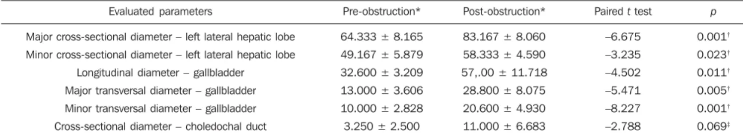
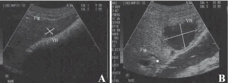
US performed seven days after the sur-gical obstruction revealed considerable and significant enlargement of the hepatic pa-renchyma (Figure 1; Table 1), as well as increased gallbladder diameter (Figure 2; Table 1). However, even with the biliary tract presenting considerable enlargement, such an enlargement was not significant in the case of the common bile duct (Figure 3; Table 1).
During laparotomy for reconstruction of the common bile duct by interposition of a tube (Monti’s procedure), noticeable hepatomegaly was observed in the six ani-mals, with the liver being felt firmer than normal, moderately pale and with a strong yellowish color. Also, increased caliber and consistency of the gallbladder and extra-hepatic biliary ducts were observed.
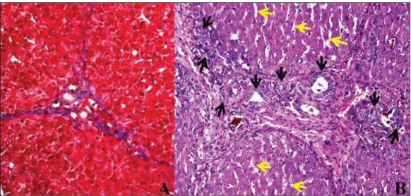
At histopathological analysis, as compar-ing pre-obstruction with post-obstruction specimens, presence of hyperplasia of the biliary ducts in the region of the portal space
Table 1 Pre- and post-obstruction sonographic morphometry values. Evaluated parameters
Major cross-sectional diameter – left lateral hepatic lobe Minor cross-sectional diameter – left lateral hepatic lobe
Longitudinal diameter – gallbladder Major transversal diameter – gallbladder Minor transversal diameter – gallbladder Cross-sectional diameter – choledochal duct
Pre-obstruction*
64.333 ± 8.165 49.167 ± 5.879 32.600 ± 3.209 13.000 ± 3.606 10.000 ± 2.828 3.250 ± 2.500
Post-obstruction*
83.167 ± 8.060 58.333 ± 4.590 57,.00 ± 11.718 28.800 ± 8.075 20.600 ± 4.930 11.000 ± 6.683
Paired t test
–6.675 –3.235 –4.502 –5.471 –8.227 –2.788
p
0.001† 0.023† 0.011† 0.005† 0.001† 0.069‡
* Mean ± standard deviation. †Significant. ‡Non-significant.
Figure 1. Sonographic images showing left lateral hepatic lobe (lines) of pigs experimentally submitted to biliary obstruction. On A, observe the pre-procedural normal longitudinal diameters; On B, observe the increased post-procedural longitudinal diameters. Also, observe the increased echogenicity of the hepatic parenchyma (asterisk) and dilation of intrahepatic biliary ducts on B (arrows).
and dilation of the hepatic sinusoids was observed among other changes (Figure 4).
DISCUSSION
For the present study, pigs (Sus scrofa domestica) were utilized, because of their resistance, easy management and with good possibilities of standardization, fre-quently being utilized in experimental stud-ies, and considered as appropriate animal models for the study of diseases related to the human life style(22,23). In the present
study, the authors have observed that pigs
represent an ideal type of experimental model since they develop obstructive jaun-dice within a short period of time.
The option for the videolaparoscopic surgery was justified for being the most uti-lized technique to approach biliary ducts, besides other advantages such as better acceptance by patients, smaller incisions, better aesthetic results, less postoperative pain, besides shorter hospital stay and low incidence of complications(2,19,24). In such a
context, a more efficient surgical recovery of the animals was observed, since manipu-lating the animals for wound dressing is
more practical and agile and the surgical wound healing is faster with less stress for the animals. Thus, the fast recovery al-lowed for the wellbeing of the animals and greater dedication of the team to the changes resulting from the obstructive procedure.
The experimental induction of extrahe-patic cholestasis is not always a successful procedure(1). However, seven days after the
obstructive procedure, the animals pre-sented with signs compatible with those described for cholestasis in humans and animals(1,2,10,19). Thus it is possible to accept
that both in humans and animals, when Figure 2. Sonographic images showing the gallbladder (lines) of a pig experimentally submitted to surgical biliary obstruction. On A, pre-obstruction cross-sectional diameters; on B, post-obstruction cross-cross-sectional diameters. On B, one observes increased gallbladder diameter, as well as increased echogenicity of the hepatic parenchyma (asterisk). (Fig., liver; VB, gallbladder).
Figure 3. Sonographic images showing choledochal duct (lines) in a pig experimentally submitted to surgical biliary obstruction. On A, pre-obstruction longi-tudinal diameters; on B, post-obstruction longilongi-tudinal diameters. On B, one observes increased diameter of the choledochal duct, as well as increased echogenicity of the hepatic parenchyma (asterisk).
there is increase in the concentration of bilirubin in the blood circulation, deposi-tion of such substance on tissues (jaundice) and glomerular excretion (bilirubinuria) are observed, while the decrease or absence of bilirubin excretion in the intestinal lumen causes changes in the color of the feces (acholia) which become lighter than nor-mal(3,6,25). Therefore, the authors believe
that pigs can be utilized as experimental surgical models for observation of obstruc-tive jaundice.
Extrahepatic biliary tract
There is currently a wide variety of im-aging techniques for evaluation of the bil-iary tract(4,6,9). Among such techniques, US
is preferred by most professionals in the initial investigation on patients with sus-pected biliary conditions(4,6,8,9). The
utiliza-tion of such technique is indicated in most abdominal diseases, and is totally non-in-vasive and essential for establishing diag-nosis and approach in many situa-tions(5,26,27). It has the advantage of being
accessible, rapid, and innocuous, besides its bedside capability. Furthermore, it is a low cost method, does not rely on ionizing radiation or contrast utilization and does not require patient(4,26) or animal(5)
seda-tion. In the present study, there were no dif-ficulties in performing the ultrasonography scan on the animals, with easy and clear vi-sualization of the liver and biliary tract.
Regardless of the gastrointestinal, he-patic or biliary function, US is appropriate for evaluation of both parenchymal organs and hollow viscera filled with fluid(26,27).
Biliary system disorders can be easily de-tected at US(28,29). Because of its cost and
convenience in association with high sen-sitivity and specificity, US is considered the best screening method in cases of lithiasis, detecting 95% of the calculi, and is also useful for visualizing choledochal dilata-tion(4,6,9,26), besides allowing, many times,
the characterization of diseases developing outside the gallbladder(4). According to
Zeman et al.(30), peripheral biliary dilatation
can be diagnosed four hours after biliary obstruction, even before significant eleva-tion of bilirubin serum levels and onset of jaundice. However, US presents limitations in the analysis of organs with gas con-tent(26,27). Factors such as obesity and
me-teorism, among others, may impair a proper analysis and identification of the gallblad-der, even in the presence of calculi(4), and
fail in diagnosing calculi in the common bile duct, particularly in its terminal por-tion, where there is greater interposition of the duodenal gas content(5,9), thus the
rel-evance of fasting prior the performance of ultrasonography. In the present study, wa-ter and food fasting was essential for a good visualization of the hepatic tissue and bil-iary tract.
A healthy gallbladder presents non echogenic content, with no echoes in its interior and thin, regular walls(4). Thus,
ul-trasonography allows the delineation of the anatomic texture of the gallbladder walls, with evaluation of parameters such as shape, size, motility and thickness of the wall, as well as the appearance of its con-tent. It also allows the demonstration of
dilated intra- and extrahepatic biliary ducts, helping in the differentiation between obstructive and non-obstructive jaun-dice(4,5,8,9,27). In the present study, the
pres-ence of calculi was not observed within the gallbladder, however, the increase in its diameter was observed as the pre- and post-obstruction images were compared.
Significant dilation of extrahepatic bil-iary ducts, with noticeable perception at ul-trasonography has important diagnostic and prognostic value(4,5,30,31). Thus, although
statistically significant difference has not been found in the diameter of the common bile duct in the present study, a consider-able increase in diameter was observed above the obstructive ligation site, which demonstrates that the procedure was effec-tive, confirming the biliary obstruction.
The dilation of the biliary tract evolves in a retrograde way after the complete ob-struction of the common bile duct, so the observation of gallbladder enlargement and dilation of the ducts is frequent, particularly at their terminal portion(4,30,31). According
to Nyland et al.(32), gallbladder and
com-mon bile duct enlargement is first seen, followed by extrahepatic bile duct dilation in the period between 24 and 48 hours; with distended intrahepatic ducts becom-ing visible five to seven days after complete biliary obstruction. However, as the biliary ducts dilate, their caliber may exceed the diameter of the portal vein branches, being visualized as parallel structures, showing the classical double duct sign(6,27). Thus, the
identification of the obstructive site is based on the identification of the region above Figure 4. Photomicrography of
which the duct is dilated and below which the caliber is normal or non-identifiable.
The clear distension of the gallbladder is one of the first signs of complete biliary obstruction(30–32). Mwanza et al.(10), in a
study involving obstruction of the common bile duct in dogs, have observed that the distension of the gallbladder was clearly seen within the first week after the ligation. Such a fact was also observed in the present study, since after seven days from the ob-struction of the common bile duct a signifi-cant alteration could be clearly observed, with such distension also occurring be-cause of the established cholestasis(3,10,32).
However, Santo(2), evaluating the caliber of
the main biliary duct in 67 human patients with choledocholithiasis, has verified that only 42 patients presented choledochal di-lation. Liu et al.(33) have reported that the
association of clinical, laboratory and sonographic criteria determine a sensitiv-ity of 96% to 98% in the diagnosis of cho-ledocholithiasis.
Thus, in dubious cases or those requir-ing more accurate information, or even for differential diagnosis, sonographic must be correlated with other diagnostic imaging methods and/or laboratory analyses(6,8,11).
In the present study, the analysis of the bio-chemical profile of the animals was per-formed in the two periods, revealing the changes in serum levels which are directly related to biliary obstruction.
Liver
When there is an obstruction of the common bile duct, the drainage of the bile into the bowel does not happen, and it gradually accumulates in the ducts, bile canaliculi and hepatocytes which, conse-quently, dilate, inducing the increase in size of the liver. Such increase, on its turn, causes compression of hepatic cells and structures, which, in association with the degenerative processes caused by cholestasis, may progress to cellular death and, secondarily, to hepatic cirrhosis(3). In
the present experiment, besides hyperpla-sia of bile ducts, the authors have observed collagen fibers stating to accumulate form-ing nodules characterizform-ing the presence of early-stage hepatic cirrhosis.
According to Sullivan(9), at early stages
of the cirrhotic process, the human cirrhotic
liver is enlarged, with a relative enlarge-ment of the lateral aspect of the left lateral lobe, a fact also observed in the present study, as when comparing the liver diam-eters measured before and after the obstruc-tive procedure, the authors observed a sig-nificant increase in the volume of the ani-mals’ organ, later confirmed by histopatho-logical analysis.
The determination of liver size is a com-mon procedure in pediatrics routine, both for detecting hepatomegaly and for moni-toring the progress of diseases or hepatic response to treatment(26,28,29). In case of
sus-picion of hepatomegaly, in vivo liver mea-surements can be performed by means of clinical and/or imaging methods such as radiography, scintigraphy, US and CT(26). In
the present study, significant results were obtained with ultrasonography as the mo-ments of the experiment were compared, so such method may be utilized for evaluation of cholestasis.
According to Sullivan et al.(34), the
clini-cal determination of the liver size is often inaccurate, and all studies relying um such parameters should be put under suspicion. Walk(35) also describes the evaluations by
means of radiography as unsatisfactory. Thus, the utilization of more reliable meth-ods for liver measurements is recom-mended, as a large liver is easily detectable, but smaller abnormalities may be underes-timated(34). In such a context, US is
consid-ered a simple and quantitative method for assessment of liver size, being the first imaging study requested for such pur-pose(4,26), also in those cases of cholestasis
associated with biliary obstruction(4,5).
The knowledge of the normal hepatic anatomy and of the pathological imaging findings of the lesions, is essential for their detection(8). At US, a healthy liver is seen
with a smooth contour, and the hepatic parenchyma with a uniform and homoge-neous echotexture, presenting echogenicity that is equal or slightly higher as compared with the spleen(5,11). US is a valuable
method for evaluating the internal architec-ture of the liver(36), allowing a detailed
study of the parenchyma, evaluating di-mensions, shape, contour, borders, changes in echogenicity and appearance of vessels and hepatic structures, as well as its rela-tionship with adjacent structures(5,26,37).
However, one should be careful, as there are diseases which normally cause in-creased echogenicity of the hepatic paren-chyma, without changes in the liver dimen-sions, which remain normal, such in fatty infiltration, steroid hepatopathy, diabetes mellitus, lymphoma and some toxic hepatopathies, while in cirrhosis and chronic cholangiohepatitis the liver gener-ally presents reduced dimensions and ir-regular contour(3,5). Thus, the diffuse and
increased echogenicity and insufficient definition of the portal vessels visualized on the animals’ liver parenchyma can be attributed to cholestasis(4,11).
In certain cases, the sonographic ap-pearance may be nonspecific(28), as
ob-served by Mwanza et al.(10) who, by
ob-structing the common bile duct in dogs, reported little change in echogenicity of the liver parenchyma, even with jaundice, bio-chemical alterations and noticeable disten-sion of the gallbladder at the first week after ligation. According to Sullivan(9), US,
CT or MRI rarely detect early-stage diffuse hepatic diseases, diagnosing them only in cases where noticeable changes are ob-served in dimensions, density, and signal intensity, i.e., in advanced stages of the disease. In the present study, seven days of biliary obstruction were enough for the animals to develop changes which could be visualized at US, biochemical alterations and noticeable clinical signs of jaundice.
As a subjective and individual analysis is considered, differences may be observed in the interpretation of US findings. The diagnosis may be influenced by different factor such as the observer experience, type of apparatus and settings of depth, gain and contrast resolution. Concomitantly with hepatic alterations, several diseases may lead to alterations in other organs utilized for comparison of echogenicity and echotexture. Such alterations constitute additional factors that might impair the diagnosis(5,37). In such cases, the diagnosis
can be substantially considered when the clinical signs are consistent(28) or by
asso-ciation with other diagnostic methods.
CONCLUSIONS
to conclude that the pigs represent appro-priate experimental models in the study of obstructive jaundice, as findings were ob-served at ultrasonography, biochemical and histopathological analysis. US allowed the visualization of significant hepatomegaly and cholecystomegaly, besides consider-able enlargement of the common bile duct in the animals, demonstrating to be a sen-sitive and relevant method in the diagno-sis of alterations resulting from extrahe-patic biliary obstruction in such animals. Additionally, changes caused by chole-stasis such as insufficient definition of the portal vessels and increased and diffuse echogenicity were perceptible.
REFERENCES
1. Prado IB, Santos MHH, Lopasso FP, et al. Cholestasis in a murine experimental model: le-sions include hepatocyte ischemic necrosis. Rev Hosp Clín Fac Med S Paulo. 2003;58:27–32. 2. Santo MA. Litíase na via biliar principal: análise
do tratamento cirúrgico por videolapascopia [tese]. São Paulo, SP: Faculdade de Medicina – Universidade de São Paulo; 2000.
3. Cotran RS, Kumar V, Robbins SL. Robbins: pathologic basis of disease. 7th ed. Philadelphia, PA: Elsevier; 2005.
4. Chammas MC, Marcelino ASZ, Saito OC, et al. Vesícula biliar. Ductos biliares. In: Lopes AC. Tratado de clínica médica. São Paulo, SP: Roca; 2006. p. 1217–32.
5. Nyland TG, Mattoon JS, Herrgesell EJ, et al. Fí-gado. In: Nyland TG, Mattoon JS. Ultra-som diag-nóstico em pequenos animais. 2ª ed. São Paulo, SP: Roca; 2005. p. 95–130.
6. Franchi-Teixeira AR, Antoniali F, Boin IFSF, et al. Icterícia obstrutiva: diagnóstico laboratorial e de imagem. Medicina (Ribeirão Preto). 1997;30: 198–208.
7. Dähnert W. Radiologia: manual de revisão. 3ª ed. Rio de Janeiro, RJ: Revinter; 2001.
8. Gunderman RB. Fundamentos de radiologia: apresentação clínica, fisiopatologia, técnicas de imagens. 2ª ed. Rio de Janeiro, RJ: Guanabara Koogan; 2007.
9. Sullivan LM. O fígado, o sistema biliar e o
pân-creas. In: Juhl JH, Crummy AB, Kuhlman JE, edi-tores. Paul & Juhl: interpretação radiológica. 7ª ed. Rio de Janeiro, RJ: Guanabara Koogan; 2000. p. 433–63.
10. Mwanza T, Miyamoto T, Okumura M, et al. Ultrasonographic evaluation of portal vein hemo-dynamics in experimentally bile duct ligated dogs. Jpn J Vet Res. 1998;45:199–206. 11. Biller DS, Kantrowitz B, Miyabayashi T.
Ultra-sonography of diffuse liver disease. A review. J Vet Intern Med. 1992;6:71–6.
12. Borges VFA, Diniz ALD, Cotrim HP, et al. Dop-plerfluxometria da veia hepática em pacientes com esteatose não alcoólica. Radiol Bras. 2011; 44:1–6.
13. Matsuoka MW, Oliveira IRS, Widman A, et al. Contribuição da ultrassonografia para o diagnós-tico das alterações histopatológicas presentes na hepatite C crônica, com ênfase na esteatose he-pática – Parte I. Radiol Bras. 2011;44:141–6. 14. Burke LMB, Vachiranubhap B, Tannaphai P, et
al. Realce por contraste de lesões hepáticas em pacientes com cirrose: estudo cruzado compara-tivo de dois agentes de contraste para RM reali-zado em uma única instituição. Resultados pre-liminares. Radiol Bras. 2011;44:147–50. 15. Barbosa ABR, Souza LRMF, Pereira RS, et al.
Espessamento parietal da vesícula biliar no exame ultrassonográfico: como interpretar? Radiol Bras. 2011;44:381–7.
16. Gössling PAM, Alves GRT, Silva RVA, et al. Bi-lioma espontâneo: relato de caso e revisão da li-teratura. Radiol Bras. 2012;45:59–60. 17. Souza LRMF, Rodrigues FB, Tostes LV, et al.
Avaliação por imagem das lesões císticas con-gênitas das vias biliares. Radiol Bras. 2012;45: 113–7.
18. Guimarães Filho A, Carneiro Neto LA, Palheta MS, et al. Doença de Caroli complicada com abs-cesso hepático: relato de caso. Radiol Bras. 2012; 45:362–4.
19. Crema E, Silva AA, Lenza RM, et al. Excluded-loop hepatojejunal anastomosis with use of laparoscopy in late management of iatrogenic ligature of the bile duct. Surg Laparosc Endosc Percutan Tech. 2002;12:110–4.
20. Walsh RM, Henderson JM, Vogt DP, et al. Long-term outcome of biliary reconstruction for bile duct injuries from laparoscopic cholecystecto-mies. Surgery. 2007;142:450–7.
21. Monti PR, Lara RC, Dutra MA, et al. New tech-niques for construction of efferent conduits based on the Mitrofanoff principle. Urology. 1997;49: 112–5.
22. Almond GW. Research applications using pigs. Vet Clin North Am Food Anim Pract. 1996;12: 707–16.
23. Bustard LK, McClellan RO. Use of pigs in bio-medical research. Nature. 1965;208:531–5.
24. Machado MAC, Herman P, Makdissi FF, et al. Ressecções hepáticas por videolaparoscopia: uti-lidade da técnica de Hemi-Pringle. Rev Bras Vi-deocir. 2005;3:56–9.
25. Lassen ED. Avaliação laboratorial do fígado. In: Thrall MA. Hematologia e bioquímica clínica veterinária. São Paulo, SP: Roca; 2007. p. 335–43. 26. Rocha SMS, Oliveira IRS, Widman A, et al. He-patometria ultra-sonográfica em crianças: proposta de novo método. Radiol Bras. 2003;36:63–70.
27. Cerri GG, Vogueira LAA. Ultra-sonografia em gastroenterologia. In: Mincis M. Gastroenterolo-gia e hepatoloGastroenterolo-gia: diagnóstico e tratamento. 3ª ed. São Paulo, SP: Lemos Editorial; 2002. p. 45–53. 28. Nyland TG, Hager DA. Sonography of the liver, gallbladder, and spleen. Vet Clin North Am Small Anim Pract. 1985;15:1123–48.
29. Nyland TG, Hager DA, Herring DS. Sonography of the liver, gallbladder, and spleen. Semin Vet Med Surg (Small Anim). 1989;4:13–31.
30. Zeman RK, Taylor KJ, Rosenfield AT, et al. Acute experimental biliary obstruction in the dog: sonographic findings and clinical implications. AJR Am J Roentgenol. 1981;136:965–7.
31. Léveillé R, Biller DS, Shiroma JT. Sonographic evaluation of the common bile duct in cats. J Vet Intern Med. 1996;10:296–9.
32. Nyland TG, Gillett NA. Sonographic evaluation of experimental bile duct ligation in the dog. Vet Radiol. 1982;23:252–60.
33. Liu TH, Consorti ET, Kawashima A, et al. The efficacy of magnetic resonance cholangiography for the evaluation of patients with suspected cho-ledocholithiasis before laparoscopic cholecystec-tomy. Am J Surg 1999;178:480–4.
34. Sullivan S, Krasner N, Williams R. The clinical estimation of liver size: a comparison of tech-niques and an analysis of the source of error. Br Med J. 1976;30:1042–3.
35. Walk L. Quantitative method to determine the liver size. Radiologe. 1978;18:354–5.
36. Lamb CR. Ultrasonography of the liver and bil-iary tract. Probl Vet Med. 1991;3:555–73.