Incidental enchondromas at knee magnetic resonance imaging:
intraobserver and interobserver agreement and prevalence
of imaging findings
*
Encondromas incidentais nos exames de ressonância magnética do joelho: concordância intraobservador e interobservador e prevalência das características de imagem
Sandra Akemi Nakamura1, Mário Müller Lorenzato2, Edgard Eduard Engel3, Maurício Eiji de Almeida Santos Yamashita4, Marcello Henrique Nogueira-Barbosa5
Objective: To evaluate intra- and interobserver agreement in the identification of incidental enchondromas at knee magnetic resonance imaging, and to assess the prevalence of imaging findings. Materials and Methods: Retrospective study reviewing 326 knee magnetic resonance images acquired in the period between November 2009 and September 2010. The images were independently and blindly analyzed by two specialists in musculoskeletal radiology, with the objective of identifying incidental enchondromas, presence of foci with signal similar to bone marrow and foci of signal absence suggestive of calcifications within the enchondromas. Inter- and intraobserver agreements were analyzed. Results: Eleven lesions compatible with enchondromas (3.3%) were identified. The interobserver agreement for the presence of enchondroma was high. Prevalence of foci of bone marrow signal inside the enchondromas was of 54.55%, and foci suggestive of calcification corresponded to 36.36%. The intraobserver agreement for foci of bone marrow signal in enchondromas was perfect, and interobserver agreement was high. Conclusion: The prevalence of incidental enchondromas in the current study was compatible with data in the literature. Excellent agreement was observed in the identification of enchondromas and in the assessment of imaging findings. A higher prevalence of fat signal foci was observed as compared with signal absence suggestive of calcifications.
Keywords: Enchondromas; Magnetic resonance imaging; Knee; Bone marrow.
Objetivo: Avaliar a confiabilidade intra e interobservador na identificação de encondromas incidentais na ressonância magnética de joelho e estudar a prevalência das características de imagens destas lesões. Materiais e Métodos: Estudo retrospectivo, com revisão de 326 ressonâncias magnéticas do joelho realizadas entre novembro de 2009 e setembro de 2010. As imagens foram analisadas por dois médicos especialistas em radiologia musculoesquelética, de forma independente e às cegas, visando a identificar encondromas incidentais, presença de focos com sinal semelhante à medula óssea e focos de ausência de sinal sugestivos de calcificações no interior dos encondromas. Foram realizadas análises das concordâncias inter e intraobservador. Resultados: Foram identificadas 11 lesões compatíveis com en-condromas (3,3%). A concordância interobservador para presença de encondroma foi alta. A prevalência de focos de sinal de medula óssea no interior dos encondromas foi 54,55% e de focos sugestivos de calcificação foi 36,36%. A concordância intraobservador para focos de sinal de medula óssea nos encondromas foi perfeita, e a concordância interobservador foi alta. Conclusão: A prevalência de encondromas incidentais no trabalho atual foi consistente com a literatura. Foi observada excelente concordância no estudo de confiabilidade da identificação de encondromas e de suas características, notando-se maior prevalência de focos com sinal de gordura do que de calcificações.
Unitermos: Encondromas; Ressonância magnética; Joelho; Medula óssea.
Abstract
Resumo
* Study developed at Hospital das Clínicas da Faculdade de Medicina de Ribeirão Preto da Universidade de São Paulo (HC-FMRPUSP), Ribeirão Preto, SP, Brazil.
1. MD, Radiologist, Fellow of Musculoskeletal Radiology, Hospital das Clínicas da Faculdade de Medicina de Ribeirão Preto da Universidade de São Paulo (HC-FMRPUSP), Ribeirão Preto, SP, Brazil.
2. MD, Radiologist specialized in Musculoskeletal Radiology, Assistant, Division of Radiology, Center of Imaging Sciences and Medical Physics, Faculdade de Medicina de Ribeirão Preto da Uni-versidade de São Paulo (FMRPUSP), Ribeirão Preto, SP, Brazil. 3. PhD, Professor, Department of Biomechanics, Medicine and Locomotor System Rehabilitation, Faculdade de Medicina
Nakamura SA, Lorenzato MM, Engel EE, Yamashita MEAS, Nogueira-Barbosa MH. Incidental enchondromas at knee magnetic resonance imaging: intraobserver and interobserver agreement and prevalence of imaging findings. Radiol Bras. 2013 Mai/Jun;46(3):129–133.
de Ribeirão Preto da Universidade de São Paulo (FMRPUSP), Ri-beirão Preto, SP, Brazil.
4. Assistant Physician, Service of Pathology, Hospital das Clínicas da Faculdade de Medicina de Ribeirão Preto da Univer-sidade de São Paulo (HC-FMRPUSP), Ribeirão Preto, SP, Brazil. 5. PhD, Professor, Center of Imaging Sciences and Medical Physics, Faculdade de Medicina de Ribeirão Preto da Universi-dade de São Paulo (FMRPUSP), Ribeirão Preto, SP, Brazil.
Mailing Address: Dra. Sandra Akemi Nakamura. Av. Bandei-rantes, 3900, Campus Universitário. Ribeirão Preto, SP, Brazil, 14048-900. E-mail: [email protected]. Received October 19, 2012. Accepted after revision March 15, 2013.
INTRODUCTION
Enchondroma is a benign neoplasm of the intramedullary bone composed of hya-line cartilage. It is the second most common benign bone neoplasia just after osteochon-droma. It is believed that enchondromas represent 12% to 24% of all benign bone tumors and 3% to 10% of all bone tu-mors(13).
In the study developed by Walden et al., 449 consecutive routine knee MRI studies were evaluated, and the authors observed the prevalence of incidental enchondromas in 2.9% of the images(13). Such authors sug-gest that the recognition of incidental en-chondromas is important to avoid confu-sion with other diseases.
The typical MRI finding of enchon-droma is described as a lesion with lobu-lated contours, characterized by the pres-ence of hypersignal on fluid-sensitive im-ages, and with heterogeneous signal inten-sity on T1-weighted images(14). The in-creased signal intensity of enchondromas on fluid-sensitive images is due to the great amount of fluid present in the hyaline car-tilage that forms such lesions. However, the literature does not emphasize the character-istics of findings of enchondromas on T1-weighted images.
The identification of foci of bone tissue or bone marrow within enchondromas is well documented on histopathological studies(15,16). In a retrospective study, Murphey et al. found the presence of foci of hypersignal within lesions on T1-weighted images of enchondromas and low-grade chondrosarcomas, and have cor-related such MRI image with histopatho-logical findings(17).
The present study aimed at assessing the reproducibility of the identification of in-cidental enchondromas and evaluating the prevalence of such lesions as well as the prevalence of MRI findings of incidental enchondromas.
MATERIALS AND METHODS
The present retrospective study was ap-proved by the Committee for Ethics in Research of Hospital das Clínicas da Facul-dade de Medicina de Ribeirão Preto da Uni-versidade de São Paulo, and reviewed con-secutive knee MRI studies performed in the period from November 2009 to September
2010, regardless the clinical complaints and age of the patients. The images were independently and blindly analyzed by each one of two radiologists at two different moments with a three-month interval be-tween the first and the second readings. One of the radiologists has ten-year expe-rience in musculoskeletal radiology, and the other, two-year experience following a post-graduation in musculoskeletal radiol-ogy.
The MR images were acquired in a high-field 1.5 T equipment, with T1-weighted sequences (TR/TE, 500/20) in sagittal plane, and intermediate weighting sequences with fat suppression in axial/sagittal/coronal planes (TR/TE, 2000–3000/50).
The criteria adopted by the radiologists to diagnose an incidental enchondroma by MRI were the same used in the study de-veloped by Walden et al.(13). It were consid-ered incidental enchondromas bone mar-row focal changes that met the following criteria: focal geographic areas of bone marrow replacement on T1-weighted im-ages, and with high signal intensity on fluid-sensitive images with fat suppression, and lobulated contours on both T1 and in-termediate weightings. Focal alterations with the mentioned characteristics, but with subchondral location, were not classified as enchondromas, since they might corre-spond to subchondral cysts or intraosseous ganglion (mucous) cysts.
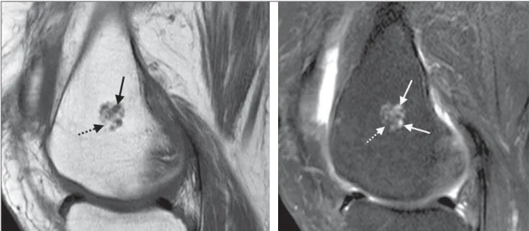
The two radiologists analyzed each im-age and recorded the respective findings of foci of signal similar to bone marrow sig-nal or foci of sigsig-nal suggesting the presence of calcifications within lesions classified as incidental enchondromas. The criterion utilized to characterize foci of bone marrow within incidental enchondromas was the presence of areas of hypersignal similar to bone marrow signal on T1-weighted im-ages, with signal loss similar to the decrease in bone marrow signal on intermediate weighting images with fat suppression (Figure 1). The criterion utilized to charac-terize the presence of calcifications within the lesions corresponded to foci of signal absence on T1-weighted sequences and on intermediate weighting sequences with fat suppression (Figure 2).
The largest diameter of each focal alter-ation compatible with incidental
enchon-droma was measured with a digital caliper on a workstation with the software Clear Canvas version 2.0.
Analyses were performed to evaluate the intra and interobserver agreement in the identification of incidental enchondromas and also foci of bone marrow and calcifi-cations within the lesions at MRI. The sta-tistical analysis was based on the kappa index (κ). A coefficient κ = 1 corresponds to a perfect agreement. A coefficient κ equal to zero indicates a randomly expected agree-ment. Negative values occur in cases where the agreement is poorer than randomly ex-pected but, according to Agresti(18), this is rarely observed. Landis et al.(19) provide the following classification for coefficient κ: absent agreement (κ < 0), neglectable agreement (0.00 < κ < 0.20), reasonable agreement (0.21 < κ < 0.40), moderate agreement (0.41 < κ < 0.60), substantial agreement (0.61 < κ < 0.80), almost perfect agreement (0.81 < κ < 1.00).
The authors have also evaluated the re-lationship between findings of foci of sig-nal similar to bone marrow within the en-chondromas and lesion size, sex and age of patients; and between presence of calcifi-cations within the lesions and the already mentioned variables. For the variable sex, the Fisher’s exact test was utilized to evalu-ate the association between two cevalu-ategori- categori-cal variables; and for the variables lesion size and patient’s age, the non-parametric Wilcoxon-Mann-Whitney test was utilized. In the cases of disagreement in relation to the presence or not of areas of fat signal within the incidental enchondromas, fur-ther evaluation was performed by a third observer with 16-year experience in mus-culoskeletal radiology. Such a third evalu-ation allowed the calculevalu-ation of the final prevalence of areas of bone marrow signal within the incidental enchondromas in the study sample.
RESULTS
44.79% (n = 146)], 56.13% (n = 183) of the images depicted right knees, and 43.87% (n = 143), left knees. The patients’ ages ranged between 4 and 85 years (mean = 40.25 years) in the total sample, and
be-tween 26 and 85 years (mean = 46.5 years) in the group of patients with enchondroma. Among the 11 enchondromas, six were found in male patients, five in the right knee. One patient presented two lesions in
different bones (femur and tibia), detected on a single study.
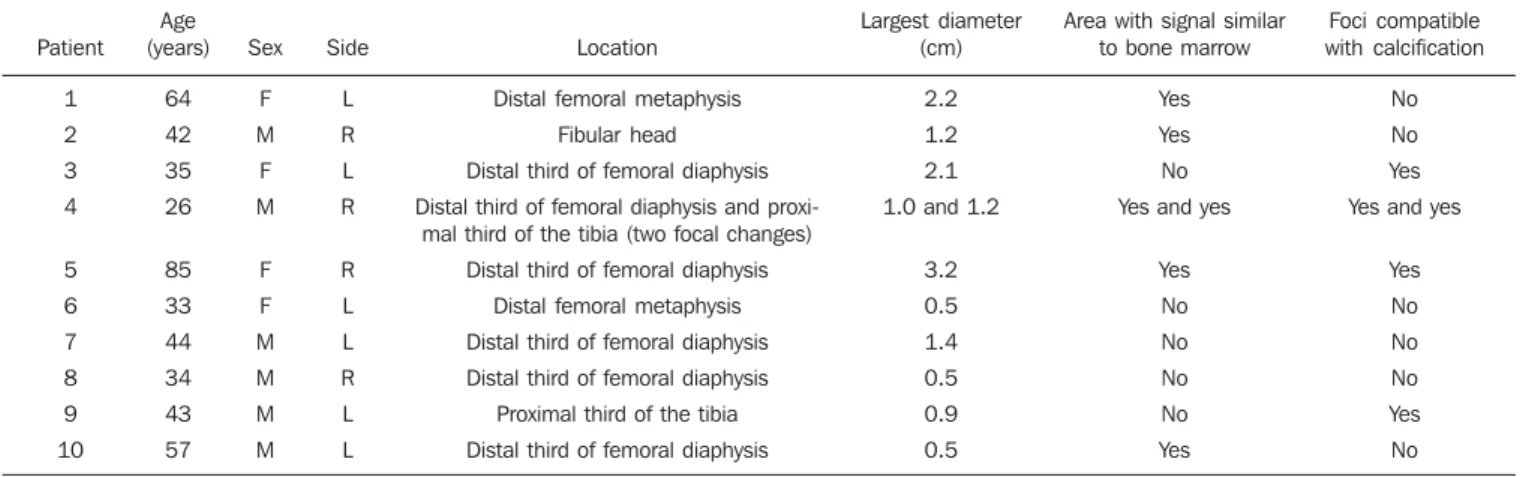
Table 1 describes the characteristics of the 11 enchondromas found in the ten pa-tients. Eight lesions were located in the Figure 1. Female, 64-year-old patient. Magnetic resonance imaging demonstrating typical incidentally found enchondroma. A: On the sagittal T1-weighted image, the lesion presents foci of intermediate signal intensity, some of them with a lobular aspect (arrow), intermingled with areas of bone marrow and fat-like signal (dashed arrow). B: On the intermediate-weighted image with fat suppression, the lesion presents foci of hypersignal with lobular aspect (arrows). Areas of hypersignal on T1-weighted images are confirmed as fat and present signal suppression (dashed arrow).
distal femur, two in the proximal tibia, and one in the fibular head. The lesions dimen-sions ranged from 0.5 to 3.2 cm (mean = 1.34 cm). Seven lesions were ≥ 1 cm.
As regards presence of calcification sig-nal within the enchondromas, intra and interobserver agreements were of 100% and, as regards fat signal within the lesion, indicating the presence of foci of bone marrow, the interobserver agreement was of 82%, and the intraobserver agreement, 100%.
In the whole sample of knee MRI stud-ies, 22 lesions of other nature were found, as follows: osteochondromas (n = 5; 1.53%); bone infarction (n = 3; 0.92%); os-teosarcomas (n = 3; 0.92%); fibrocystic changes (n =1; 0.30%), multiple subchon-dral cystic lesions in a patient with chronic hemarthropathy (n = 1; 0.30%), multiple lesions of paracoccidioidomycosis (n = 1; 0.30%), intramedullary fibrous lesion (n = 1; 0.30%), aneurysmal bone cyst (n = 1; 0.30%), chondrosarcoma (n = 1; 0.30%), fibrous cortical defect/cortical desmoid (n = 1; 0.30%), lesion suspicious for metasta-sis (n = 1; 0.30%), pigmented villonodular synovitis (n = 1; 0.30%) and synovial sar-coma (n = 1; 0.30%).
DISCUSSION
The histopathological identification of foci of bone tissue or bone marrow within enchondromas has previously been de-scribed in the literature(15–17). Cartilage lob-ules may be separated by normal bone marrow, or may be partially encased by
mature lamellar bone(16). This last charac-teristic is thought to reflect endochondral ossification in the periphery of the cartilage lobule(16).
The differentiation between low-grade chondrosarcomas and enchondromas rep-resents a challenge to radiologists, ortho-pedists and pathologists, since the treat-ment for each of these conditions is quite different, and their clinical, radiological and histopathological characteristics are very similar to each other(20–23). The pres-ence of bone tissue within the histopatho-logically identified lesion is potentially useful to differentiate enchondromas from low-grade intramedullary chondrosarco-mas(15,16,23), but the applicability of such a concept in cases of MRI is still to be deter-mined. In fact, the presence of foci of high signal intensity on MRI T1-weighted se-quences, suggesting the presence of bone marrow within cartilaginous lesions, has not shown to be useful to differentiate such histological lesion types in at least two pre-vious studies(17,24). The coexistence of both histological types in a single lesion could explain the identification of foci of bone marrow signal also in chondrosarcomas. Another hypothesis is that adjacent areas of healthy bone might have been invaded or involved by a low-grade chondrosarcoma. At MRI T1-weighted sequences, small foci of high signal intensity may be present in enchondromas as a result of the presence of engulfed normal medullary fat(25).
According to Walden et al., the preva-lence of incidentally found enchondromas at knee MRI was of 2.9%, most of times
lo-cated in the distal femur (2.0%)(13). In the study of Walden et al., the prevalence of lesions in the distal tibia corresponded to 0.7%, and 0.2% in the fibula. The preva-lence of incidentally found enchondromas in the present study (3.3%) was, therefore, compatible with data in the literature. In the present study, the prevalence of enchondro-mas in the distal femur (2.4%) was slightly higher than the prevalence observed by Walden et al(13). Enchondromas may also be incidentally found with a relative frequency at routine shoulder studies (2.1%), in most of times located in the contiguous region of the physis(26). In that study(26), the finding of areas of bone marrow within the lesion or intermingled with cartilage has not been investigated.
As compared with the prevalence of neoplasms such as chondrosarcomas and osteosarcomas in the general population, the high prevalence of such tumors in the present series is due to the fact that the authors’ institution is a reference center for bone tumors.
The interobserver agreement in the clas-sification of incidental enchondromas ac-cording to the criteria developed by Walden et al. was high, with a coefficient κ of 0.84 [confidence interval (CI) 95%: 0.65–1.00] in the first reading, and 1.00 (CI 95%: 1.00–1.00) in the second reading per-formed three months after the first one. No other study evaluating this reproducibility is found in the literature.
The prevalence of foci of fat or bone marrow within enchondromas was of 54.55% (6 of 11 lesions). The intraobserver Table 1 Cases of magnetic resonance imaging studies with focal alterations compatible with incidental enchondromas.
Patient 1 2 3 4 5 6 7 8 9 10 Age (years) 64 42 35 26 85 33 44 34 43 57 Sex F M F M F F M M M M Side L R L R R L L R L L Location
Distal femoral metaphysis
Fibular head
Distal third of femoral diaphysis
Distal third of femoral diaphysis and proxi-mal third of the tibia (two focal changes)
Distal third of femoral diaphysis
Distal femoral metaphysis
Distal third of femoral diaphysis
Distal third of femoral diaphysis
Proximal third of the tibia
Distal third of femoral diaphysis
Largest diameter (cm)
2.2
1.2
2.1
1.0 and 1.2
3.2 0.5 1.4 0.5 0.9 0.5
Area with signal similar to bone marrow
Yes
Yes
No
Yes and yes
Yes No No No No Yes Foci compatible with calcification No No Yes
Yes and yes
Yes No No No Yes No
agreement for this finding was perfect, with κ = 1.00 (CI 95%: 1.00–1.00), and the interobserver agreement was κ = 0.82 (CI 95%: 0.48–1.00).
The prevalence of calcifications within enchondromas was of 36.36% (4 of 11 le-sions). Both intra- and interobserver agree-ments in relation to the finding of foci of calcification within enchondromas accord-ing to the criterion of low signal intensity at MRI were perfect, with κ = 1.00 (CI 95%: 1.00–1.00).
No significant relationship was ob-served between lesion size, patient’s sex/ age and presence of foci of bone marrow within enchondromas, likewise for calcifi-cations represented by foci of signal ab-sence at MRI.
The present study has identified a high prevalence of foci of fat and calcification within enchondromas at MRI, and good reproducibility of such findings. The find-ing of areas with bone marrow signal inter-mingled with typically cartilaginous re-gions presented a higher prevalence than the finding suggestive of calcifications.
Some limitations of the present study must be mentioned. One of them – and im-portant – is its retrospective nature. Other significant limitation is related to the fact that there was no histopathological evi-dence of the enchondromas, but the authors believe that such a limitation does not com-promise the present study results, since the enchondromas were classified according criteria habitually accepted as reliable to characterize such lesions at MRI(13,17,26).
CONCLUSION
The prevalence of incidentally found enchondromas in the present study (3.3%) is compatible with data in the literature. In the present sample, the authors have
ob-served a high prevalence of foci of fat and calcification within the lesions at MRI, and excellent agreement in the evaluation of these findings reproducibility. The finding of areas with bone marrow signal inter-mingled with typically cartilaginous re-gions had a higher prevalence than the find-ing suggestive of calcifications.
REFERENCES
1. Nogueira-Barbosa MH, Sá JL, Trad CS, et al. Res-sonância magnética na avaliação das reações pe-riosteais. Radiol Bras. 2010;43:266–71. 2. Ribeiro DS, Araújo Neto C, D’Almeida F, et al.
Achados de imagem das alterações musculoes-queléticas associadas ao lúpus eritematoso sistê-mico. Radiol Bras. 2011;44:52–8.
3. Simão MN, Nogueira-Barbosa MH. Ressonância magnética na avaliação das variações anatômicas meniscais e da anatomia ligamentar paramenis-cal: potenciais causas de erro de interpretação. Radiol Bras. 2011;44:117–22.
4. Tavares Júnior WC, Faria FM, Figueiredo R, et al. Fadiga óssea: causa de dor em joelhos na os-teoartrite. Radiol Bras. 2012;45:273–8. 5. Nunes RB, Amaral DT, Oliveira VS. Propedêutica
radiológica do impacto femoroacetabular em tem-pos de tomografia computadorizada e ressonân-cia magnética: o que o radiologista precisa saber. Radiol Bras. 2011;44:249–55.
6. Grassi CG, Diniz FV, Garcia MRT, et al. Aspec-tos de imagem na tendinite calcária pré-vertebral. Radiol Bras. 2011;44:327–30.
7. Moura MVT. Interposição de fragmento perios-teal na fratura da placa epifisária femoral distal: estudo por ressonância magnética. Radiol Bras. 2012;45:184–6.
8. Nogueira-Barbosa MH, Savarese LG, Herrero CFPS, et al. Raízes nervosas redundantes da cauda equina: revisão da literatura. Radiol Bras. 2012;45:155–9.
9. Simão MN, Helms CA, Richardson WJ. Achados de ressonância magnética em cistos epidurais de origem discal em pacientes não operados e após microdiscectomia. Radiol Bras. 2012;45:205–9. 10. Jacob Jr C, Barbosa DM, Batista PR, et al. Fratura toracolombar do tipo explosão: o que o radiolo-gista deve conhecer. Radiol Bras. 2012;45:101–4.
11. Chojniak R, Grigio HR, Bitencourt AGV, et al. Biópsia percutânea por agulha grossa de tumores de partes moles guiada por tomografia computa-dorizada: resultados e correlação com análise da peça cirúrgica. Radiol Bras. 2012;45:259–62.
12. Cotta AC, Melo RT, Castro RCR, et al. Dificul-dades diagnósticas no osteoma osteoide do coto-velo: estudo clínico, radiológico e histopatológico. Radiol Bras. 2012;45:13–9.
13. Walden MJ, Murphey MD, Vidal JA. Incidental enchondromas of the knee. AJR Am J Roentgenol. 2008;190:1611–5.
14. Kaplan PA, Dussault R, Helms CA, et al. Muscu-loskeletal MRI. Philadelphia, PA: WB Saunders; 2001.
15. Mirra JM, Gold R, Downs J, et al. A new histo-logic approach to the differentiation of enchon-droma and chondrosarcoma of the bones. A clini-copathologic analysis of 51 cases. Clin Orthop Relat Res. 1985;(201):214–37.
16. McCarthy EF, Tyler WK. Distinguishing enchon-droma from low-grade central chondrosarcoma. Pathol Case Rev. 2001;6:8–13.
17. Murphey MD, Flemming DJ, Boyea SR, et al. Enchondroma versus chodrosarcoma in appen-dicular skeleton: differentiating features. Radio-graphics. 1998;18:1213–37.
18. Agresti A. Categorical data analysis. New York, NY: John Wiley; 1990.
19. Landis JR, Koch GG. The measurement of ob-server agreement for categorical data. Biometrics. 1977;33:159–74.
20. Ferrer-Santacreu EM, Ortiz-Cruz EJ, González-López JM, et al. Enchondroma versus low-grade chondrosarcoma in appendicular skeleton: clini-cal and radiologiclini-cal criteria. J Oncol. 2012;2012: 437958.
21. Shariat Torbaghan S, Ashouri M, Jalayer Naderi N, et al. Histopathologic differentiation between enchondroma and well-differentiated chondrosa-rcoma: evaluating the efficacy of diagnostic his-tologic structures. J Dent Res Dent Clin Dent Prospects. 2011;5:98–101.
22. Donthineni R, Ofluo—lu O. Solitary enchondro-mas of long bones: pattern of referral and out-come. Acta Orthop Traumatol Turc. 2010;44: 397–402.
23. Vanel D, Kreshak J, Larousserie F, et al. Enchon-droma vs. chondrosarcoma: a simple, easy-to-use, new magnetic resonance sign. Eur J Radiol. 2012 Jan 5. [Epub ahead of print].
24. Choi BB, Jee WH, Sunwoo HJ, et al. MR differ-entiation of low-grade chondrosarcoma from en-chondroma. Clin Imaging. 2013;37:542–7. 25. Douis H, Saifuddin A. The imaging of
cartilagi-nous bone tumours. I. Benign lesions. Skeletal Radiol. 2012;41:1195–212.