UTERINE EMBOLIZATION FOR MANAGEMENT OF SYMPTOMATIC
FIBROIDS: QUALITY-OF-LIFE IMPACT*
Nestor Kisilevzky1
OBJECTIVE: To determine the impact on the quality of life in patients with symptomatic uterine fibroids submitted to uterine artery embolization. MATERIALS AND METHODS: Forty patients with symptomatic fi-broids submitted to embolization have answered a quality-of-life questionnaire before and 12 weeks after the procedure. RESULTS: Mean score for symptoms severity before the procedure was 62.07 ± 6.34 and decreased with statistical significance to 20.42 ± 3.81 after the procedure. Similarly, scores for quality of life have improved from 40.26 ± 2.98 before the procedure to 85.06 ± 2.57 after the procedure, which again was statistically significant. CONCLUSION: Uterine embolization results in evident symptoms relief and significant improvement in the quality of life of patients with symptomatic fibroids.
Keywords: Therapeutic embolization; Uterus; Leiomyoma; Quality of life; Interventional radiology.
Embolização uterina para tratamento de miomas sintomáticos: impacto na qualidade de vida.
OBJETIVO: Verificar a mudança na qualidade de vida de pacientes portadoras de miomatose uterina sinto-mática submetidas a tratamento por embolização. MATERIAIS E MÉTODOS: Quarenta mulheres portadoras de miomatose uterina sintomática que foram tratadas com a técnica de embolização responderam a um questionário de qualidade de vida antes e 12 semanas após o procedimento. RESULTADOS: Verificou-se que o escore médio relacionado com a gravidade dos sintomas nas 40 pacientes antes da embolização foi de 62,07 ± 6,34 e se modificou, com significância estatística após o tratamento, quando se verificou escore médio de 20,42 ± 3,81. Da mesma forma, comprovou-se a melhora na qualidade de vida pela modificação dos escores antes e depois do tratamento, o que também apresentou significância estatística, passando de 40,26 ± 2,98 para 85,06 ± 2,57. CONCLUSÃO: A embolização uterina provoca alívio evidente dos sinto-mas relacionados com a miomatose e proporciona melhora substancial da qualidade de vida das pacientes. Unitermos: Embolização terapêutica; Útero; Leiomioma; Qualidade de vida; Radiologia intervencionista. Abstract
Resumo
* Study developed at Unidade de Radiologia Intervencionista do Hospital Santa Catarina, São Paulo, SP, Brazil.
1. Master in Surgery – Universidade Estadual de Campinas (Unicamp), Campinas, SP, Interventional Radiologist at Endovas-cular Clínica Médica Ltda., São Paulo, SP, Brazil.
Mailing address: Dr. Nestor Kisilevzky. Endovascular Clínica Médica Ltda. Praça Oswaldo Cruz, 124, conj. 31, Paraíso. São Paulo, SP, Brazil 04004-070. E-mail: [email protected]
Received November 13, 2006. Accepted after revision April 17, 2007.
INTRODUCTION
Uterine myomas, also called leiomy-omas or fibroids, are the most common benign gynecologic tumors, and may be present in up to 40% of women in child-bearing age(1).
Most frequently, myomatosis affects nulliparous, obese, black women, or those with family history of myomatosis or hyperestrogenic syndrome(2).
Despite the absolutely benign nature of myomas, complaints of uncomfortable symptoms such as menorrhagia (abnormal menstrual bleeding), dysmenorrhea (pain-ful menstruation), a felling of pressure in the pelvis, increase in the urinary
fre-quency, pain, infertility or increase in ab-dominal volume and palpable pelvic mass are very frequent(3).
Clinical presentation is variable and is particularly related to the size, location and number of myomas, but certainly myoma-tosis symptoms, when present, result in a remarkable impairment of the patients’ quality of life.
The most frequent symptom is an abnor-mal uterine bleeding (menorrhagia) that usually is characterized by increased men-strual duration and blood flow volume, sometimes leading to anemia(4). Generally,
women complain of a progression in the intensity, that is to say, a gradual increase in the menstrual blood flow obliging them to more frequent tampon or pad changes, or even to the use of diapers during the period. It is in these circumstances where women report that they avoid leaving home, scheduling professional or social activities because of the discomfort caused by the menstruation, or in order to avoid embarrassing or upsetting situations.
Until recently, only two therapeutic modalities were available for treating symptomatic myomatosis: the surgical ap-proach or hormone therapy.
Definitely, hysterectomy is the most commonly performed procedure for the management of myomatosis. It is estimated that myomatosis accounts for one third of almost 400,000 hysterectomies performed in the United States of America(5).Despite
the advantage of being definitely curative, hysterectomy is a formal surgical procedure that requires hospital stay for some days and a variable period of postoperative re-covery and convalescence. Also, hysterec-tomy may be associated to a considerable blood loss, ureter injury, prolapse and other complications(6). Besides, it eliminates any
possibility of fertility, which represents a deep loss for nulliparous women.
290
by the prestigious journal The Lancet, em 1995, suggesting that this was a highly ef-ficient method for the management of uter-ine myomatosis symptoms(7).
Since that time, numerous clinical ex-periments have been reported worldwide, demonstrating the validity of this promis-ing percutaneous procedure(8–15).
Uterine embolization is a minimally invasive modality of interventional radiol-ogy that consists in intentionally blocking the arteries that supply blood to the fi-broids, causing their ischemia and shrink-age, resulting in the resolution of their symptoms. For this purpose, a thin catheter is inserted under local anesthesia, by means of puncture of the femoral artery in the in-guinal region, and, under fluoroscopic vi-sion generated by a digital angiography apparatus, the catheter is inserted and ad-vanced to the uterine arteries. Gelatin
microspheres measuring about 500-µ is
then injected through the angiographic catheter until stasis of the branches that supply blood to the fibroids(16).
Since we started applying this technique in Brazil, in 1999, we have already treated 450 patients. Technical and clinical out-comes from this initial experiment have been previously published(17).
The present study reports outcomes from a group of patients submitted to uter-ine embolization, utilizing a system for evaluation and comparison of their quality of life before and after the treatment.
MATERIALS AND METHODS
The present study evaluated data ob-tained from images and questionnaires answered by 40 women with symptomatic uterine myomatosis who were submitted to uterine embolization in the period between July/2004 and June/2006, in the Division of Interventional Radiology at Hospital Santa Catarina in São Paulo, SP, Brazil.
The analysis included data from women with complaints resulting from the pres-ence of uterine fibroids who, during the preoperative clinical evaluation and the period of postoperative clinical follow-up had answered a questionnaire about qual-ity of life related to the disease treated. The mean age of patients was 38 years (age range = 22 – 46 years). As regards the
pa-tients’ race, 31 women were Caucasian, five were black, and four Asian. The pa-tients’ gestational history showed that 28 of them were multiparous, and 12 were nul-liparous. Amongst the patients included in the present study, 28 reported a job or pro-fessional activity, and 12 defined their ac-tivity as “housewives”. The main complaint leading to the treatment was increased menstrual bleeding, with or without anemia in 29 patients, and pain or compressive symptoms secondary to uterine increase in 11. The diagnosis of uterine myomatosis was based on data collected from the clini-cal history, cliniclini-cal examination and pelvic magnetic resonance imaging or ultrasound. All of the pre- and postoperative image studies were performed in an ambulatorial setting in different divisions of the hospi-tal where the patients were submitted to the treatment. All the patients presented with increased uterine volume (mean = 666 cm³, ranging between 245 cm³ and 1.930 cm³). The method included puncture of the right femoral artery, angiographic study of uter-ine arteries, and embolization with
cali-brated 500–700-µ spheres (Embosferas®).
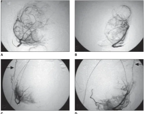
The procedures were documented by means of pre- and post-embolization angiographic studies (Figure 1).
All the patients remained hospitalized 24 hours for rest and observation.
At the time of the preprocedural clini-cal evaluation, all the patients answered a questionnaire for evaluation of their qual-ity of life related to the presence of fibroids (Chart 1). This questionnaire was specifi-cally developed for this purpose in the Georgetown University, Washington, USA(18), and translated into Portuguese by
a professional translator. It includes 37 questions, and is divided into two parts. The first one includes eight questions about the intensity or severity of symptoms re-ported by the patient. Each of these ques-tions offer five opques-tions corresponding to intensity: “no”, “little”, “reasonable”, “much”, “very much”, with a correspond-ing scorcorrespond-ing from 1 to 5. The final scorcorrespond-ing is converted into a corrected score by means of a mathematical formula. The sec-ond part of the questionnaire includes 29
Figure 1. Standard angiographic documentation of uterine embolization procedure: A: Selective cath-eterization of right uterine artery. Intrauterine branches can be observed delimitating nodular areas. B: Left-sided selective catheterization with angiographic aspect similar to the right side. C: After emboliza-tion, disappearance of the branches that supply blood to the fibroids, with main intrauterine branches preserved. Also, a preserved right ovarian artery is observed with ascending course (arrow). D: After embolization, main branches and left ovarian artery are preserved (arrow).
A
C D
(To be continued on page 292)
Chart 1 Quality-of-life questionnaire utilized in the present study.
QUALITY-OF-LIFE QUESTIONNAIRE RELATED TO UTERINE FIBROIDS.
The following items correspond to common symptoms experienced by women affected by uterine fibroids. Please, consider each of these symptoms as being related to the presence of fibroids or to your menstrual cycle. Each of the questions is related to the level and intensity of anguish or discomfort you have experienced as a result of these symptoms during the last three months.
There is no correct or wrong answer. Simply choose an option which best fits your symptoms that has led you to seek for the treatment (indicate by marking X on the appropriate site).
During the last three months, what has been the level of discomfort caused by...
1. Heavy bleeding during menstruation.
2. Clots in the menstrual flow.
3. Alteration in the period duration as compared to previous periods.
4. Alteration in the interval between periods as compared to the past.
5. Feeling of pressure or tension in the pelvis.
6. Frequent necessity to urinate in the daytime.
7. Frequent necessity to urinate at night.
8. Feeling of fatigue.
No 1 1 1 1 1 1 1 1 Little 2 2 2 2 2 2 2 2 Reasonable 3 3 3 3 3 3 3 3 Much 4 4 4 4 4 4 4 4 Very much 5 5 5 5 5 5 5 5
The questions below relate to the impact caused by the fibroids symptoms intensity on your life. Please, consider each of these questions as being exclu-sively related to the presence of fibroids during the last three months.
There is no correct or wrong answer. Simply choose an option which best fits your symptoms that has led you to seek for the treatment (indicate by marking X on the appropriate site). If no option fits your experience, select “at no time” (1).
Most of time 4 4 4 4 4 4 4 4 4 4 4 4 4 4 During the last three months, with which frequency symptoms caused by
fibroids... At no moment 1 1 1 1 1 1 1 1 1 1 1 1 1 1 Few times 2 2 2 2 2 2 2 2 2 2 2 2 2 2 Some times 3 3 3 3 3 3 3 3 3 3 3 3 3 3 All the time 5 5 5 5 5 5 5 5 5 5 5 5 5 5 9. Have caused anxiety because of the unpredictability of the amount and duration
of your menstrual period?
10. Have caused preoccupation with a travel?
11. Have affected your physical activities?
12. Have caused tiredness?
13. Have made you to reduce the amount of time spent on exercises or physical activities?
14. Have made you feel like you were not in control of your own life?
15. Have caused a fear that you might stain your underwear?
16. Have made you feel less productive?
17. Have caused somnolence in the daytime?
18. Have cause embarrassment because of overweight?
19. Have made you feel difficulty in your daily activities?
20. Have affected your social activities?
21. Have made you feel uncomfortable with the size of your abdomen?
292
23. Have made you feel sad, discouraged or desperate?
24. Have made you feel depressed?
25. Have made you “feel like a wet rag”?
26. Have caused preoccupation with your health?
27. Have made you to plan your activities more carefully?
28. Have made you feel bothered to carry pads and additional cloths to prevent accidents?
29. Have made you experience embarrassing situations?
30. Have caused uncertainty about the future?
31. Have caused irritation?
32. Have caused a fear that you might stain your external clothes?
33. Have affected the size of your clothes during your periods?
34. Have made you feel like you were not in the control of your health?
35. Have made you feel tiredness like the energy had been sucked out of you?
36. Have caused a decrease in your sexual desire?
37. Have made you to avoid having sexual relations?
Most of time 4 4 4 4 4 4 4 4 4 4 4 4 4 4 4 During the last three months, with which frequency symptoms caused by
fibroids... At no moment 1 1 1 1 1 1 1 1 1 1 1 1 1 1 1 Few times 2 2 2 2 2 2 2 2 2 2 2 2 2 2 2 Some times 3 3 3 3 3 3 3 3 3 3 3 3 3 3 3 All the time 5 5 5 5 5 5 5 5 5 5 5 5 5 5 5 Chart 1 Quality-of-life questionnaire utilized in the present study. (Continued).
Calculation of quality of life related to the presence of fibroids
1 – Calculation of the symptoms severity (higher value = higher severity — scale 1 to 100)
Scale
Symptom severity
Summation – items
Sum up the values obtained in 1 to 8
Possible values < e >
8; 40
Possible options
32
Formula:
Converted score = (current score – < possible score) × 100 Possible options
2 – Calculation by category and general quality of life
Scale Preoccupation Activity Mood/energy Self-control Embarrassment Sexuality
Total quality of life
Results summation items
9 + 15 + 22 + 28 + 32
10 + 11 + 13 + 19 + 20 + 27 + 29 12 + 17 + 23 + 24 + 25 + 31 + 35 14 + 16 + 26 + 30 + 34
18 + 21 + 33 36 + 37
Summation
Possible values < e >
5; 25 7; 35 7; 35 5; 25 3; 15 2; 10 29; 145 Possible options 20 28 28 20 12 8 116
Formula for calculation of quality of life — higher value = higher quality of life — scale 1 to 100
questions about the frequency in which the fibroids symptoms affect aspects of the patients’ daily lives. These questions are divided into groups corresponding to six aspects: preoccupation, activity, mood/en-ergy, self-control, embarrassment, and sexuality. Each of these questions offers five answering options to measure fre-quency: “at no time”, “few times”, “some-times”, “most of time” and “all the time”, with a corresponding scoring from 1 to 5. The final score is converted into a corrected score by means of a mathematical formula. In the first part, the questionnaire pre-sents the evaluation of symptoms intensity stratified from 0 to 100, meaning that 100 corresponds to the highest intensity or se-verity of the patients complaint. The sec-ond part evaluates the quality-of-life itself, i.e. the health condition expressed by a stratified scoring from 0 to 100, where 100 corresponds to the best level of quality-of-life in general, and for each of the specific aspects investigated by the questionnaire. During the 12-week follow-up period following the procedure, the patients were submitted to a pelvic magnetic resonance imaging for evaluating the uterine size as compared to the similar study previously performed. Also, they were asked to, again, answer the quality-of-life questionnaire.
Data obtained from questionnaires an-swers as well as those concerning the
uter-ine volumes evaluated by magnetic reso-nance imaging were transcribed into a Microsoft Excel worksheet for statistical analysis. Initially, all of the variables were descriptively analyzed. As for quantitative variables, this analysis was performed through observation of minimum and maximum values, as well as calculation of median and standard deviation. As for qualitative variables, absolute and relative frequencies were calculated. The paired Student’s t test was utilized for analysis of premoment x postmoment equality hypoth-esis; when the data normality assumption was rejected, the non-parametric Wilcoxon test was utilized. The significance level utilized for these tests was 5%.
The present study was submitted to the Committee for Ethics in Research of do Hospital Santa Catarina, whose approval (Process CEP019/06) has established that the study was conducted in compliance with the Resolution 196/96 of Conselho Nacional de Saúde (National Council of Health).
RESULTS
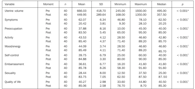
All of the statistical variables evaluated before and after the procedure are shown on Table 1.
Mean uterine volume measured by post-embolization magnetic resonance imaging
was 450 cm³, corresponding to a statisti-cally significant decrease in volume of 32.5% (Figures 2 and 3).
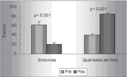
The mean score related to symptoms intensity reported in the preprocedural quality-of-life questionnaires was 62.07. The postprocedural quality-of-life ques-tionnaires demonstrated a statistically sig-nificant decrease in the mean score to 20.42, representing a 67.1% improvement in symptoms.
The analysis of total quality of life (health condition) related to the patients’ myomatosis showed postprocedural score of 40.26, that changed substantially after the treatment, achieving 85.06. This was statistically significant, representing an improvement of 52.6% in the patients’ quality of life (Figure 4).
All the items evaluated by the question-naire demonstrated changes after the treat-ment (Figure 5).
Mean score related to “preoccupation” in the preprocedural questionnaires was 37.87. In the post procedural question-naires this score changed to 83.5, showing a statistically significant difference, and corresponding to an improvement of 54.6% in this item.
Mean score related to “activity” in the preprocedural questionnaires was 43.53. In the postprocedural questionnaires, this score changed to 85.49, showing a
statis-Table 1 Mean, standard deviation, minimum, maximum values and median of variables in pre- and postprocedural evaluation of 40 patients.
Variable Uterine volume Symptoms Preoccupation Activity Mood/energy Self-control Embarrassment Sexuality
Quality of life
Moment Pre Post Pre Post Pre Post Pre Post Pre Post Pre Post Pre Post Pre Post Pre Post n 40 40 40 40 40 40 40 40 40 40 40 40 40 40 40 40 40 40 Mean 666.03 449.95 62.07 20.42 37.88 83.50 43.53 85.40 44.09 85.49 39.75 84.88 36.61 85.78 28.44 83.75 40.27 85.06 SD 418.70 289.64 6.34 3.81 8.16 5.45 4.12 4.37 3.74 4.11 6.79 3.30 6.77 8.26 8.00 7.05 2.98 2.58 Minimum 245.00 168.00 46.80 9.30 10.00 65.00 28.50 71.40 28.50 71.40 25.00 80.00 16.20 58.30 12.50 62.50 33.60 76.70 Maximum 1930.00 1350.00 78.10 28.10 45.00 95.00 46.60 89.20 46.60 89.20 60.00 90.00 41.60 91.60 37.50 87.50 44.80 8.70 Median 495.00 357.50 62.50 20.25 40.00 85.00 42.80 85.70 46.60 85.70 40.00 85.00 41.60 91.60 25.00 87.50 40.50 85.30 p < 0.001*
< 0.001†
< 0.001†
< 0.001†
< 0.001†
< 0.001†
< 0.001†
< 0.001†
< 0.001†
294
Figure 3. Pre-embolization (A) and postembolization magnetic resonance imaging (sagittal view) dem-onstrating complete ischemia of subserous and significant decrease in uterine volume.
A B
the postprocedural questionnaires, this score changed to 83.55, showing a statis-tically significant difference, and corre-sponding to an improvement of 65.97%.
DISCUSSION
Since the publication of the first scien-tific study on uterine embolization in 1995, much has been learned about this theme. The huge amount of papers published and studies presented in international con-gresses in the last ten years constitute un-equivocal scientific evidence that uterine embolization is an effective and safe method for treating symptomatic fibroids, repre-senting a dominant therapy for symptom-atic myomatosis. Up to the present mo-ment, it is estimated that more than 200,000 patients have already been treated world-wide by means of uterine embolization.
Besides being safe and effective in the management of myomatosis symptoms, the method has already proved to present some additional advantages.
Considering that this is a minimally in-vasive, percutaneous method performed under local anesthesia, it allows a rapid clinical recuperation and quick recovery of normal daily activities of the patients. A study developed in Canada and published in 2003, including more than 550 women, demonstrated that 82% of patients submit-ted to uterine embolization have a single-day hospital stay(19).
Another study developed in the United States of America and published in 2004 tically significant difference, and
corre-sponding to an improvement of 49% in this item.
Mean score related to “mood/energy” in the preprocedural questionnaires was 44.01. In the postprocedural question-naires, this score changed to 85.39, show-ing a statistically significant difference, and corresponding to an improvement of 48.52% in this aspect.
Mean score related to “self-control” in the preprocedural questionnaires was 39.75. In the postprocedural question-naires, this score changed to 84.87, show-ing a statistically significant difference, and corresponding to an improvement of 53.31% in this item.
Mean score related to “embarrassment” in the preprocedural questionnaires was 36.60. In the postprocedural question-naires, this score changed to 85.78, show-ing a statistically significant difference, and
corresponding to an improvement of 57.33% in this item.
Mean score related to “sexuality” in the preprocedural questionnaires was 28.43. In
Figure 2. Statistical representation, comparing preprocedural and postprocedural uterine volumes.
reported that 94% of patients submitted to uterine embolization lost less than ten working days, and that about 90% of women fully recovered their activities within two - three weeks following the pro-cedure(20).
Uterine embolization advantages come more evident in a comparison be-tween results from hysterectomy and uter-ine embolization. A randomized study de-veloped in Spain, comparing results from hysterectomy and uterine embolization, has evidenced that embolization results in shorter hospital stay, quicker clinical recov-ery, and lower incidence of complica-tions(21).
Besides the mentioned advantages, the embolization impact on the patients´ qual-ity of life must be taken into consideration. The questionnaire utilized in the present study was based on a survey involving both healthy women and other affected by symp-tomatic myomatosis. So, the idea was to create a simple tool for evaluating the im-pairment to the quality of life from the point-of-view of the patients. This study is the first to report the application of this type of questionnaire to Brazilian women. Be-sides being evident per se, the results are very similar to those presented by interna-tional studies.
A North-American study involving has shown a 35% improvement in symptoms and quality of life in 64 patients(22).
The largest multicentric study ever de-veloped in the world including more than 2 thousand patients submitted to uterine embolization has demonstrated that the symptoms intensity score evaluated by the quality-of-life changed from 59 before the procedure to 20 after the treatment. The same study has shown that the patients´ quality of life improved from 47 to 87 points(23).
Uterine embolization benefits and ad-vantages are translated into a very high rate of satisfaction reported by patients submit-ted to this treatment. A recently published Dutch study has shown that, 36% of 158 women submitted to embolization declared to be “satisfied”, and 57% “very satisfied” with this modality of treatment(24).
CONCLUSION
Uterine embolization is a minimally invasive and effective method for alleviat-ing the uncomfortable symptoms caused by fibroids. The utilization of a quality-of-life questionnaire has resulted in a simple and effective tool for demonstrating the im-provement in symptoms and in quality of life as a whole.
Uterine embolization becomes a highly significant method for treating women who wish to preserve their uteri, or those who need to quickly recover their normal activi-ties after the treatment.
REFERENCES
1. Cramer SF, Patel A. The frequency of uterine leiomyomas. Am J Clin Pathol 1990;94:435–438. 2. Brosens IA, Lunenfeld B, Donnez J. Pathogenesis and medical management of uterine fibroids. London, UK: Parthenon Publishing Group, 1999. 3. Buttram VC Jr, Reiter RC. Uterine leiomyomata: etiology, symptomatology, and management. Fertil Steril 1981;36:433–445.
4. American College of Obstetricians and Gynecolo-gists. An educational aid to obstetrician-gynecolo-gist: uterine leiomyomata. ACOG Technical Bul-letin 1994;192:863–870.
5. Lepine LA, Hillis SD, Marchbanks PA, et al. Hyste-rectomy surveillance – United States, 1980–1993. MMWR CDC Surveill Summ 1997;46:1–15. 6. Harris WJ. Complications of hysterectomy. Clin
Obstet Gynecol 1997;40:928–938.
7. Ravina JH, Herbreteau D, Ciraru-Vigneron N, et al. Arterial embolisation to treat uterine myomata. Lancet 1995;346:671–672.
8. Ravina JH, Merland JJ, Herbreteau D, Houdart E, Bouret JM, Madelenat P. Preoperative emboliza-tion of uterine fibroma. Preliminary results (10 cases). Presse Med 1994;23:1540.
9. Goodwin SC, McLucas B, Lee M, et al. Uterine artery embolization for the treatment of uterine leiomyomata: midterm results. J Vasc Interv Radiol 1999;10:1159–1165.
10. Spies JB, Scialli AR, Jha RC, et al. Initial results from uterine fibroid embolization for symptom-atic leiomyomata. J Vasc Interv Radiol 1999;10: 1149–1157.
11. Walker W, Green A, Sutton C. Bilateral uterine artery embolisation for myomata: results, compli-cations and failures. Min Invas Ther Allied Technol 1999;8:449–454.
12. Worthington-Kirsch RL, Popky GL, Hutchins FL Jr. Uterine arterial embolization for the manage-ment of leiomyomas: quality-of-life assessmanage-ment and clinical response. Radiology 1998;208:625– 629.
13. Hutchins FL, Worthington-Kirsch RL, Berkowitz
296
RP. Selective uterine artery embolization as pri-mary treatment for symptomatic leiomyomata uteri. J Am Assoc Gynecol Laparosc 1999;6:279– 284.
14. Spies JB, Ascher SA, Roth AR, Kim J, Levy EB, Gomez-Jorge J. Uterine artery embolization for leiomyomata. Obstet Gynecol 2001;98:29–34. 15. Pelage JP, LeDref O, Soyer P, et al. Fibroid-related
menorrhagia: treatment with superselective em-bolization of the uterine arteries and midterm follow-up. Radiology 2000;215:428–431. 16. Spies JB, Benenati JF, Worthington-Kirsch RL.
Initial experience with use of tris-acryl gelatin microspheres for uterine artery embolization for leiomyomata. J Vasc Interv Radiol 2001;12:1059– 1063.
17. Kisilevzky N. Embolização uterina para tratamento de mioma sintomático: experiência inicial e
re-visão da literatura. Radiol Bras 2003;36:129–140. 18. Spies JB, Coyne K, Guaou-Guaou N, Boyle D, Skyrnaz-Murphy K, Gonzalves SM. The UFS-QOL, a new disease-specific symptom and health-related quality of life questionnaire for leiomyo-mata. Obstet Gynecol 2002;99:290–300. 19. Pron G, Mocarski E, Bennett J, et al. Tolerance,
hospital stay, and recovery after uterine artery embolization for fibroids: the Ontario Uterine Fibroid Embolization Trial. J Vasc Interv Radiol 2003;14:1219–1222.
20. Bruno J, Sterbis K, Flick P, et al. Recovery after uterine artery embolization for leiomyomas: a detailed analysis of its duration and severity. J Vasc Interv Radiol 2004;15:801–807. 21. Pinto I, Chimeno P, Romo A, et al. Uterine
fi-broids: uterine artery embolization versus ab-dominal hysterectomy for treatment – a
prospec-tive, randomized, and controlled clinical trial. Radiology 2003;226:425–431.
22. Smith WJ, Upton E, Shuster EJ, Klein AJ, Schwartz ML. Patient satisfaction and disease specific quality of life after uterine artery embo-lization. Am J Obstet Gynecol 2004;190:1697– 1703; discussion 1703–1706.
23. Spies JB, Myers ER, Worthington-Kirsch R, Mulgund J, Goodwin S, Mauro M; FIBROID Registry Investigators. The FIBROID Registry: symptom and quality-of-life status 1 year after therapy. Obstet Gynecol 2005;106:1309–1318. 24. Lohle PNM, Boekkooi FP, Smeets AJ, et al.