Predictive factors for pelvic magnetic resonance in
response to arterial embolization of a uterine
leiomyoma
Eduardo Zlotnik,I,IIMarcos de Lorenzo Messina,II Felipe Nasser,IBreno Boueri Affonso,II Ronaldo Hueb Baroni,INelson Wolosker,I,IIEdmund Chada BaracatII
IHospital Israelita Albert Einstein, Interventional Radiology Department, Sa˜o Paulo/SP, Brazil.IIHospital das Clı´nicas da Faculdade de Medicina da Universidade de Universidade de Sa˜o Paulo, Gynecology Clinic, Sa˜o Paulo/SP, Brazil.
OBJECTIVE:Minimally invasive methods are used as alternatives to treat leiomyomas and include uterine artery embolization, which has emerged as a safe, effective method. This study aims to evaluate the magnetic resonance imaging predictors for a reduction in leiomyoma volume in patients undergoing uterine artery embolization.
METHODS: This prospective longitudinal study was performed at a university hospital. We followed 50 symptomatic premenopausal women with uterine leiomyomas who underwent uterine artery embolization. We examined 179 leiomyomas among these patients. Magnetic resonance imaging was performed one month before and six months after uterine artery embolization. Two radiologists who specialized in abdominal imaging independently interpreted the images. Main Outcome Measures: The magnetic resonance imaging parameters were the uterus and leiomyomas volumes, their localizations, contrast perfusion pattern and node-to-muscle ratio.
RESULTS:Six months after treatment, the average uterine volume reduction was 38.91%, and the leiomyomas were reduced by 55.23%. When the leiomyomas were submucosal and/or had a higher node-to-muscle ratio in the T2 images, the volume reduction was even greater (greater than 50%). Other parameters showed no association.
CONCLUSIONS: We conclude that symptomatic uterine leiomyomas in patients undergoing uterine artery embolization exhibit volume reductions greater than 50% by magnetic resonance imaging when the leiomyomas are submucosal and/or had a high node-to-muscle ratio in the T2 images.
KEYWORDS: Uterine Leiomyoma; Uterine Artery Embolization; Magnetic Resonance Imaging.
Zlotnik E, Messina ML, Nasser F, Affonso BB, Baroni RH, Wolosker N, et al. Predictive factors for pelvic magnetic resonance in response to arterial embolization of a uterine leiomyoma. Clinics. 2014;69(3):185-189.
Received for publication onApril 14, 2013;First review completed onMay 9, 2013;Accepted for publication onAugust 16, 2013 E-mail: [email protected]
Tel.: 55 11 2151-5202
& INTRODUCTION
Uterine leiomyomas have become an important public health problem. Uterine leimyomas adversely affect a woman’s quality of life due to their high prevalence, symptomatology and adverse effects on fertility (1).
The objective of leiomyoma treatment is symptom control and/or the re-establishment of reproductive capacity, if the patient desires. Uterine artery embolization (UAE) has emerged as an alternative therapy. UAE was initially used
to prepare for surgery, and its safety and effectiveness (2-4) have led to its use as a first-choice treatment in many cases. To treat leiomyoma patients with UAE, an accurate diagnosis is essential. In particular, the care provider must know the number and size of leiomyomas and which ones will have the best response.
Ultrasound is the most commonly used test for leio-myoma diagnosis due to its precision in identifying tumors, ease of use and low operating cost. However, other imaging methods are necessary when either the number of leiomyo-mas or their volume is very large or very small because measuring each leiomyoma under these circumstances is difficult (5-6). In these situations, magnetic resonance imaging (MRI) offers greater diagnostic precision, greater accuracy and interobserver variation independence (7).
Most studies that have evaluated leiomyoma treatment with UAE assessed the development of leiomyomas based on the largest tumor, which may introduce bias into the Copyrightß2014CLINICS– This is an Open Access article distributed under
the terms of the Creative Commons Attribution Non-Commercial License (http:// creativecommons.org/licenses/by-nc/3.0/) which permits unrestricted non-commercial use, distribution, and reproduction in any medium, provided the original work is properly cited.
No potential conflict of interest was reported.
treatment response analysis (8-12). Few authors have evaluated all of a patient’s tumors (13-14), and those studies that did had fewer than 31 patients in the study. Several studies have used MRI to evaluate leiomyoma progression, but few (13-15) have analyzed the tumor’s pretreatment characteristics as predictive factors for treatment outcome. These studies used the T2 signal and the node location. The objective of this study was to use MRI to evaluate the factors that predict leiomyoma reduction in patients undergoing UAE.
& MATERIALS AND METHODS
This prospective longitudinal study included 50 women with leiomyomas who underwent UAE between September 2008 and August 2009. All patients underwent similar treatments following the same protocol. This study was approved and performed according to the hospital’s Research Ethics Committee.
The inclusion criteria were an ultrasound diagnosis of uterine leiomyoma, the presence of symptoms (menorrhagia and/or pelvic pain) as the primary complaint and an indication for UAE (i.e., desire to keep the uterus and/or contraindications for surgery). The exclusion criteria were malignant genital neoplasms, pelvic inflammatory disease, allergy to iodinated contrast, coagulopathy, renal failure, vasculitis, pelvic irradiation, pregnancy and a subserosal leiomyoma with a pedicle smaller than 50% of the diameter of the fibroid.
The patients were 27 to 44 years old, with an average age of 36.3 years. Of the 50 patients, 18 (36%) had been pregnant; 10 patients (20%) had had one pregnancy; and eight (16%) had two or more pregnancies. Twenty-one patients were white (42%), 13 (26%) were multiethnic and 16 (32%) were black.
The patients underwent UAE with spinal anesthesia and were kept under continuous analgesia for 24 hours. They underwent an MR exam using a high-field scanner (Siemens Magnetom 3-Tesla, Siemens, Germany). The exam was divided into two stages. The first stage occurred one month before UAE, and the second occurred six months after the procedure. The exams were performed within 10 days after menstruation.
For these two stages, a standard imaging protocol for evaluating the female pelvis was used, along with a diffusion-weighted sequence and dynamic sequences after paramagnetic contrast to evaluate the perfusion pattern of myomatous lesions. The images were interpreted indepen-dently by two radiologists who specialize in abdominal imaging. The pre- and postprocedure exams were analyzed randomly, nonsequentially and anonymously to minimize bias.
Parameters that may have been related to a greater than 50% reduction in the initial volume were evaluated. The adopted models considered a binary response, specifically whether there was a reduction of greater than 50% in the volume.
The leiomyomas were evaluated individually. MR was used to evaluate the morphology, including the radiological dimension(s) of the leiomyoma and uterus, and the volume, which was measured using the formula for an ellipse. The leiomyoma fibroids were counted, and their locations in the myometrium were classified as submucosal, intramural or
subserosal. Perfusion and the characterization of the T2 signal were also evaluated.
The contrast perfusion pattern was evaluated in the T1 images using the PP ratio. The peak high-contrast phase (HCP) and precontrast phase (PCP) are parameters for this relationship, and the PP ratio = [(HCP fibroid-PCP fibroid)/ PCP fibroid]X100 (16).
Other morphological changes were observed in T2-weighted sequences, and the images were evaluated when tissue hydration was sufficient. The T2 signal intensity for the leiomyoma fibroids and striated muscle, which does not change with the procedure, and the T2 ratio (i.e., signal intensity of the fibroid divided by the signal intensity for the muscle) were evaluated. The T2 signal intensities for leiomyomas may be different due to the patient’s biotype. Comparing fibroids from the same patient, which are interdependent with other fibroids in the uterus, using a common denominator for each patient’s leiomyomas becomes necessary. Thus, the T2 fibroid-to-muscle ratio allowed for the adjustment of these differences and was significant in a multivariate analysis. The same ratio was used by Sipola et al. (16), who demonstrated its usefulness in predicting greater leiomyoma reduction.
The numeric variables are described as means, standard deviations (SD) and confidence intervals or as medians and interquartile ranges when the values were not normally distributed. The qualitative variables are described as absolute frequencies and percentages. The comparisons between pre- and postembolization in relation to the uterine volume and T2-weighted muscle values were analyzed using the Wilcoxon test.
Univariate and multivariate analyses were conducted to identify the effects of several factors on the percent reduction in the fibroid volume. These models were used to assess the reliability of the measurements taken from different fibroids from the same patient. For both methods, variables that showed a p,0.20 in the univariate analysis were included in the multivariate models. The level of significance adopted was 0.05. All analyses were performed using SPSS (version 17.0, SPSS, Inc., Chicago, Ill).
& RESULTS
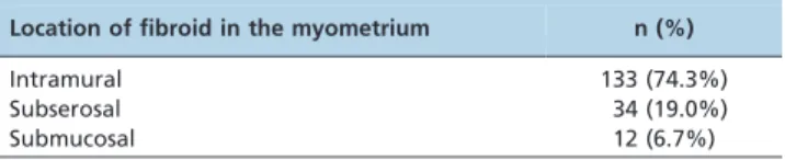
There were 179 fibroids among the 50 patients. The patients had a median of three fibroids, with a range of one to eight, and most fibroids were intramural. The locations of the fibroids are presented in Table 1.
The median uterine volume was 517 cm3(312.2 to 648.8
cm3) prior to the procedure and was reduced to 256.5 cm3
(184.0 to 464.0 cm3) after embolization (p,0.001). The uterine volume reduction in the 50 patients ranged from 0.67 to 93.53%, with an average of 38.91% (95% CI = 33.41-44.41).
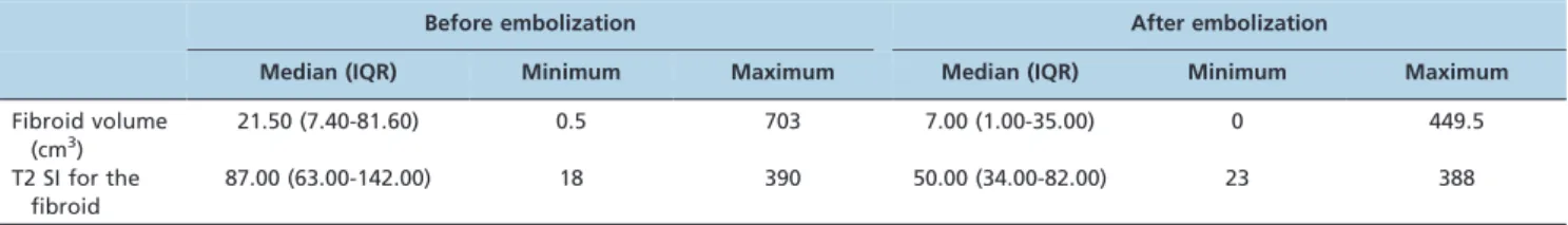
The majority of the leiomyoma fibroids (97.7%) showed a volume reduction after embolization and a reduction in the T2 signal intensity (Table 2). The percent reduction in
Table 1 -Fibroid classification according to the location in the myometrium.
Location of fibroid in the myometrium n (%)
Intramural 133 (74.3%)
Subserosal 34 (19.0%)
fibroid volume ranged from 40.63 to 100%, with a mean of 55.23% and a SD of 31.80%.
The number of fibroids and the uterine and fibroid volumes before and after embolization were not associated with a reduction in the fibroid volume (Table 3). The location of the fibroid in the myometrium, T2 fibroid signal and PP and T2 node-to-muscle ratios were the variables selected for the multivariate model. In this mode, the PP ratio and the T2 fibroid signal were no longer significant. The multivariate model showed that there was a greater chance that the fibroid volume decreased by more than 50% as the T2 fibroid-to-muscle ratio increased (OR: 1.528; 95% CI: 1.186-2.149;p= 0.015).
Regarding fibroids located in the myometrium, when the fibroid location was submucosal, a greater than 50% reduction in its volume was observed.
& DISCUSSION
The patients were monitored for six months based on studies that showed that the leiomyomas were significantly reduced during this period (8,13,15).
In the present study and studies by Gabriel-Cox et al. (12) and Sipola et al. (16), the patient’s age did not correlate with a reduction in size. In a study by Jha et al. (13), increased age was a predictor of failure. Emphasizing the difficulty in comparing the mean ages of the patients across studies is important; in this study, the mean age was 36.3 years, whereas in the previously mentioned studies, the mean ages were 45, 48 and 45 years, respectively. These data become more important when comparing the long-term results from studies involving patients who are close to menopause, when there is a greater likelihood that the symptoms will resolve (17).
The mean uterine volume (517.7 cm3) in this study was similar to that described by Gabriel-Cox et al. (12) in a multicenter study. The present study did not find differ-ences in volume reduction for different uterine volumes, which is comparable to the results reported by Gabriel-Cox et al. (12) and Prollius et al. (18). The leiomyomas were reduced by 55.23%, and the uterus was reduced by 38.91%, compared with the 60% and 40% reductions, respectively, reported in the literature (14,19).
There was no association between leiomyoma size and volume reduction. Thus, in terms of size reduction, patients with large leiomyomas and patients with smaller leiomyo-mas are both good candidates for UAE. Other authors found similar results based on leiomyoma size (11,14,16,20). When the leiomyomas were submucosal, there was an increased reduction in leiomyoma volume in this study. Jha et al. (13) and de Souza and Williams (21) showed that a greater volume reduction occured when the leiomyoma was sub-mucosally located within the uterus compared with other locations. Spies et al. found that leiomyomas located in the submucosa were associated with a greater volume reduction after UAE compared with subserosal leiomyomas (21). However, this difference did not persist over 12 months. In another study (18), the location of the leiomyoma did not have a significant effect on its size reduction; however, in this sample population, only two of 47 fibroids were submucosal. In the present study, there was a trend toward a greater leiomyoma size reduction in the fibroids with a high T2 signal, which is consistent with the findings of de Souza and Williams (21), Oguchi et al. (22), Burn et al. (14) and Harman et al. (15); however, Sipola et al. (16) did not find a significant association (p= 0.08) when analyzing this
vari-able. The greatest T2 signal and greatest response could be related to the leiomyoma’s cellularity and vascularization Table 2 -Measurement of the fibroid volume and T2 signal intensity before and after embolization.
Before embolization After embolization
Median (IQR) Minimum Maximum Median (IQR) Minimum Maximum
Fibroid volume (cm3)
21.50 (7.40-81.60) 0.5 703 7.00 (1.00-35.00) 0 449.5
T2 SI for the fibroid
87.00 (63.00-142.00) 18 390 50.00 (34.00-82.00) 23 388
IQR = interquartile range. SI = signal intensity.
Table 3 -Univariate and multivariate analyses for factors associated with a volume reduction greater than 50%.
Univariate Multivariate
OR CI (95%) p-value OR CI (95%) p-value
Uterine volume pre-embolization 1.000 (0.999; 1.001) 0.725
Fibroid location in the myometrium
Intramural/submucosal 0.099 (0.010; 0.952) 0.045 0.132 (0.016; 1.095) 0.061
Subserosal/submucosal 0.051 (0.005; 0.526) 0.012 0.056 (0.006; 0.504) 0.010*
Fibroid volume (tertile)
Up to 10 cm3/greater than 56 cm3 1.206 (0.561; 2.594) 0.631
Between 10 and 56 cm3/greater than 56 cm3 1.386 (0.654; 2.937) 0.394
T2 fibroid/muscle ratio 1.262 (1.063; 1.500) 0.008 1.528 (1.086; 2.149) 0.015*
PP ratio 1.005 (0.998; 1.012) 0.167 1.005 (0.998; 1.013) 0.174
T2 fibroid signal intensity 1.004 (0.999; 1.009) 0.080 0.997 (0.988; 1.006) 0.463
Four or fewer fibroids/more than four fibroids 0.637 (0.292; 1.391) 0.258
*
(22), but Yamashita et al. (23) stated that some degenerated leiomyomas had a higher T2 signal intensity and then greater volume reduction. Therefore, factors other than degeneration and cellularity, or perhaps the overlap of these factors, may explain the variability of these results.
The value of the PP ratio, which measures contrast perfusion, was controversial in our study. This ratio was not associated with a reduced leiomyoma volume in the multivariate analysis. This outcome is supported by results from Burn et al. (14) and de Souza and Williams (21). Fibroid behavior may follow a different pattern depending on the vascularization of these fibroids and the adjacent myometrium; however, this theory is still under study (21). In the analysis of variables that may be related to leiomyoma volume reduction, there was no association with uterine volume, leiomyoma volume, number of fibroids or T2 signal intensity. In a study by Sipola et al. (16), there was also no relationship between volume reduction and leiomyoma volume before embolization. Therefore, the present study showed a greater reduction for higher T2 ratios. Unlike the present study, the PP ratio described and evaluated here did not affect treatment outcomes (16).
In this study, the main outcome variable was leiomyoma volume reduction. The clinical improvement related to greater volume reduction is still controversial and does not necessarily correlate with an improvement in clinical symptoms (21). Additionally, reducing the size of the submucosal area may be more important for symptomatol-ogy than the size of the leiomyoma (9). Other authors showed that size reduction was associated with clinical outcomes, such as clinical failure, hysterectomy (24) and symptom recurrence (25). Leiomyoma volume reduction is an important treatment goal and affects a patient’s progress. Despite the known interdependence of multiple leiom-yomas within a patient, most authors have evaluated leiomyoma progress on the basis of the largest tumor, whether by analyzing vascular factors (25-26), genetic factors (27) or growth factors (28). This method may constitute a bias in the analysis of treatment response (8-12). Thus, the aforementioned variables (i.e., age and leiomyoma location) in addition to the use of the dominant fibroid may complicate comparisons among studies.
Studying the therapeutic alternatives to the conservative treatment of uterine leiomyoma and the diagnosis of and indications for these alternatives will lead to a better understanding of these parameters. Therefore, when coun-seling and informing patients about conservative and alternative methods for uterine leiomyoma treatment, emphasizing that some patients will require new interven-tions is important. When assessing these interveninterven-tions, care providers should try to understand which leiomyomas will respond the best. Our study reinforces this tendency and provides new challenges for medical science because the specialist’s and patient’s understanding of the method are not widespread. New studies could be performed to confirm these findings and suggest which parameters would provide gynecologists and patients with the best knowledge.
In conclusion, uterine leiomyomas from symptomatic patients who undergo UAE had a volume reduction greater than 50% when MRI determined that the fibroids were submucosal and/or there was a high T2 fibroid/muscle ratio.
& AUTHOR CONTRIBUTIONS
This study was a multi-specialty collaboration. Zlotnik E, Messina ML and Baracat EC are gynecologists; they evaluated the patients and conducted this study with help from Wolosker N, a vascular surgeon. Affonso BB and Nasser F are interventional radiologists, and Baroni RH is from the imaging department. All authors participated in the study and reviewed this paper.
& REFERENCES
1. Wu J, Wechter M, Geller E, Nguyen T, Visco A. Hysterectomy rates in the United States, 2003. Obstet Gynecol. 2007;110(5):1091-5, http://dx. doi.org/10.1097/01.AOG.0000285997.38553.4b.
2. Walker W, Bratby M. Magnetic resonance imaging analysis of fibroid location in women achieving pregnancy after uterine artery emboliza-tion. Cardiovasc Intervent Radiol. 2007;30(5):876-81, http://dx.doi.org/ 10.1007/s00270-007-9118-2.
3. Volkers N, Hehenkamp W, Birnie E, Ankum W, Reekers J. Uterine artery embolization versus hysterectomy in the treatment of symptomatic uterine fibroids: 2 years outcome from randomized EMMY trial. Am J Obstet Gynecol. 2007;196(6):519.e1-11.
4. Kim D, Lee H, Lee M, Kim H, Cho J, Cha S. Long-term results of symptomatic fibroids treated with uterine artery embolization: In conjuction with MR evaluation. Eur J Radiol. 2010;73(2):339-44. 5. Omary R, Vasireddy S, Chrisman H, Ryu R, Pereles F, Carr J, et al. The
effect of pelvic MR imaging on the diagnosis and treatment of women with presumed symptomatic uterine fibroids. J Vasc Radiol. 2002;13(11): 1149-53, http://dx.doi.org/10.1016/S1051-0443(07)61957-5.
6. Dueholm M, Lundorf E, Sorensen J, Ledetoug S, Olesen F, Lauresen H. Reproducibility of evaluation of the uterus by transvaginal sonography, hysterographic examination, hysteroscopy and magnetic resonance imaging. Hum Reprod. 2002;17(1):195-200, http://dx.doi.org/10.1093/ humrep/17.1.195.
7. Cura M, Cura A, Bugnone A. Role of magnetic resonanse imaging in patient selection for uterine artery embolization. Acta Radiol. 2006;47(10):1105-14, http://dx.doi.org/10.1080/02841850600965047. 8. Van dKS, Hehenkamp W, Volkers N, Birnie E, Ankum W, Reekers J.
Uterine artery embolization vs hysterectomy in the treatment of symptomatic uterine fibroids: 5-year outcome from the randomized EMMY trial. Am J Obstet Gynecol. 2010;203(2):105e.1-13.
9. Spies J, Myers E, Worthington-Kirsh R, Malgund J, Goodwin S, Mauro M. The fibroid registry: symptom and quality-of-life status 1 year after therapy. Obstet gynecol 2005;106(6):1309-18, http://dx.doi.org/10.1097/ 01.AOG.0000188386.53878.49.
10. Spies J, Cooper J, Worthington-Kirsch R, Lipman J, Mills B, Benenati J. Outcome of uterine embolization and hysterectomy for leiomyomas: results of a multicenter study. Am J Obstet Gynecol 2004;191(1):22-31, http://dx.doi.org/10.1016/j.ajog.2004.03.037.
11. Pron G, Cohen M, Soucie J, Garvin G, Vanderburgh L, Bell S. The Ontario Uterine Fibroid Embolization Trial. Parte 1. Baseline patient character-istics, fibroid burden, and impact on life. Fertil Steril. 2003;79(1):112-9, http://dx.doi.org/10.1016/S0015-0282(02)04539-9.
12. Gabriel-Cox K, Jacobson G, Armstrong M, Hung Y, Learman L. Predictors of hysterectomy after artery uterine embolization for leiomyoma. Am J Obstet Gynecol. 2007;196(6):588e1-e6.
13. Jha R, Ascher S, Imaoka I, Spies J. Symptomatic fibroleiomyomata: MR imaging of the uterus before and after uterine arterial embolization. Radiology. 2000;217(1):228-35, http://dx.doi.org/10.1148/radiology.217. 1.r00se49228.
14. Burn P, McCall J, Chinn R, Vashisht A, Smith J, Healy J. Uterine fibroleiomyoma: MR imaging appearances before and after embolization of uterine arteries. Radiology. 2000;21(3):720-34.
15. Harman M, Zeteroglu S, Arslan H, Sengul M, Etlik O. Predictive value of magnetic resonanse imaging signal and contrast-enhancement characteis-tics on post-embolization volume reduction of uterine fibroids. Acta Radiol. 2006;47(4):427-35, http://dx.doi.org/10.1080/02841850600557117. 16. Sipola P, Ruuskanen A, Yawu L, Husso M, Vanninen R, Hippela¨inen M, et al. Preinterventional quantitative magnetic resonance imaging predicts uterus and leiomyoma size reduction after uterine artery embolization. J Magn Reson Imaging. 2010;31(3):617-24, http://dx.doi.org/10.1002/ jmri.22063.
17. Tropeano G, Di SC, Amoroso S, Gualano M, Bonomo L, Scambia G. Long-term effects of uterine fibroid embolization on ovarian reserve; a prospective cohort study. Fertil Steril. 2010;94(6):2296-300, http://dx.doi. org/10.1016/j.fertnstert.2009.12.007.
18. Prollius A, de Vries C, Loggenberg E, Du Plessis A, Nel M, Wessels PH. Uterine artery embolisation for symptomatic fibroids: the efect of the large uterus in outcome. BJOG 2004;111(3):239-42, http://dx.doi.org/10. 1046/j.1471-0528.2003.00019.x.
19. Bradley L. Uterine fibroid embolization: a viable alternative to hyster-ectomy. Am J Obstet Gynecol. 2009;201(2):127-3.
with successful symptom and imaging outcome. Radiology. 2002; 222(1):45-52, http://dx.doi.org/10.1148/radiol.2221010661.
21. De Souza N, Williams A. Uterine arterial embolization for leiomyomas: perfusion and volume changes at MR imaging and relation to clinical outcome. Radiology. 2002;222(2):367-74, http://dx.doi.org/10.1148/radiol. 2222010584.
22. Oguchi O, Mori A, Kobayashi Y, Horiuchi A, Nikaido T, Fujii S. Prediction of histopathologic features and proliferative activity of uterine leiomyoma by magnetic resonance imaging prior to GnRH analogue therapy: correlation between T2-weighted images and effect of GnRH analogue. J Obstet Gynaecol. 1995;21(2):107-17.
23. Yamashita Y, Torashima M, Takahashi M, Tanaka N, Katabuchi H, Miyazaki K, et al. Hyperintense uterine leiomyoma at T2-weighted MR imaging: differentiation with dynamic enhanced MR imaging and clinical implications. Radiology. 1993;189(3):721-5.
24. Goodwin S, McLucas B, Lee M, Chen G, Perrella R, Vedantham S, et al. Uterine artery embolization for the treatment of uterine leiomyomata
midterm results. J Vasc Interv Radiol. 1999;10(9):1159-65, http://dx.doi. org/10.1016/S1051-0443(99)70213-7.
25. Pelage J, Guaou N, Jha R, Ascher S, Spies J. Uterine fibroid tumors: long-term MR imaging outcome after embolization. Radiology. 2004;230(3): 803-9, http://dx.doi.org/10.1148/radiol.2303030111.
26. Walocha J, Litwin J, Miodonski A. Vascular system of intramural leiomyomata revealed by corrosion casting and scanning electron microscopy. Hum Reprod. 2003;18(5):1088-93, http://dx.doi.org/10. 1093/humrep/deg213.
27. Borsari R, Bozzini N, Junqueira C, Soares JJ, Hila´rio S, Baracat E. Genic expression of the uterine leiomyoma in reproductive-aged women after treatment with goserelin. Fertil Steril. 2010;94(3):1072-7, http://dx.doi. org/10.1016/j.fertnstert.2009.03.112.