Sao Paulo Med J. 2016; 134(4):355-8 355
CASE REPORT
DOI: 10.1590/1516-3180.2016.00081203Intrauterine thrombosis of umbilical artery – case report
Trombose intrauterina de artéria umbilical – relato de caso
Gustavo Henrique de Oliveira
I, Cristiane de Moraes Dias
II, Denise Cristina Mós Vaz-Oliani
III, Antonio Hélio Oliani
IVHospital da Criança e Maternidade (HCM), Faculdade de Medicina de São José do Rio Preto (FAMERP) and Instituto de Medicina
Reprodutiva e Fetal SS (IMR), São José do Rio Preto, SP, Brazil
ABSTRACT
CONTEXT: Umbilical cord thrombosis is related to greater fetal and perinatal morbidity and mortality. It is usually associated with umbilical cord abnormalities that lead to mechanical compression with conse-quent vascular ectasia. Its correct diagnosis and clinical management remains a challenge that has not yet been resolved.
CASE REPORT: This study reports a case of umbilical artery thrombosis that occurred in the second half of a pregnancy. The umbilical cord was long, thin and overly twisted and the fetus presented severe intrauter-ine growth restriction. The clinical and histopathological indings from this case are described.
CONCLUSIONS: This case report emphasizes the diiculty in diagnosing and clinically managing abnor-malities of intrauterine life with a high chance of perinatal complications.
RESUMO
CONTEXTO: A trombose do cordão umbilical está relacionada com o aumento da morbimortalidade fetal e perinatal. É geralmente associada a alterações do cordão umbilical que levam à compressão mecânica com consequente ectasia vascular. Seu correto diagnóstico e manejo clínico é um desaio que não está ainda bem esclarecido.
RELATO DE CASO: Neste relato se descreve caso de trombose da artéria umbilical de ocorrência na se-gunda metade da gravidez associada a cordão umbilical longo, ino, excessivamente retorcido, associado a feto com restrição de crescimento intrauterino grave. São descritos seus achados clínicos e histopatoló-gicos correlacionados.
CONCLUSÃO: Este relato de caso reforça a diiculdade diagnóstica e de manejo clínico em alteração da vida intrauterina com grande possibilidade de complicações perinatais.
IMD, MSc. Visiting Professor, Interdepartmental Centre for Fetal Medicine, Faculdade de Medicina de São José do Rio Preto (FAMERP), and Attending Physician, Instituto de Medicina Reprodutiva e Fetal SS (IMR), São José do Rio Preto, SP, Brazil.
IIMD. Member of the Interdepartmental Centre for Fetal Medicine, Faculdade de Medicina de São José do Rio Preto (FAMERP), and Attending Physician, Instituto de Medicina Reprodutiva e Fetal SS (IMR), São José do Rio Preto, SP, Brazil. IIIMD, MSc, PhD. Coordinator, Centre for Fetal Medicine, Faculdade de Medicina de São José do Rio Preto (FAMERP), and Adjunct Professor, Department of Gynecology and Obstetrics, São José do Rio Preto, SP, Brazil.
IVMD, MSc, PhD. Head, Department of Gynecology and Obstetrics, Faculdade de Medicina de São José do Rio Preto (FAMERP), and Technical Director, Instituto de Medicina Reprodutiva e Fetal SS (IMR), São José do Rio Preto, SP, Brazil.
KEY WORDS: Thrombosis. Prenatal diagnosis. Fetal growth retardation. Ultrasonography, prenatal. Embryonic and fetal development.
PALAVRAS-CHAVE: Trombose. Diagnóstico pré-natal. Retardo do crescimento fetal. Ultrassonograia pré-natal.
CASE REPORT | Oliveira GH, Dias CM, Vaz-Oliani DCM, Oliani AH
356 Sao Paulo Med J. 2016; 134(4):355-8 INTRODUCTION
Vascular thrombosis of the umbilical cord has been described as an abnormality that increases fetal morbidity and mortality during intrauterine life and the perinatal period. Its incidence is estimated to be one case in every 1300 gestations and up to one in every 250 deliveries when only high-risk pregnancies are taken into consideration.1 he main causes of this phenomenon
are abnormalities of the umbilical cord, such as: excessive twist-ing, presence of a true knot, very long or very short cord, loops around the body or cervical region, marginal or velamentous pla-cental insertion and very thin cord with little Wharton jelly. Such situations may lead to vascular ectasia followed by thrombosis with a high risk of impairment of fetal wellbeing.2,3 In this article,
we report a case of umbilical artery thrombosis that was diag-nosed during the prenatal period with development of signiicant intrauterine growth restriction.
CASE REPORT
he patient was 30 years of age, in her second pregnancy, without any relevant personal or family history, and had been attending prenatal care for low-risk pregnancies. Her previous pregnancy had run its course without any complications, with a neonate born at term with adequate weight.
At 32 weeks of gestation in the second pregnancy, the uter-ine height was observed in a roututer-ine consultation to be less than what would be expected for the gestational age. he patient was referred for ultrasonography, from which the fetal weight was diagnosed as below the 5th percentile for the gestational age, with
a single umbilical artery and presence of a cervical umbilical cord loop. Doppler low analysis did not show any abnormalities in this examination.
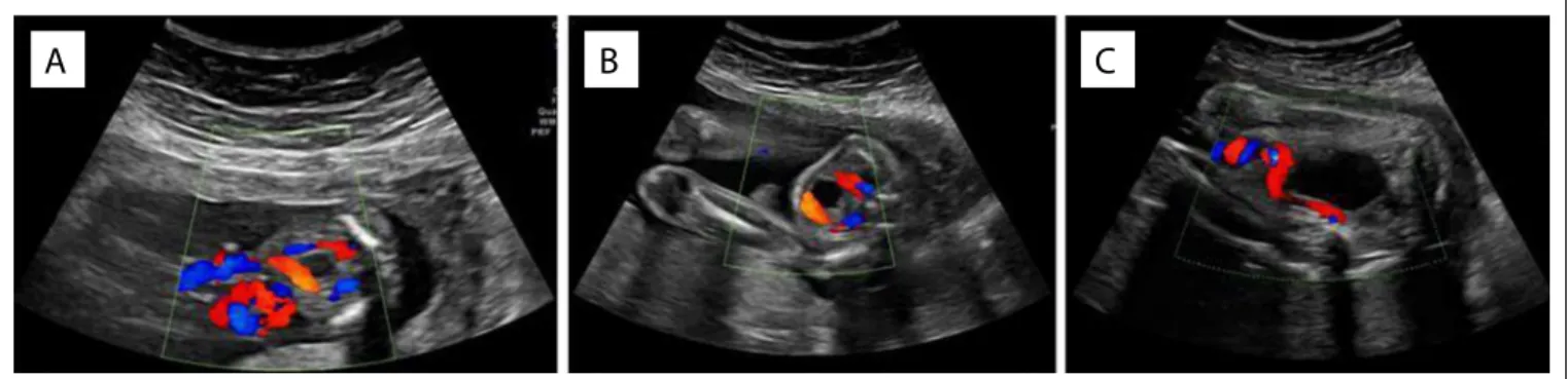
Retrospective analysis on ultrasound imaging on the patient that had been produced during this pregnancy showed that there were no abnormalities of growth, fetal structure or umbilical cord up to the 22nd week (Figures 1A, B and C). he hypothesis of
spontaneous intrauterine umbilical artery thrombosis was raised.
Weekly serial ultrasound imaging was started and fetal pulmo-nary maturation treatment was instituted using corticosteroids.
At 34 weeks of gestation, cessation of fetal growth was observed and also a slight decrease in amniotic luid volume. Doppler velocimetry on the umbilical artery showed that the pul-satility index value was close to the 90th percentile for the
gesta-tional age, with adequate venous lows. It was decided to imple-ment early delivery by means of caesarean section. It was observed during the procedure that the amniotic luid had the appearance of meconium and that the umbilical cord was thin and approxi-mately 80 cm in length, with excessive twisting (Figure 2) and two tight cervical loops.
he newborn was male, with irst-minute Apgar of 9 and ith-minute Apgar of 10, weighing 1080 g. His gestational age from the somatic Capurro method was 34 weeks. He was referred to the neonate intensive care unit only because of low weight. He presented good postnatal evolution and was discharged from hospital on the 37th day of life. he histological analysis showed
massive thrombosis in one of the umbilical arteries.
Figure 2. Imaging in which the long, thin, excessively twisted umbilical cord was identiied.
Figure 1. Imaging of the case at 18 weeks (1a) and 22 weeks (1b), and then at 32 weeks (1c), when the “disappearance” of one of the umbilical arteries was noticed.
Intrauterine thrombosis of umbilical artery – case report | CASE REPORT
Sao Paulo Med J. 2016; 134(4):355-8 357 DISCUSSION
Over recent decades, there have been great advances in prena-tal care, especially through the evolution of ultrasound imag-ing and better understandimag-ing of embryology and fetal life. Concomitantly, pregnant women have become more demand-ing, such that they require their obstetrician and prenatal doc-tor (who are oten faced with threats and lawsuits) to provide safe and precise follow-up.4 However, situations without any precise
diagnosis or clear management are frequently encountered. It is known that umbilical cord thrombosis is associated with adverse fetal and perinatal outcomes, but its diagnosis and follow-up constitutes a clinical challenge.
Umbilical cord abnormalities such as an overly long cord (more than 70 cm) or short cord (less than 35 cm), excessive twisting (more than 0.3 cm/loop, reduced diameter (less than 8.0 mm), anomalous placental insertions and presence of true knots and loops are well-established risk factors for gestational complications and impair-ment of fetal wellbeing. hese abnormalities lead to mechani-cal compression or blood ectasia in the fetal vascular path.5-10
Moreover, such abnormalities are related to the etiology of umbili-cal cord thrombosis, and the risk of their occurrence is generally three times higher in the presence of cord thrombosis.11 In a study
on autopsies conducted on 139 fetuses ater spontaneous intrauter-ine death, vascular thrombosis of the umbilical cord was identiied in 20% of the cases, mainly when there was excessive twisting of the umbilical cord and reduction of its diameter.6 In situations of
pres-ence of umbilical cord abnormalities, intensive fetal monitoring during delivery and histological analysis on the umbilical cord are recommended, especially in cases of intrauterine death.12
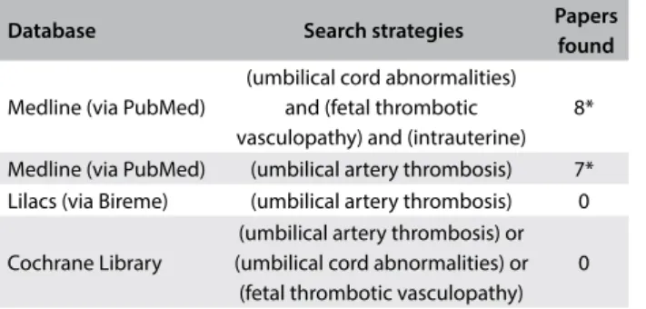
A few studies on umbilical cord thrombosis have already been conducted. he systematized results from the main database in the literature are presented in Table 1. here seems to be slight male predominance. Venous thrombosis alone is the most com-mon occurrence, found in approximately 70% of the cases, fol-lowed by concomitant arterial and venous thrombosis in 20% of
the cases and arterial thrombosis alone in 10% of the cases.1 It is
believed that the incidence of umbilical artery thrombosis alone is very small, ranging from 0.0025% to 0.045% of gestations.1,3
Although venous thrombosis is more frequent, it is believed that adverse outcomes are more common in arterial thrombo-sis.1 In the main published papers on this subject, associations
with growth restriction, fetal death, meconium in the amniotic luid, acute fetal distress during labor and higher rates of emer-gency caesarian sections have been noted.2,3,6,13 he most
com-mon abnormalities of the umbilical cord associated with umbili-cal artery thrombosis are: short or long cord, excessive twisting and anomalous placental insertions.3,11
CONCLUSIONS
In the case presented here, which occurred in a low-risk preg-nancy in which up to the 22nd week there had been no
suspi-cion of abnormality, spontaneous intrauterine umbilical artery thrombosis was detected in the third trimester of pregnancy and was associated with a thin and long umbilical cord, with excessive twisting and cervical loops. Severe intrauterine growth restric-tion and deteriorarestric-tion of fetal wellbeing were also observed. he diagnosis only became possible through making compari-sons with the patient’s initial ultrasound screenings, in which both umbilical arteries were clearly identiied, with subsequent conirmation through histological analysis. Unfortunately, ind-ings such as cord length abnormality and cord twisting are not commonly identiied. In the presence of umbilical cord abnor-malities, the risk of complications during intrauterine life, at the time of delivery and during the perinatal period, needs to be taken into consideration. Although the procedure in such cases is not well established, regular monitoring of fetal wellbeing, implementation of pulmonary maturation and early delivery in the event of fetal deterioration are recommended. Fetal monitor-ing is important because of the risk of adverse outcomes.
REFERENCES
1. Heifetz SA. Thrombosis of the umbilical cord: analysis of 52 cases and literature review. Pediatr Pathol. 1988;8(1):37-54.
2. Tantbirojn P, Saleemuddin A, Sirois K, et al. Gross abnormalities of the umbilical cord: related placental histology and clinical signiicance. Placenta. 2009;30(12):1083-8.
3. Sato Y, Benirschke K. Umbilical arterial thrombosis with vascular wall necrosis: clinicopathologic indings of 11 cases. Placenta. 2006;27(6-7):715-8.
4. MacLennan A, Nelson KB, Hankins G, Speer M. Who will deliver our grandchildren? Implications of cerebral palsy litigation. JAMA. 2005;294(13):1688-90.
5. Avagliano L, Marconi AM, Candiani M, Barbera A, Bulfamante G. Thrombosis of the umbilical vessels revisited. An observational study of 317 consecutive autopsies at a single institution. Hum Pathol. 2010;41(7):971-9.
Database Search strategies Papers found
Medline (via PubMed)
(umbilical cord abnormalities) and (fetal thrombotic vasculopathy) and (intrauterine)
8*
Medline (via PubMed) (umbilical artery thrombosis) 7*
Lilacs (via Bireme) (umbilical artery thrombosis) 0
Cochrane Library
(umbilical artery thrombosis) or (umbilical cord abnormalities) or
(fetal thrombotic vasculopathy) 0
*Two studies were available through both the irst and the second database searches.
CASE REPORT | Oliveira GH, Dias CM, Vaz-Oliani DCM, Oliani AH
358 Sao Paulo Med J. 2016; 134(4):355-8
6. Peng HQ, Levitin-Smith M, Rochelson B, Kahn E. Umbilical cord stricture and overcoiling are common causes of fetal demise. Pediatr Dev Pathol. 2006;9(1):14-9.
7. Baergen RN. Cord abnormalities, structural lesions, and cord “accidents”. Semin Diagn Pathol. 2007;24(1):23-32.
8. Chan JS, Baergen RN. Gross umbilical cord complications are associated with placental lesions of circulatory stasis and fetal hypoxia. Pediatr Dev Pathol. 2012;15(6):487-94.
9. Machin GA, Ackerman J, Gilbert-Barness E. Abnormal umbilical cord coiling is associated with adverse perinatal outcomes. Pediatr Dev Pathol. 2000;3(5):462-71.
10. Baergen RN, Malicki D, Behling C, Benirschke K. Morbidity, mortality, and placental pathology in excessively long umbilical cords: retrospective study. Pediatr Dev Pathol. 2001;4(2):144-53.
11. Redline RW. Clinical and pathological umbilical cord abnormalities in fetal thrombotic vasculopathy. Hum Pathol. 2004;35(12):1494-8. 12. Hasegawa J, Matsuoka R, Ichizuka K, Sekizawa A, Okai T. Ultrasound
diagnosis and management of umbilical cord abnormalities. Taiwan J Obstet Gynecol. 2009;48(1):23-7.
13. Klaritsch P, Haeusler M, Karpf E, Schlembach D, Lang U. Spontaneous intrauterine umbilical artery thrombosis leading to severe fetal growth restriction. Placenta. 2008;29(4):374-7.
Sources of funding: None
Conlict of interests: None
Date of irst submission: January 20, 2016
Last received: March 1, 2016
Accepted: March 12, 2016
Address for correspondence:
Gustavo Henrique de Oliveira Rua Angeolino Caselli, 360
Redentora — São José do Rio Preto (SP) — Brasil CEP 15015-010