Prevalence of electrocardiographic abnormalities in primary
care patients according to sex and age group. A retrospective
observational study
Julia Pereira Afonso dos Santos
I, Antonio Luiz Pinho Ribeiro
II, Diomildo Andrade-Junior
III, Milena Soriano Marcolino
IVDepartment of Internal Medicine and Telehealth Network of Minas Gerais, University Hospital, Medical School, Universidade Federal de
Minas Gerais, Belo Horizonte (MG), Brazil
INTRODUCTION
Cardiovascular diseases are the leading cause of death worldwide1 and have a high socioeco-nomic impact.2 he high mortality and morbidity associated with these diseases makes diagno-sis and management of these conditions essential in clinical practice.
Electrocardiograms (ECGs) are important examinations for assessing cardiac disease. Prior knowledge of the prevalence of abnormalities in the population studied is useful for inter-preting ECG indings.3 Additionally, electrocardiographic abnormalities are independently asso-ciated with the incidence of coronary heart disease and with poor cardiac disease outcomes.4
he prevalence of electrocardiographic abnormalities varies with age and sex.3,5,6 Recently, many studies have focused on the unique aspects of cardiac disease in women, in order to opti-mize its diagnosis and treatment.4,7,8 In this regard, the present study may contribute to the lit-erature on the subject through highlighting the diferences in ECG indings between men and women in separate age groups, in a large sample of Brazilian primary care patients, and through discussing the particularities of female ECGs in relation to male ones.
OBJECTIVE
he aim of this study was to analyze the prevalences of self-reported comorbidities and electro-cardiographic abnormalities according to age and sex among Brazilian primary care patients. Some speciic aspects of women’s ECGs in relation to men’s ones are also discussed.
METHODS
his retrospective observational study included all ECGs that were recorded in primary care units and then analyzed by cardiologists of the Telehealth Network of Minas Gerais (TNMG),
IMedical Student, Department of Internal
Medicine, Medical School, Universidade Federal de Minas Gerais, Belo Horizonte (MG), Brazil.
IIMD, PhD. Full Professor, Department of Internal
Medicine, Medical School, Universidade Federal de Minas Gerais, Belo Horizonte (MG), Brazil.
IIIMD. Physician, Department of Internal
Medicine, Medical School, Universidade Federal de Minas Gerais, Belo Horizonte (MG), Brazil.
IVMD, MSc, PhD. Associate Professor, Department
of Internal Medicine, Medical School, Universidade Federal de Minas Gerais, Belo Horizonte (MG), Brazil.
KEY WORDS: Electrocardiography. Sex.
Age groups. Primary health care.
ABSTRACT
BACKGROUND: Knowledge of the prevalence of electrocardiographic abnormalities in a population is useful for interpreting the indings. The aim here was to assess the prevalence of electrocardiographic abnormalities and self-reported comorbidities and cardiovascular risk factors according to sex and age group among Brazilian primary care patients.
DESIGN AND SETTING: Observational retrospective study on consecutive primary care patients in 658 cities in the state of Minas Gerais, Brazil, whose digital electrocardiograms (ECGs) were sent for analysis to the team of the Telehealth Network of Minas Gerais (TNMG).
METHODS: All ECGs analyzed by the TNMG team in 2011 were included. Clinical data were self-reported and electrocardiographic abnormalities were stratiied according to sex and age group.
RESULTS: A total of 264,324 patients underwent ECG examinations. Comorbidities and cardiovascular risk factors were more frequent among women, except for smoking. Atrial ibrillation and lutter, premature beats, intraventricular blocks, complete right bundle branch block and left ventricular hypertrophy were more frequent among men, and nonspeciic ventricular repolarization abnormalities and complete left bundle branch block among women.
a Brazilian large-scale public telehealth service, from January 1 to December 31, 2011. During this period, the service provided support to primary care practitioners in 658 municipalities in the state of Minas Gerais, among which 85% have fewer than 14,000 inhabitants. It performed teleconsultations and remote interpre-tation of diagnostic tests, including ECG analysis.9
Digital 12-lead electrocardiograms were produced using tele-electrocardiograph machines made by Tecnologia Eletrônica Brasileira (TEB; São Paulo, Brazil) or Micromed Biotechnology (Brasília, Brazil) and were sent over the internet to an analysis cen-ter, from which the examinations were immediately forwarded to a team of cardiologists, who analyzed the ECGs using standardized criteria.10 he team of cardiologists was composed of ten cardiol-ogists who had been trained and were experienced in ECG analy-sis. heir ECG analyses were also subject to periodic auditing and feedback.9 Only one individual reviewed each ECG.
he clinical data were self-reported and were gathered imme-diately before the patients were subjected to the ECG exam. A stan-dard questionnaire was used, which sought data including age, sex, medications in use, comorbidities (hypertension, diabetes, obesity, dyslipidemia, chronic kidney disease, chronic obstructive pulmo-nary disease and coropulmo-nary artery disease), prior acute myocardial infarction, smoking and family history of coronary heart disease. For the purpose of this study, all consecutive ECGs from January 1, 2011, to December 31, 2011, were analyzed. Electrocardiograms with technical issues such as interference or errors in the place-ment of electrodes were excluded. he proportion of atrial lutter was considered along with the proportion of atrial ibrillation, as has also been done in other epidemiological studies.11 he prev-alence of electrocardiographic abnormalities was evaluated and stratiied according to sex and age groups. he age groups encom-passed every two decades of life: from 0 to 19.9 years of age; 20 to 39.9; 40 to 59.9; 60 to 79.9; and greater than or equal to 80 years. Rankings of the most common abnormalities according to age group and sex were elaborated and a table of the inal ranking according to sex and age group was constructed.
he IBM SPSS statistics sotware for Windows version 20.0 (2011 release; IBM Corporation, Armonk, NY, USA) was used for the statistical analyses. Categorical variables were reported as counts and percentages; continuous variables were reported as means and standard deviations (SD) or medians with interquartile range (IQR), as appropriate. his study was approved by the Research Ethics Committee of the Federal University of Minas Gerais.
RESULTS
Over the course of this study, ECG recordings from 264,324 pri-mary care patients were analyzed by the TNMG cardiology team; 58.7% of the patients were women. he patients’ mean age was 51 ± 19 years; 7.2% of them were between zero and 19.9 years of
age; 21.3% between 20 and 39.9 years; 37.6% between 40 and 59.9 years; 28.2% between 60 and 79.9 years; and 5.0% greater than 80 years. In 0.7% of the examinations, the patient’s age was not included. he youngest group was excluded from further evalu-ation here.
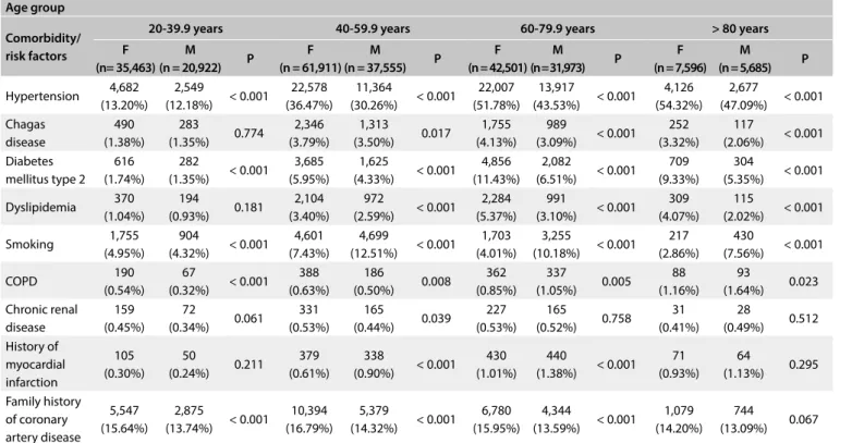
Table 1 shows the prevalences of self-reported comorbidities.
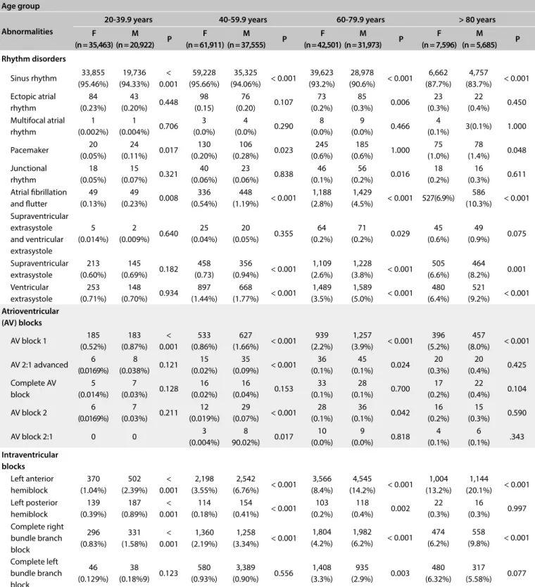
Tables 2A and 2B show the prevalences of electrocardiographic
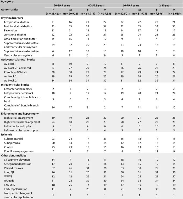
abnormalities according to sex and age groups. Table 3 shows the
ranking of the prevalences of electrocardiographic abnormalities according to sex and age groups.
Hypertension was the most frequent comorbidity, except in the group from 20 to 39.9 years of age, followed by a family his-tory of coronary artery disease and smoking. In the group from 20 to 39.9 years of age, a family history of coronary artery disease was the most frequent risk factor for cardiovascular disease. From the age of 60 years, diabetes mellitus began to show signiicant prevalence: 11.4% and 6.5% respectively among men and women between 60 and 79.9 years of age and 9.3% and 5.3% among those aged 80 years and over. In general, the prevalence of comorbidities was higher in women of all age groups. he most common elec-trocardiographic abnormalities of all were nonspeciic ventricu-lar repoventricu-larization abnormalities, with prevalences ranging from 9.2% in women aged 20 to 39.9 years to 38.0% in those aged 80 and over (P = 0.008).
In the age group from 20 to 39.9 years, 80.6% of the tests in males and 70.7% in females were normal. he main electrocar-diographic abnormality in women was let anterior hemiblock (LAH)12 (1.0%), followed by complete right bundle branch block (RBBB) (0.8%). In men, early repolarization pattern (ERP) (4.1%) and LAH (2.4%) were the most prevalent.
Between 40 and 59.9 years of age, 66.1% and 59.9% of the examinations among women and men respectively were normal. Among women, the most common abnormalities remained sim-ilar to those of the younger age group described above, despite increases in their prevalence (3.6% for LAH and 2.2% for RBBB). Among men, these indings became predominant (6.8% and 3.3%, respectively) and the prevalences of let atrium enlargement and ventricle hypertrophy increased (3.3% and 4.1%, respectively).
In the age group from 60 to 79.9 years, 46.7% of females and 40.8% of males presented normal results from the tests. Let ven-tricular hypertrophy became the second most prevalent abnormal result, following LAH (4.8% in women, 7.0% in men). Let bundle branch block (LBBB) (3.3% and 2.9%, respectively), irst-degree atrioventricular block (AVB) (2.2% and 3.9%) and atrial ibrilla-tion and lutter (2.8% and 4.5%) became more frequent.
(10.3%). In women, let ventricular hypertrophy remained a major result (8.7%), as did RBBB (6.2%), LBBB (6.3%) and LAH (13.2%). LAH was present in over 20% of examinations on males and irst-de-gree AVB in 8.0%.
DISCUSSION
In this study, on a large sample of primary care patients, elec-trocardiographic abnormalities were relatively common indings, even in the younger age groups. In the age group from 20 to 39.9 years, 19.4% of the women and 29.3% of the men had at least one abnormal result. he prevalence of abnormalities increased with age and was higher among males in all age groups. Atrial ibril-lation and lutter, premature beats, intraventricular block, com-plete right bundle branch block and let ventricle hypertrophy were more frequent among men. Women had higher prevalences of nonspeciic ventricular repolarization abnormalities and com-plete let bundle branch block.
Most examinations (87.1%) were conducted on patients aged between 20 and 79.9 years. Women presented a higher proportion of self-reported comorbidities, except for smoking. his reinforces the indings in the literature on this subject, which indicate that women care more about their health and therefore tend to be more aware of their medical conditions.12,13
With regard to comorbidities and cardiovascular risk factors, hypertension was the most common one (34.2% and 28.9% in women and men, respectively) from 20 years of age onwards,
followed by family history of coronary artery disease (16.0% and 13.6% in women and men). he prevalence of hyperten-sion in the population aged 60-79.9 years in the present analysis (48.2%) was similar to what was found among subjects from 60 to 70 years of age (48.6%) in a cross-sectional study that inves-tigated hypertension in the population of a Brazilian state cap-ital.14 In another study, in which household surveys were con-ducted in 15 Brazilian state capitals and in the federal district, the prevalence of self-reported hypertension among individuals aged 25-39 years (7.4% to 15.7%) was similar to what was found in the present study in the age group of 20-40 years.15 he Brazilian Longitudinal Study of Adult Health (ELSA-Brasil) also had simi-lar igures.16 his suggests that our sample may be representative of the Brazilian population.
Sex differences regarding hypertension are well known, from epidemiology to pathophysiology to target organ damage. Women have higher awareness, treatment and control rates and lower prevalence of left ventricular hypertrophy (LVH).7 This was seen in the sample of the present study: while reports of disease were higher in females, , males had higher prevalence of LVH in all age groups.
Self-reported diabetes was more frequent among females, mostly in individuals over 60 years of age. In the literature, slightly higher prevalence of diabetes in males has been reported world-wide. Nonetheless, studies from the Caribbean and from south-ern Africa showed higher prevalence of diabetes in women than
Table 1. Reported comorbidities and risk factors, according to sex and age group (n = 264,324) Age group
Comorbidity/ risk factors
20-39.9 years 40-59.9 years 60-79.9 years > 80 years
F (n= 35,463)
M
(n = 20,922) P
F (n = 61,911)
M
(n = 37,555) P
F (n = 42,501)
M
(n = 31,973) P
F (n = 7,596)
M
(n = 5,685) P
Hypertension 4,682 (13.20%)
2,549
(12.18%) < 0.001
22,578 (36.47%)
11,364
(30.26%) < 0.001
22,007 (51.78%)
13,917
(43.53%) < 0.001
4,126 (54.32%)
2,677
(47.09%) < 0.001 Chagas
disease
490 (1.38%)
283
(1.35%) 0.774
2,346 (3.79%)
1,313
(3.50%) 0.017
1,755 (4.13%)
989
(3.09%) < 0.001 252 (3.32%)
117
(2.06%) < 0.001 Diabetes
mellitus type 2 616 (1.74%)
282
(1.35%) < 0.001
3,685 (5.95%)
1,625
(4.33%) < 0.001
4,856 (11.43%)
2,082
(6.51%) < 0.001 709 (9.33%)
304
(5.35%) < 0.001
Dyslipidemia 370 (1.04%)
194
(0.93%) 0.181
2,104 (3.40%)
972
(2.59%) < 0.001
2,284 (5.37%)
991
(3.10%) < 0.001 309 (4.07%)
115
(2.02%) < 0.001
Smoking 1,755 (4.95%)
904
(4.32%) < 0.001
4,601 (7.43%)
4,699
(12.51%) < 0.001
1,703 (4.01%)
3,255
(10.18%) < 0.001 217 (2.86%)
430
(7.56%) < 0.001
COPD 190 (0.54%)
67
(0.32%) < 0.001 388 (0.63%)
186
(0.50%) 0.008
362 (0.85%)
337
(1.05%) 0.005
88 (1.16%)
93
(1.64%) 0.023 Chronic renal
disease
159 (0.45%)
72
(0.34%) 0.061
331 (0.53%)
165
(0.44%) 0.039
227 (0.53%)
165
(0.52%) 0.758
31 (0.41%)
28
(0.49%) 0.512 History of
myocardial infarction
105 (0.30%)
50
(0.24%) 0.211
379 (0.61%)
338
(0.90%) < 0.001 430 (1.01%)
440
(1.38%) < 0.001 71 (0.93%)
64
(1.13%) 0.295
Family history of coronary artery disease
5,547 (15.64%)
2,875
(13.74%) < 0.001
10,394 (16.79%)
5,379
(14.32%) < 0.001
6,780 (15.95%)
4,344
(13.59%) < 0.001
1,079 (14.20%)
744
(13.09%) 0.067
in men, which was a pattern similar to the one found in the pres-ent study. his was possibly due to higher rates of obesity among females from such developing regions, since obesity is one of the greatest risk factors for diabetes.17
here were fewer smokers aged between 20 and 39.9 years than in the age groups of 40-59.9 and 60-79.9 years. his corroborates the results from several studies that have demonstrated reduc-tions in smoking rates over recent decades, mainly inluenced by
Table 2A. Electrocardiograms abnormalities according to sex and age group: rhythm abnormalities, atrioventricular block and intraventricular conduction defects (n = 264,324)
Age group
Abnormalities
20-39.9 years 40-59.9 years 60-79.9 years > 80 years
F (n = 35,463)
M (n = 20,922) P
F (n = 61,911)
M
(n = 37,555) P
F (n = 42,501)
M
(n = 31,973) P
F (n = 7,596)
M
(n = 5,685) P
Rhythm disorders
Sinus rhythm 33,855 (95.46%) 19,736 (94.33%) < 0.001 59,228 (95.66%) 35,325
(94.06%) < 0.001
39,623 (93.2%)
28,978
(90.6%) < 0.001 6,662 (87.7%)
4,757
(83.7%) < 0.001 Ectopic atrial rhythm 84 (0.23%) 43 (0.20%) 0.448 98 (0.15) 76 (0.20) 0.107 73 (0.2%) 85 (0.3%) 0.006 23 (0.3%) 22 (0.4%) 0.450 Multifocal atrial rhythm 1 (0.002%) 1 (0.004%) 0.706 3 (0.0%) 4 (0.0%) 0.290 8 (0.0%) 9 (0.0%) 0.466 4
(0.1%) 3(0.1%) 1.000
Pacemaker 20 (0.05%) 24 (0.11%) 0.017 130 (0.20%) 106 (0.28%) 0.023 245 (0.6%) 185 (0.6%) 1.000 75 (1.0%) 78 (1.4%) 0.048 Junctional rhythm 18 (0.05%) 15 (0.07%) 0.321 40 (0.06%) 23 (0.06%) 0.838 46 (0.1%) 56 (0.2%) 0.016 18 (0.2%) 16 (0.3%) 0.611 Atrial ibrillation and lutter 49 (0.13%) 49 (0.23%) 0.008 336 (0.54%) 448
(1.19%) < 0.001 1,188 (2.8%)
1,429
(4.5%) < 0.001 527(6.9%) 586
(10.3%) < 0.001 Supraventricular extrasystole and ventricular extrasystole 5 (0.014%) 2 (0.009%) 0.640 25 (0.04%) 20 (0.05%) 0.355 64 (0.2%) 71 (0.2%) 0.029 45 (0.6%) 49 (0.9%) 0.075 Supraventricular extrasystole 213 (0.60%) 145 (0.69%) 0.182 458 (0.73) 356
(0.94%) < 0.001 1,109 (2.6%)
1,228
(3.8%) < 0.001 505 (6.6%) 464 (8.2%) 0.001 Ventricular extrasystole 253 (0.71%) 148 (0.70%) 0.934 897 (1.44%) 668
(1.77%) < 0.001 1,489 (3.5%)
1,589
(5.0%) < 0.001 480 (6.4%)
521
(9.2%) < 0.001
Atrioventricular (AV) blocks
AV block 1 185 (0.52%) 183 (0.87%) < 0.001 533 (0.86%) 627
(1.66%) < 0.001 939 (2.2%)
1,257
(3.9%) < 0.001 396 (5.2%)
457
(8.0%) < 0.001
AV 2:1 advanced 6 (0.0169%) 8 (0.038%) 0.121 15 (0.02%) 35
(0.09%) < 0.001 36 (0.1%) 45 (0.1%) 0.024 20 (0.3%) 20 (0.4%) 0.425 Complete AV block 5 (0.014%) 7 (0.03%) 0.128 16 (0.02%) 16 (0.04%) 0.153 33 (0.1%) 28 (0.1%) 0.700 17 (0.2%) 22 (0.4%) 0.104
AV block 2 6 (0.0169%) 7 (0.03%) 0.211 12 (0.019%) 29
(0.07%) < 0.001 28 (0.1%) 36 (0.1%) 0.042 16 (0.2%) 15 (0.3%) 0.590
AV block 2:1 0 0 3
(0.004%) 8 90.02%) 0.017 10 (0.0%) 9 (0.0%) 0.818 4 (0.1%) 6 (0.1%) .343 Intraventricular blocks Left anterior hemiblock 370 (1.04%) 502 (2.39%) < 0.001 2,198 (3.55%) 2,542
(6.76%) < 0.001 3,566 (8.4%)
4,545
(14.2%) < 0.001 1,004 (13.2%)
1,144
(20.1%) < 0.001 Left posterior hemiblock 139 (0.39%) 187 (0.89%) < 0.001 114 (0.18%) 154
(0.41%) < 0.001 103 (0.2%) 118 (0.4%) 0.002 22 (0.3%) 16 (0.3%) 0.997 Complete right bundle branch block 296 (0.83%) 331 (1.58%) < 0.001 1,360 (2.19%) 1,258
(3.34%) < 0.001 1,804 (4.2%)
1,982
(6.2%) < 0.001 474 (6.2%)
558
(9.8%) < 0.001
Complete left bundle branch block 46 (0.129%) 38 (0.18%9) 0.123 580 (0.93%) 3,389 (0.90%) 0.556 1,408 (3.3%) 935 (2.9%) 0.003 480 (6.32%) 317 (5.58%) 0.077
tobacco control initiatives such as tax increases on these prod-ucts and creation of restrictions on public smoking, among other equally efective measures.
Octogenarians reported lower frequency of Chagas disease, diabetes mellitus, smoking and dyslipidemia than did younger
subjects, thus indicating that people who reach older age groups usually have fewer comorbidities and cardiovascular risk factors, which may be related to survival bias.
he prevalence of chronic kidney disease (CKD) is very likely to be underestimated: about 0.5% among women and men over 60 years.
Table 2B. Electrocardiograms abnormalities according to sex and age group: enlargement and hypertrophy, ischemia and other abnormalities (n = 264,324)
Age group
Abnormalities
20-39.9 years 40-59.9 years 60-79.9 years > 80 years
F (n = 35,463)
M
(n = 20,922) P
F (n = 61,911)
M
(n = 37,555) P
F (n = 42,501)
M
(n = 31,973) P
F (n = 7,596)
M
(n = 5,685) P Enlargement and hypertrophy Right atrial enlargement 38 (0.10%) 28 (0.13%) 0.371 81 (0.13%) 82 (0.21%) 0.001 103 (0.2%) 86 (0.3%) 0.509 16 (0.2%) 16 (0.3%) 0.477 Right ventricular enlargement 11 (0.03%) 30
(0.14%) < 0.001 16 (0.02%)
36
(0.09%) < 0.001 32 (0.1%) 45 (0.1%) 0.007 11 (0.1%) 10 (0.2%) 0.665 Left atrial hypertrophy 218 (0.61%) 238
(1.13%) < 0.001 1,157 (1.86%)
1,245
(3.31%) < 0.001 1,470 (3.5%)
1,785
(5.6%) < 0.001 357 (4.7%) 308 (5.4%) 0.064 Left ventricular hypertrophy 152 (0.42%) 412
(1.96%) < 0.001 1,055 (1.70%)
1,526
(4.06%) < 0.001 2,036 (4.8%)
2,250
(7.0%) < 0.001 658 (8.7%) 521 (9.2%) 0.324 Ischemia Subendocardial 16 (0.04%) 13 (0.06%) 0.278 135 (0.21%) 6 (0.25%) 0.233 233 (0.5%) 141 (0.4%) 0.042 79 (1.0%) 40 (0.7%) 0.051 Subepicardial 37 (0.10%) 51
(0.24%) < 0.001 273 (0.44%)
276
(0.73%) < 0.001 390 (0.9%) 333 (1.0%) 0.089 80 (1.1%) 51 (0.9%) 0.376
Q wave 10 (0.02%) 14 (0.06%) 0.031 136 (0.21%) 194
(0.51%) < 0.001 189 (0.4%)
298
(0.9%) < 0.001 51 (0.7%)
56
(1.0%) 0.049 Poor R wave
progression
195 (0.549%)
286
(1.366%) < 0.001 829 (1.33%)
922
(2.45%) < 0.001 1,279 (3.0%)
1,642
(5.1%) < 0.001 354 (4.7%)
381
(6.7%) < 0.001
Other abnormalities ST segment elevation 79 (0.22%) 490
(2.34%) < 0.001 136 (0.21%)
537
(1.42%) < 0.001 120 (0.3%)
262
(0.8%) < 0.001 36 (0.5%) 42 (0.7%) 0.051 ST segment depression 44 (0.12%) 27 (0.12%) 0.872 304 (0.49%) 165 (0.43%) 0.249 382 (0.9%) 278 (0.9%) 0.692 114 (1.5%) 53 (0.9%) 0.004
Peaked T waves 3 (0.008%) 9 (0.043%) 0.007 9 (0.01%) 24
(0.06%) < 0.001 5 (0.0%)
26
(0.1%) < 0.001 7 (0.1%)
7
(0.1%) 0.600
Long QT 7 (0.019%) 3 (0.01%) 0.642 21 (0.03%) 12 (0.03%) 0.869 26 (0.1%) 15 (0.0%) 0.433 5 (0.1%) 6 (0.1%) 0.546 WPWS 86 (0.24%) 66 (0.31%) 0.107 95 (0.15%) 79 (0.21) 0.037 48 (0.1%) 51 (0.2%) 0.103 9 (0.1%) 4 (0.1%) 0.418
Brugada pattern 5 (0.014%) 8 (0.038%) 0.068 4 (0.006%) 13 (0.034%) 0.001 4 (0.0%) 7
(0.0%) 0.224 0 0
Low QRS 39 (0.109%) 13 (0.062%) 0.071 152 (0.24%) 83 (0.22%) 0.440 132 (0.3%) 139 (0.4%) 0.006 40 (0.5%) 29 (0.5%) 0.998 Early repolarization 112 (0.31%) 851
(4.06%) < 0.001 106 (0.17%)
710
(1.89%) < 0.001 78 (0.2%)
287
(0.9%) < 0.001 6 (0.1%)
27
(0.5%) < 0.001 Nonspeciic ventricular repolarization abnormalities 3,278 (9.24%) 2,152
(10.28%) < 0.001
12,578 (20.3%)
6,919
(18.42%) < 0.001
12,994 (30.6%)
8,784
(27.5%) < 0.001 2,890 (38.0%) 2,035 (35.8%) 0.008 Normal 28,599 (80.64%) 14,787
(70.67%) < 0.001
40,945 (66.13%)
22,481
(59.86%) < 0.001
19,843 (46.7%)
13,031
(40.8%) < 0.001 2,232 (29.4%)
1,378
(24.2%) < 0.001
A study in Juiz de Fora, a city in the same Brazilian state, showed that the prevalence in the same age group was 25.2%. It is possible that many patients were not aware of their condition, which thus emphasizes the need for screening, especially among individuals with high blood pressure and diabetes, which are the leading risk factors for CKD.18
Diferences between the sexes regarding the cardiovascular sys-tem result from diferences in gene expression from the sex chro-mosomes. his can also be further modiied through the inluence of sex-related hormones and other environmental factors, thereby resulting in sex-speciic gene expression.8 hus, electrocardiographic
F = female examinations; M = male examinations; WPWS = Wolf-Parkinson-White syndrome.
Table 3. Ranking of electrocardiograms abnormalities according to sex and age group (n = 264,324) Age group
Abnormalities
20-39.9 years 40-59.9 years 60-79.9 years ≥ 80 years F
(n = 35,463) M (n = 20,922)
F (n = 61,911)
M (n = 37,555)
F (n = 42,501)
M (n = 31,973)
F (n = 7,596)
M (n = 5,685) Rhythm disorders
Ectopic atrial rhythm 13 16 21 22 22 22 20 21
Multifocal atrial rhythm 33 33 33 34 32 33 33 33
Pacemaker 21 21 18 18 14 17 15 12
Junctional rhythm 22 22 24 27 25 24 23 25
Atrial ibrillation and lutter 15 15 11 12 9 8 4 3 Supraventricular extrasystole
and ventricular extrasystole 29 32 25 28 23 23 17 16 Supraventricular extrasystole 6 12 10 13 10 10 5 7
Ventricular extrasystole 4 11 6 9 5 7 7 6
Atrioventricular (AV) blocks
AV block 1 8 10 9 10 11 9 9 8
AV block 2:1 advanced 27 27 29 24 26 26 22 23
Complete AV block 30 30 27 29 27 29 24 22
AV block 2 28 29 30 25 29 28 26 27
AV block 2:1 34 34 34 32 31 32 32 31
Intraventricular blocks
Left anterior hemiblock 2 3 2 3 2 2 2 2
Left posterior hemiblock 10 9 19 17 19 20 21 24 Complete right bundle branch
block 3 6 3 5 4 4 8 4
Complete left bundle branch
block 16 17 8 2 7 11 6 10
Enlargement and hypertrophy
Right atrial enlargement 19 19 23 20 20 21 25 26 Right ventricular enlargement 24 18 28 23 28 27 27 28
Left atrial hypertrophy 5 8 4 6 6 5 10 11
Left ventricular hypertrophy 9 5 5 4 3 3 3 5
Ischemia
Subendocardial 23 24 17 33 15 18 14 18
Subepicardial 20 14 13 14 12 12 13 15
Q wave 25 23 15 15 16 13 16 13
Poor R wave progression 7 7 7 7 8 6 11 9
Other abnormalities
ST segment elevation 14 4 16 11 18 16 19 17
St segment depression 17 20 12 16 13 15 12 14
Peaked T waves 32 26 31 26 33 30 29 29
Long QT 26 31 26 31 30 31 31 30
WPWS 12 13 22 21 24 25 28 32
Brugada 31 28 32 30 34 34 34 34
Low QRS 18 25 14 19 17 19 18 19
Early repolarization 11 2 20 8 21 14 30 20
Nonspeciic changes of
abnormalities may show primary diferences between men and women. In the present study, 33.9% of the women and 40.1% of the men aged 40-59.9 years presented abnormal examinations. his was similar to the indings of another Brazilian study that also evaluated such abnormalities stratiied by age, although this other study did not examine the prevalence in relation to sex and also included patients from secondary care.19
LAH was one of the most common disorders in all age groups, with increasing prevalence according to age. It may be caused by hypertension, cardiomyopathies, Chagas disease in endemic coun-tries and Lev and Lenegre disease, and may form part of a benign senile degenerative process.20 However, this abnormality has little or no correlation with poor prognosis and is poorly associated with higher numbers of comorbidities.20 he prevalence rates for LAH in the combined population aged 40-79.9 years were 5.5% for women and 10.2% for men. his was compatible with several studies that have indicated that the prevalences of let axis devi-ation (which could be an indicator of LAH) and of LAH among men are around twice as high as among women.3 One example of such indings comes from an Indian study in which diferent rates of abnormal ECG results between the sexes were observed among people aged 45-74 years: 5.7% for women and 9.6% for men. here was also strong agreement regarding the prevalence of let ventricular hypertrophy between this Indian study and the present study: 2.9% and 5.1% in the present study, versus 2.8% and 4.6% in the Indian study, in women and men respectively.6
he prevalence of atrial ibrillation was strongly associated with greater age, and it was higher in men than in women, in all age groups. Our indings regarding the prevalence of atrial ibrillation according to age and sex were similar to data from high-income countries.11 his conirms and extends the indings of a previous paper from our group,21 from a subsample of the data used in the present study that was analyzed without the Minnesota Code. Since atrial ibrillation is a major risk factor for stroke, but there is no national health pol-icy to promote primary and secondary stroke prevention among patients with atrial ibrillation (the new oral anticoagulants are not provided through the public health system and there are not enough anticoagulation clinics to control patients on warfarin),22 the data provided by the present study is very important for stakeholders.
Another very frequent inding in all age groups was RBBB, which gives rise to a threefold increased risk of cardiovascular events and has been correlated with larger numbers of comor-bidities.23 RBBB also presented increasing prevalence with age, as had already been observed in the evaluation on RBBB within the Copenhagen City Heart Study.24 Complete RBBB had higher prevalence in the present study than in the Danish study (4.0% and 2.5% in men and women respectively, versus 1.5% and 0.5%).24 One hypothesis that would explain this discrepancy is the higher number of patients with Chagas disease in Brazil.
It has been well established that men present higher frequencies of intraventricular block and RBBB than do women.25 his was also found in the present study in relation to LAH, let posterior hemiblock and RBBB, but not in relation to LBBB. A statistically signiicant dif-ference in the frequency of LBBB between men and women was only present in the age group from 60 to 79.9 years, which is understandable, given the usually late onset of LBBB.26 In this group, the prevalence was 2.9% in men and 3.3% in women. Other studies have also found similar prevalences of LBBB in both sexes3,26 but none of them fur-ther explored the slightly higher prevalence of LBBB among women.
Nonspeciic ventricular repolarization abnormalities were the most prevalent abnormalities in all age groups. his is consistent with the previously mentioned American study that evaluated electro-cardiographic disorders in 20,962 people according to sex and age.5 hese abnormalities have been correlated with signiicantly higher risk of fatal coronary heart disease,27 for which primary arrhythmia is the main mechanism.28 his ECG disorder was more prevalent among women, and this might be explained by the signiicant inlu-ence of sex hormones on the QT interval in women: whereas this component is only shortened through the inluence of testosterone in men, signiicant estrogen activity in women prolongs this interval while their progesterone acts similarly to testosterone.29 hese non-speciic repolarization abnormalities were also found to be predictors of CHD events and CHD death among postmenopausal women.30
Chagas disease is still highly prevalent in Brazil. Out of the 5.7 million people chronically infected in Latin America, 20% are in this country.31 he most common electrocardiographic indings in Chagas disease are RBBB (22.7%) and LAH (22.5%). In addi-tion to these, second and third-degree atrioventricular blocks and atrial ibrillation are also strongly associated with Chagas disease.32 In the present study, 2.9% of the patients reported having Chagas disease and, as previously described, this may explain the higher prevalence of RBBB in relation to other studies.27
Interestingly, ECG abnormalities suggestive of acute ischemia, i.e. signs of subendocardial and subepicardial injury, were 0.3% and 0.6% overall, even though the present study was on tests performed within primary care. hese cases are supposed to be attended in emergency centers. However, many of the municipalities studied here do not have any emergency units or hospitals, and therefore patients seek care for emergency conditions at primary care centers. In addition, many patients become so used to attending primary care centers that they seek help there even in emergency situations.
his study has certain limitations. he comorbidities and med-ications were self-reported, so they may have been underreported. he electrocardiographic reports followed predetermined patterns, using criteria established by the Brazilian Society of Cardiology.10 hese criteria have not yet been validated in as many popula-tion-based studies as the Minnesota code.36 However, the criteria used relect current practices in Brazil, thus ensuring the ability to generalize the results to other primary care settings in this country.
CONCLUSION
his study on a large sample of primary care patients showed that electrocardiographic abnormalities were relatively com-mon indings, even in the younger age groups. he prevalence of abnormalities increased with age and was higher in men in all age groups, even though women had higher frequency of self-reported comorbidities. Atrial ibrillation and lutter, prema-ture beats, intraventricular blocks, complete right bundle branch block and let ventricle hypertrophy were more frequent in men. Women had higher prevalence of nonspeciic ventricular repo-larization abnormalities and complete let bundle branch block.
he correlations of age and sex with electrocardiographic abnor-malities that were made through the present study may help towards increasing the predictive value of ECGs and contribute towards diagnosing and subsequently managing many common cardiovas-cular diseases within primary care. Furthermore, the indings from this study reinforce the importance of consolidating programs for prevention and screening of diseases that enhance cardiovascular risk such as hypertension, diabetes, hyperlipidemia and smoking.
REFERENCES
1. GBD 2013 Mortality and Causes of Death Collaborators. Global, regional,
and national age-sex speciic all-cause and cause-speciic mortality for
240 causes of death, 1990-2013: a systematic analysis for the Global
Burden of Disease Study 2013. Lancet. 2015;385(9963):117-71.
2. Kankeu HT, Saksena P, Xu K, Evans DB. The inancial burden from
non-communicable diseases in low- and middle-income countries:
a literature review. Health Res Policy Syst. 2013;11:31.
3. De Bacquer D, De Backer G, Kornitzer M. Prevalences of ECG indings
in large population based samples of men and women. Heart.
2000;84(6):625-33.
4. Denes P, Larson JC, Lloyd-Jones DM, Prineas RJ, Greenland P. Major
and minor ECG abnormalities in asymptomatic women and risk of
cardiovascular events and mortality. JAMA. 2007;297(9):978-85.
5. Prineas RJ, Le A, Soliman EZ, et al. United States national prevalence
of electrocardiographic abnormalities in black and white middle-age
(45- to 64-Year) and older (≥ 65-Year) adults (from the Reasons for
Geographic and Racial Diferences in Stroke Study). Am J Cardiol.
2012;109(8):1223-8.
6. Sachin Khane R, Surdi AD. Gender diferences in the prevalence of
electrocardiogram abnormalities in the elderly: a population survey
in India. Iran J Med Sci. 2012;37(2):92-9.
7. Doumas M, Papademetriou V, Faselis C, Kokkinos P. Gender diferences in
hypertension: myths and reality. Curr Hypertens Rep. 2013;15(4):321-30.
8. Garcia M, Mulvagh SL, Merz CN, Buring JE, Manson JE. Cardiovascular
Disease in Women: Clinical Perspectives. Circ Res. 2016;118(8):1273-93.
9. Alkmim MB, Figueira RM, Marcolino MS, et al. Improving patient access
to specialized health care: the Telehealth Network of Minas Gerais,
Brazil. Bull World Health Organ. 2012;90(5):373-8.
10. Sociedade Brasileira de Cardiologia. Diretrizes da Sociedade Brasileira
de Cardiologia sobre Análise e Emissão de Laudos Eletrocardiográicos
(2009 [Guidelines of Sociedade Brasileira de Cardiologia about analysis
and issuance of expert opinion in electrocardiographic (2009)]. Arq
Bras Cardiol. 2009;93(3 Supl 2):2-19.
11. Chugh SS, Havmoeller R, Narayanan K, et al. Worldwide epidemiology
of atrial ibrillation: a Global Burden of Disease 2010 Study. Circulation.
2014;129(8):837-47.
12. Bertakis KD, Azari R, Helms LJ, Callahan EJ, Robbins JA. Gender diferences
in the utilization of health care services. J Fam Pract. 2000;49(2):147-52.
13. Galdas PM, Cheater F, Marshall P. Men and health help-seeking
behaviour: literature review. J Adv Nurs. 2005;49(6):616-23.
14. Souza ARA, Costa A, Nakamura D, et al. Um estudo sobre hipertensão
arterial sistêmica na cidade de Campo Grande, MS [A study on systemic
arterial hypertension in Campo Grande, MS, Brazil]. Arq Bras Cardiol.
2007;88(4):441-6.
15. Passos VMA, Assis TD, Barreto SM. Hipertensão arterial no Brasil:
estimativa de prevalência a partir de estudos de base populacional
[Hypertension in Brazil: estimates from population-based prevalence
studies]. Epidemiol Serv Saude. 2006;15(1):35-45.
16. Chor D, Pinho Ribeiro AL, Sá Carvalho M, et al. Prevalence, Awareness,
Treatment and Inluence of Socioeconomic Variables on Control
of High Blood Pressure: Results of the ELSA-Brasil Study. PloS One.
2015;10(6):e0127382.
17. Sobers-Grannum N, Murphy MM, Nielsen A, et al. Female gender is a
social determinant of diabetes in the Caribbean: a systematic review
and meta-analysis. PloS One. 2015;10(5):e0126799.
18. Bastos RMR, Bastos MG, Ribeiro LC, Bastos RV, Teixeira MTB. Prevalência
da doença renal crônica nos estágios 3, 4 e 5 em adultos [Prevalence
of chronic kidney disease, stages 3, 4 and 5 in adults]. Rev Assoc Med
19. Giuliano ICB, Barcellos Junior CL, von Wangenheim A, Coutinho MSSA.
Emissão de laudos eletrocardiográicos a distância: experiência da rede
catarinense de telemedicina [Issuing electrocardiographic reports
remotely: experience of the telemedicine network of Santa Catarina].
Arq Bras Cardiol. 2012;99(5):1023-30.
20. Elizari MV, Acunzo RS, Ferreiro M. Hemiblocks revisited. Circulation.
2007;115(9):1154-63.
21. Marcolino MS, Palhares DM, Benjamin EJ, Ribeiro AL. Atrial ibrillation:
prevalence in a large database of primary care patients in Brazil.
Europace. 2015;17(12):1787-90.
22. Marcolino MS, Polanczyk CA, Bovendorp AC, et al. Economic evaluation
of the new oral anticoagulants for the prevention of thromboembolic
events: a cost-minimization analysis. Sao Paulo Med J. 2016;134(4):322-9.
23. Schneider JF, Thomas HE, Kreger BE, et al. Newly acquired right
bundle-branch block: The Framingham Study. Ann Intern Med. 1980;92(1):37-44.
24. Bussink BE, Holst AG, Jespersen L, et al. Right bundle branch block:
prevalence, risk factors, and outcome in the general population: results
from the Copenhagen City Heart Study. Eur Heart J. 2013;34(2):138-46.
25. Kreger BE, Anderson KM, Kannel WB. Prevalence of intraventricular
block in the general population: the Framingham Study. Am Heart J.
1989;117(4):903-10.
26. Imanishi R, Seto S, Ichimaru S, et al. Prognostic signiicance of incident
complete left bundle branch block observed over a 40-year period.
Am J Cardiol. 2006;98(5):644-8.
27. Ribeiro AL, Sabino EC, Marcolino MS, et al. Electrocardiographic
abnormalities in Trypanosoma cruzi seropositive and seronegative
former blood donors. PLoS Negl Trop Dis. 2013;7(2):e2078.
28. Kumar A, Prineas RJ, Arnold AM, et al. Prevalence, prognosis, and
implications of isolated minor nonspeciic ST-segment and T-wave
abnormalities in older adults: Cardiovascular Health Study. Circulation.
2008;118(25):2790-6.
29. Ganjehei L, Massumi A, Nazeri A, Razavi M. Cardiac arrhythmias in
women. Tex Heart Inst J. 2011;38(2):157-9.
30. Rautaharju PM, Kooperberg C, Larson JC, LaCroix A. Electrocardiographic
abnormalities that predict coronary heart disease events and mortality
in postmenopausal women: the Women’s Health Initiative. Circulation.
2006;113(4):473-80.
31. Chagas disease in Latin America: an epidemiological update based on
2010 estimates. Wkly Epidemiol Rec. 2015;90(6):33-43.
32. Marcolino MS, Palhares DM, Ferreira LR, Ribeiro AL. Electrocardiogram
and Chagas disease: a large population database of primary care
patients. Global Heart. 2015;10(3):167-72.
33. Levy D, Garrison RJ, Savage DD, Kannel WB, Castelli WP. Prognostic
implications of echocardiographically determined left ventricular mass
in the Framingham Heart Study. N Engl J Med. 1990;322(22):1561-6.
34. Kannel WB, Dannenberg AL, Levy D. Population implications of
electrocardiographic left ventricular hypertrophy. Am J Cardiol.
1987;60(17):85I-93I.
35. Gasperin CA, Germiniani H, Facin CR, Souza AM, Cunha CLP. Análise
dos critérios eletrocardiográicos para determinação de sobrecarga
ventricular esquerda [An analysis of electrocardiographic criteria
for determining left ventricular hypertrophy]. Arq Bras Cardiol.
2002;78(1):72-82.
36. Marty AT. Minnesota Code Manual of Electrocardiographic Findings.
Critical Care Medicine. 1983;11(7):583. Available from: http://
journals.lww.com/ccmjournal/Citation/1983/07000/Minnesota_
Code_Manual_of_Electrocardiographic.29.aspx. Accessed in 2017
(Sep 18).
This study was presented at the 42nd International Congress of
Electrocardiology, held in Comandatuba, Bahia, Brazil, on June 26, 2015
Sources of funding: The Telehealth Network of Minas Gerais is sponsored by the State Government of Minas Gerais, by its Health
Department (Secretaria de Estado da Saúde de Minas Gerais) and its
research agency FAPEMIG (Fundação de Amparo à Pesquisa de Minas
Gerais); and by the Brazilian Government, including the Ministry of
Health and the Ministry of Science and Technology and their research
and innovation agencies, CNPq (Conselho Nacional de Desenvolvimento
Cientíico e Tecnológico) and FINEP (Financiadora de Estudos e
Projetos). This study was sponsored by FAPEMIG, the research agency
of the state of Minas Gerais, Brazil (grant number CDS-RED-00004-14).
ALR was supported in part by CNPq (research productivity bursary
309073/2011-1), ,FAPEMIG (PPM-00428-17) and Instituto Nacional de
Ciência e Tecnologia para Avaliação de Tecnologias em Saúde (project
CNPq/465518/2014-1). DFAJ was supported by the Pro-Rectorate for
Research (Pró-Reitoria de Pesquisa, PRPq) of the Federal University of
Minas Gerais (PIBIC/CNPq). JPAS received a grant from CNPq (grant
number 180404/2014-8)
Conlict of interest: None declared
Date of irst submission: July 16, 2017
Last received: July 16, 2017
Accepted: August 29, 2017
Address for correspondence:
Milena Soriano Marcolino
Av. Professor Alfredo Balena, 190 — Sala 246
Belo Horizonte (MG) — Brasil
CEP 30130-100
E- mail: [email protected]