Cop
yright
© ABE&M t
odos os dir
eit
os r
eser
vados
.
Clinical practice guidelines for the
management of hypothyroidism
Diretrizes clínicas práticas para o manejo do hipotiroidismo
Gabriela Brenta1, Mario Vaisman2, José Augusto Sgarbi3, Liliana Maria Bergoglio4, Nathalia Carvalho de Andrada5, Pedro Pineda Bravo6, Ana Maria Orlandi7, Hans Graf8, on behalf of the Task Force on Hypothyroidism of the Latin American Thyroid Society (LATS)
ABSTRACT
Introduction: Hypothyroidism has long been known for its effects on different organ systems, leading to hypometabolism. However, subclinical hypothyroidism, its most prevalent form, has been recently related to cardiovascular risk and also to maternal-fetal complications in pregnant women. Objectives: In these clinical practice guidelines, several aspects of this ield have been discussed with the clear objectives of helping physicians treat patients with hypothyroidism, and of sharing some of our Latin American-based clinical experience. Materials and methods:
The Latin American Thyroid Society commissioned a Task Force on Hypothyroidism to develop
evidence-based clinical guidelines on hypothyroidism. A systematic review of the available li-terature, focused on the primary databases of MedLine/PubMed and Lilacs/SciELO was perfor-med. Filters to assess methodological quality were applied to select the best quality studies. The strength of recommendation on a scale from A-D was based on the Oxford Centre for Evidence- -based Medicine, Levels of Evidence 2009, allowing an unbiased opinion devoid of subjective viewpoints. The areas of interest for the studies comprised diagnosis, screening, treatment and a special section for hypothyroidism in pregnancy. Results: Several questions based on diag-nosis, screening, treatment of hypothyroidism in adult population and speciically in pregnant women were posed. Twenty six recommendations were created based on the answers to these questions. Despite the fact that evidence in some areas of hypothyroidism, such as therapy, is lacking, out of 279 references, 73% were Grade A and B, 8% Grade C and 19% Grade D. Conclu-sions: These evidence-based clinical guidelines on hypothyroidism will provide uniied criteria for management of hypothyroidism throughout Latin America. Although most of the studies referred to are from all over the world, the point of view of thyroidologists from Latin America is also given. Arq Bras Endocrinol Metab. 2013;57(4):265-99
Keywords
Hypothyroidism; subclinical hypothyroidism; clinical practice guidelines; evidence based medicine
RESUMO
Introdução: O hipotiroidismo é amplamente reconhecido por seus efeitos sobre os diferentes sistemas orgânicos, levando ao hipometabolismo. No entanto, o hipotiroidismo subclínico, sua apresentação mais prevalente, tem sido recentemente relacionado ao risco cardiovascular e também com complicações materno-fetais em gestantes. Objetivos: Nestas diretrizes clínicas, vários aspectos do hipotiroidismo foram discutidos com objetivos claros de ajudar os médicos a tratar pacientes com hipotiroidismo e de compartilhar algumas das nossas experiências clíni-cas na América Latina. Materiais e métodos: A Sociedade Latino-Americana de Tireoide formou uma Força-Tarefa para desenvolver diretrizes baseadas em evidências clínicas sobre o hipoti-roidismo. Foi realizada uma revisão sistemática da literatura existente, com foco em bancos de dados primários do MedLine/PubMed e Lilacs/SciELO. Foram feitas análises para avaliar a qua-lidade metodológica no sentido de selecionar os melhores estudos. A força de recomendação em uma escala de A-D foi baseada no Centro de Oxford para a Medicina Baseada em Evidência – Níveis de Evidência 2009 –, permitindo uma opinião imparcial, desprovida de pontos de vista
Thyroid Dysfunction Task Force. Latin American Thyroid Society
1 Dr. Cesar Milstein Hospital,
Buenos Aires, Argentina
2 Universidade Federal do
Rio de Janeiro (UFRJ), Rio de Janeiro, RJ, Brazil
3 Faculdade de Medicina de Marília
(Famena), São Paulo, SP, Brazil
4 National University of Córdoba
Clinical Hospital, Córdoba, Argentina
5 Guidelines Project, Brazilian
Medical Association, Brazil
6 University of Chile Clinical
Hospital, Santiago del Chile, Chile
7 Dr. Teodoro Alvarez Hospital,
Buenos Aires, Argentina
8 Universidade Federal do Paraná
(UFPR), Curitiba, PR, Brazil
Correspondence to: Gabriela Brenta Virrey Del Pino, 3370, 3ºA CABA 1426, Argentina [email protected]
Cop
yright
© ABE&M t
odos os dir
eit
os r
eser
vados
.
subjetivos. As áreas de interesse compreenderam estudos de diagnóstico, triagem, tratamento e uma seção especial de hipotiroidismo na gravidez. Resultados: Foram feitos vários questio-namentos relacionados ao diagnóstico, triagem e tratamento do hipotiroidismo na população adulta e, especiicamente, em mulheres grávidas. Foram elaboradas vinte e seis recomenda-ções baseadas nas respostas a essas perguntas. Apesar da falta de evidências em algumas áreas como o tratamento do hipotiroidismo, de 279 referências, 73% eram de Grau A e B, 8% de Grau C e 19% de Grau D. Conclusões: Essas diretrizes baseadas em evidências clínicas sobre o hipotiroidismo poderão [fornecer um critério consensual de como tratar o hipotiroidismo na América Latina. Apesar de a maior parte dos estudos referidos ser da experiência internacional em hipotiroidismo, o ponto de vista dos tiroidologistas da América Latina foi contemplado. Arq Bras Endocrinol Metab. 2013;57(4):265-99
Descritores
Hipotiroidismo; hipotiroidismo subclínico; diretrizes clínicas práticas; medicina baseada em evidências
INTRODUCTION
H
ypothyroidism is one of the most frequent endo-crine diseases. It is usually detected by clinicians and often now looked for by other specialists such as gynecologists and cardiologists, who are more aware of its unwanted effects. Therefore, the purpose of these clinical practice guidelines has been to develop a syste-matic statement designed to assist health care professio-nals and patients in making decisions about appropriate health care for the management of hypothyroidism. We also sought to illustrate traditional concepts regarding overt hypothyroidism and to provide an updated view of the controversies and assertions in the ield of subcli-nical hypothyroidism.The guidelines are divided into four areas, addres-sing diagnosis, screening, treatment and a special sec-tion for hypothyroidism in pregnancy. In these guideli-nes, the topic of hypothyroidism in paediatrics was not included.
The main questions posed were: How to make the diagnosis of hypothyroidism? Who should be screened for hypothyroidism? How should case inding be done? When should thyroid ultrasonography be performed? Which patients with subclinical hypothyroidism should be considered for treatment with thyroid hormones? How should patients with hypothyroidism be treated and monitored? When and how to screen hypothyroi-dism in pregnant women? How are hypothyroihypothyroi-dism and subclinical hypothyroidism deined in pregnant women? What is the role of thyroid autoimmunity in fertility and pregnancy? When and how to treat hypo-thyroidism in pregnant women? Each question was answered according to the available literature and was concluded with a series of recommendations.
Consensus process
The guidelines were developed by members of the La-tin American Thyroid Society (LATS) who took part in the Task Force on Hypothyroidism. The representatives acting on behalf of LATS were: Hans Graf, José Sgar-bi and Mario Vaisman from Brasil; Liliana Bergoglio, Gabriela Brenta and Ana Orlandi from Argentina; and Pedro Pineda Bravo from Chile. In order to prepare the manuscript, three face-to-face meetings were held. A robust exchange of e-mails was required to agree on all concepts that were included and to make modiica-tions according to suggesmodiica-tions. The clinical guidelines task force started its activities in late 2011. The grading of evidence was performed with the help of Nathalia Carvalho de Andrada, from the Guidelines Project of the Brazilian Medical Association. After several revi-sions, the initial draft was reviewed and approved by the LATS President.
Evidence
Cop
yright
© ABE&M t
odos os dir
eit
os r
eser
vados
.
therapeutic interventions. The results of the literature search provided studies that were graded by level of evidence. The strength of recommendation was based on the Oxford Centre for Evidence-based Medicine, Levels of Evidence 2009, allowing an unbiased opi-nion devoid of subjective viewpoints. As some medical conditions did not lend themselves ideally to giving formal recommendations based on studies with high or conclusive levels of evidence (recommendation gra-des A and B), being relevant clinical situations, their recommendations were retained, even when they were supported only or primarily by expert opinion (re-commendation grade D).
There is a relationship between the methodological quality of the study design (strength of evidence – le-vels 1 to 5) and the same degree of recommendation (grades A to D). After critical evaluation of the litera-ture, recommendations that always took into account the best available evidence to date for each issue were made, underscoring both certainties and uncertainties.
The areas of interest in which the evidence was dis-tributed included: Diagnosis, Differential diagnosis/ Symptom prevalence study, Therapy/Prevention, Etio-logy/Harm, and Prognosis. Based on the strength of evidence, four grades of recommendations from A to D were assigned. The original Oxford Centre for Eviden-ce-based Medicine Levels of Evidence can be found at http://www.cebm.net/index.aspx?o=1025.
With the purpose of interpreting the Oxford EBM classiication of evidence in simpliied manner, a summary of studies for diagnosis and approved by the LATS President:
For diagnosis
• Grade A: includes prospective cohort studies performed at a single center location, validated with a gold standard parameter (or a systematic review of this kind of study).
• Grade B: includes exploratory cohort studies (these studies collect information and trawl the data, e.g. using a regression analysis, to ind which factors are “signiicant”) that only seek information in a part of the population; or fol-low-up of untreated control patients in a rando-mized control trial (RCT); or studies with poor follow-up. Level B studies can be multi-centric, prospective or restrospective (or a systematic review of this kind of study). Also included are non-consecutive cohort studies, or with very
li-mited population (or a systematic review of this kind of study).
• Grade C: includes case-control studies; or ca-se-series (and poor quality prognostic cohort studies).
• Grade D: includes expert opinion without ex-plicit critical appraisal, or based on physiology, bench research or “irst principles”.
And for therapy
• Grade A: includes individual randomized con-trol trials (RCT) (with narrow Conidence In-terval) (or a systematic review of this kind of study).
• Grade B: includes individual cohort studies (including low quality RCT; e.g., < 80% fol-low-up) (or a systematic review of this kind of study). It also includes individual case-control studies (or a systematic review of this kind of study).
• Grade C: includes case-series (and poor quality cohort and case-control studies).
• Grade D: includes expert opinion without ex-plicit critical appraisal, or based on physiology, bench research or “irst principles”.
Grades of recommendation
A Consistent level 1 studies
B Consistent level 2 or 3 studies or extrapolations from level 1 studies
C Level 4 studies or extrapolations from level 2 or 3 studies
D Level 5 evidence or troublingly inconsistent or inconclusive studies of any level
“Extrapolations” are where data is used in a situation that has potentially clinically important differences than the original study situation.
di-Cop
yright
© ABE&M t
odos os dir
eit
os r
eser
vados
.
sease in the patient studied (post-test probability) can be determined. The LR is used to assess how good a diagnostic test is and to help in selecting an appropriate diagnostic test(s) or sequence of tests. LR has advanta-ges over sensitivity and speciicity because it is less likely to change with the prevalence of the disorder.
In the treatment section of the Guidelines, some calculations of the magnitude of therapeutic clinical trials results found were also run. They were expressed by the absolute risk reduction (ARR) and the Number Needed to Treat value (NNT = 1/ARR), which deines the number of patients you need to treat to prevent an outcome or unwanted event (for example: death). In medicine, a number needed to treat (NNT) value of 5 or less represents a clinically signiicant inding. Ma-thematically, a number needed to treat (NNT) of 5 is equal to a 20% improvement. A number needed to treat (NNT) of 2 is equal to a 50% improvement.
BACKGROUND ON HYPOTHYROIDISM
The function of the thyroid gland is regulated by thyroid--stimulating hormone (TSH), which is synthesized and secreted from the anterior pituitary gland. Thyroid hormones exert a negative feedback in patients with an intact hypothalamic-pituitary-thyroid axis, thereby con-trolling thyroid gland metabolism. A decrease in thyroid hormone production stimulates more TSH secretion.
Hypothyroidism is characterized by a deicient thyroid hormone production by the thyroid gland, which can be severe or moderate. Severe deicit of thyroid hormones deines overt hypothyroidism (OH). The moderate form, called subclinical hypothyroidism (SH), seldom has signs and symptoms and is deined biochemically by TSH concentration above the upper limit of the reference range, with thyroid hormone le-vels that remain within the reference ranges. SH must be deined in the absence of hypothalamic or pituitary disease and nonthyroidal illness (1) (B).
Determination of etiology
Primary overt hypothyroidism (OH) refers to reduced thyroid hormone production, which causes an increase in TSH levels. Decreased thyroid secretion can also be a result of reduced stimulation of the thyroid gland due to decreased thyrotropin-releasing hormone (TRH) or TSH action. Hypothyroidism can also be explained by a reduced action of thyroid hormones in target organs, as in the rare cases of thyroid hormone resistance (2) (D).
The most frequent cause of primary hypothyroi-dism is chronic autoimmune thyroiditis (Hashimo-to’s thyroiditis). However it can also result from the treatment of hyperthyroidism with surgery, antithyroid drugs or 131I irradiation. Several forms of thyroiditis, including postpartum, silent, subacute or cytokine-in-duced thyroiditis can also cause permanent or transitory hypothyroidism. Less frequently, it can be a consequen-ce of iniltrative or infectious disease, external radiation, thyroid dysgenesis, functional defects in thyroid hor-mone biosynthesis and release, and congenital defects in thyroid hormone biosynthesis. Iodine deiciency and iodine excess are well recognized causes of primary hypothyroidism, as are also the use of certain drugs that include antithyroid agents: lithium, natural and synthe-tic goitrogenic chemicals, tyrosine kinase inhibitors, in-terleukin-2 or interferon-a (IFN-a) etc. SH shares the same etiology as overt primary hypothyroidism.
High serum TSH levels can often be the result of inadequate replacement therapy with levothyroxine due to poor compliance, drug interactions, lack of appropriate waiting time for the determination of TSH, both from the beginning of the treatment (3 months are required) or from change of dose or between com-mercial brands of levothyroxine (L-T4) (2 months are required) (3) (D), or from changes in the course of the disease.
Central hypothyroidism has multiple causes (tu-mors, trauma, vascular, infections, iniltrative, in-lammatory or congenital). Apart from loss of functio-nal tissue, central hypothyroidism can also result from functional defects in TSH biosynthesis or release due to both mutations and from drugs such as dopamine and glucocorticoids.
Peripheral hypothyroidism can be a consequence of mutations in genes that intervene in the thyroid res-ponse in target tissues (thyroid hormone resistance), or even more rarely be due to thyroid hormone consump-tion in massive infantile hemangioma (2) (D).
Cop
yright
© ABE&M t
odos os dir
eit
os r
eser
vados
.
action, isolated pituitary resistance to thyroid hormo-ne, higher TSH values reported in obesity, and adrenal insuficiency (4) (D).
Iodine deiciency in Latin America
As a result of the recognition of iodine deiciency as the principal etiology for goiter worldwide, in the 1940s, several countries in Latin America started introducing the use of iodized salt in order to eliminate the goiter belts. Having recognized this disease as a public health problem, the World Health Organization (WHO) cre-ated a study group on endemic goiter, with the colla-boration of John Stanbury and other prestigious Latin American experts to assure a permanent surveillance on iodine nutrition and thyroid function. This decision re-sulted in positive steps taken to prevent the development of goiter, such as in Argentina, where the national law on salt iodization was inally passed in 1967 (5) (D). In 1980, all the consequences of the lack of iodine, ranging from goiter to mental retardation, were gathered under the term “iodine deiciency disorders” (IDDs) and the measurement of urinary iodine concentration (UIC) le-vels became the best tool to monitor population iodi-ne deiciency status (6) (D). In 1994, in an attempt to reduce the prevalence of iodine deiciency worldwide, the WHO recommended eliminating IDDs by iodizing all salt for human consumption (6) (D). The prevalence of goiter as it relates to iodine nutrition has been more recently studied in school children from 13 Latin Ameri-can countries with the use of the ThyroMobil model. In some of the regions, the prevalence was very low (3.1%), while in others it reached 25%, with median UIC corre-lating well with the iodine in the salt. The median iodi-ne content in the salt varied outside the recommended range of 20-40 parts per million (ppm), but was greater than 78 ppm in 83.1% of all samples, demonstrating a great achievement in the elimination of iodine deiciency in most of the studied countries (7) (B). From 2005 on, the WHO determined that each country should make its own reports on the iodine deiciency situation every 3 years. By 2007, the number of countries with iodine de-iciency (median UIC < 100 μg/l) had reduced from 54 in 2003 to 47. Nevertheless, underexposure to iodine is still an issue (as is overexposure) (8) (B).
Biochemical evaluation
Serum TSH is the irst-line diagnostic test for primary hypothyroidism. This is due to the inverse log-linear
relationship between the concentrations of TSH and free T4, which determines that small linear decreases in free T4 concentrations are associated with an expo-nential increase in TSH concentrations (9) (B). Addi-tionally, the TSH assay is accurate, widely available, safe and relatively inexpensive. Third-generation TSH im-munometric assays (IA) have very high sensitivity and speciicity (10,11) (B,D).
TSH determination is essential for the diagnosis of hypothyroidism because clinical symptoms are not speciic. There is no pathognomonic sign to clearly distinguish a patient with hypothyroidism. The asso-ciation of bradycardia with delayed ankle relex and coarse and dry skin presents a positive likelihood ratio (the likelihood ratio – + LR – that a given test result would be expected in a patient with the target disor-der compared to the likelihood that the same result would be expected in a patient without that disorder) of only 3.75 (+LR = 3.75, 95% CI 1.65-8.52) (12,13) (B,B) (14)(C), increasing the diagnosis (calculated as from 2% (pre-test disease prevalence) to 5% in overt hypothyroidism (OH) and from 10% (pre-test disea-se prevalence) to 25% in subclinical hypothyroidism (SH) (15) (B). Table 1 lists symptoms with the best positive likelihood ratios for clinical diagnosis of hypo-thyroidism (16) (B).
Diagnostic accuracy is based on the comparison of a diagnostic test with its gold standard. However, for hypothyroidism, no gold standard is available. Al-though the introduction of ultrasensitive TSH tests has improved the study of thyroid disease, in hyperthyroi-dism in particular, estimating the diagnostic accuracy of TSH for the detection of hypothyroidism still has its limitations.
Cop
yright
© ABE&M t
odos os dir
eit
os r
eser
vados
.
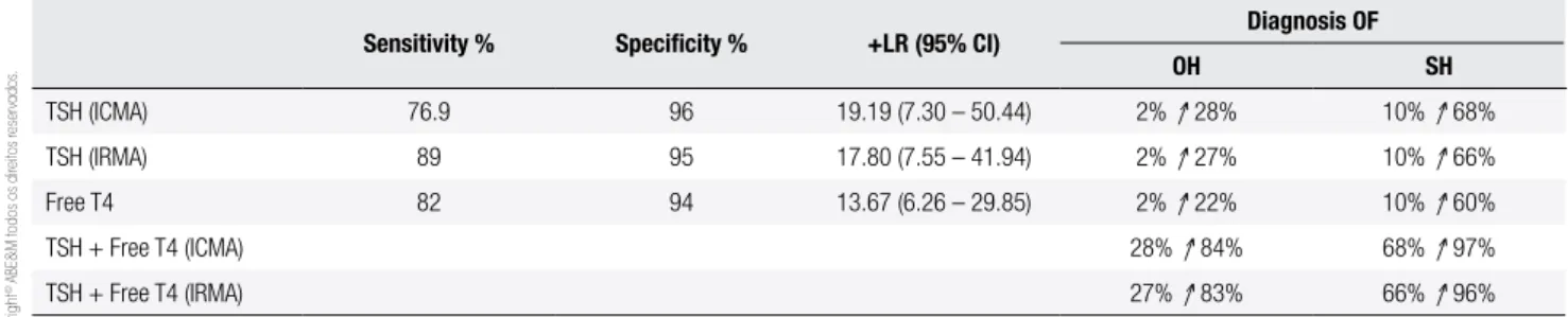
With regards to the determination of isolated free T4 in ambulatory patients, sensitivity and speciicity have been estimated at 82% and 94% respectively, in-creasing the probability of disease detection to 22% in overt hypothyroidism and to 60% in SH. The associa-tion of both TSH and free T4 increases the diagnostic accuracy for overt hypothyroidism from 28% to 84% (ICMA) and from 27% to 83% (IRMA). For SH ho-wever, given its higher prevalence, there is an increase in diagnostic accuracy with both tests, from 68% to 97% (ICMA) and from 66% to 96% (IRMA) (17) (B) (Table 2).
In hospitalized patients, a special situation presen-ted itself (18,19) (B). The use of drugs such as dopa-mine or glucorticoids that reduce TSH serum levels or other drugs that interfere with levothyroxine absorp-tion, together with the lack of correct administration of levothyroxine and the presence of nonthyroidal disease (NTI), all explain why thyroid function testing in the admitted patient is often hampered. Free T4 testing be-comes less reliable during hospitalization, especially in the critically ill patient (20) (D).
With a TSH cutoff level of above 10 mU/L, the probability of disease detection increases from 2%
(pre-test disease prevalence) to 34%. (+LR = 24.75; 95% CI 9.47-64.67) (21) (B). If serum TSH is within normal range, it is very dificult to sustain the diagnosis of hypo-thyroidism unless a hypothalamic-pituitary cause is in-volved. However, in those cases, clinical manifestations will prevail. If TSH levels are high but free T4 levels are normal, unless there is a nonthyroidal illness syndrome present, it most probably indicates SH (22) (D).
As stated above, the reason for maintaining circula-ting T4 values within the reference range in SH is the high sensitivity of the pituitary thyrotroph for even very small decreases of serum T4. The best method to assess free T4 is ultrailtration or dialysis equilibrium/liquid chromatography – tandem mass spectrometry (UF/ DE/LC-MS/MS) which display a stronger log/linear relationship with TSH compared with immunoassay (IA) methods (23) (B), however, for practical reasons, free T4 has to rely on IA methods that “estimate” its serum levels.
Some authors have proposed dividing SH into a mild form (serum TSH concentrations of 4·5-9 mU/L) and a severe form (TSH ≥ 10 mU/L) in regards to its respective clinical consequences (24) (D).
Analysis of the upper limit of normal range TSH values according to age and speciic populations
The normal range of TSH values in thyroid disease-free individuals has been traditionally accepted as 0.45-4.5 mU/L. This corresponds to 2.5th to 97.5th percentile of the distribution curve of TSH in most populations. This reference range is considered as “experimental” or population based (22,24) (D,D). This concept is based on epidemiologic studies such as the National Health and Nutritional Examination Survey (NHANES) III, where the reference range for TSH concentration was found to be 0.45-4.12 mU/l, with a median value of 1.4 mU/L (25) (B).
Table 2. Diagnosis of overt hypothyroidism (OH) and subclinical hypothyroidism (SH) in ambulatory patients
Sensitivity % Speciicity % +LR (95% CI) Diagnosis OF
OH SH
TSH (ICMA) 76.9 96 19.19 (7.30 – 50.44) 2% ↑ 28% 10% ↑ 68%
TSH (IRMA) 89 95 17.80 (7.55 – 41.94) 2% ↑ 27% 10% ↑ 66%
Free T4 82 94 13.67 (6.26 – 29.85) 2% ↑ 22% 10% ↑ 60%
TSH + Free T4 (ICMA) 28% ↑ 84% 68% ↑ 97%
TSH + Free T4 (IRMA) 27% ↑ 83% 66% ↑ 96%
Adapted from reference 17 (B). Positive likelihood ratio (+LR).
Table 1. Symptoms of hypothyroidism and correlation for diagnosis
Symptom Frequency (%) Likelihood Ratio (+ LR) (95% Conidence interval)
Deeper voice 14 7.1 (2.0-24.7)
Hoarse voice 21 5.2 (2.1-12.6)
Puffy eyes 36 4.0 (2.2-7.3)
Constipation 20 3.6 (1.6-8.1)
Coldness 39 3.5 (2.0-6.0)
Poor memory 39 2.6 (1.6-4.2)
Cop
yright
© ABE&M t
odos os dir
eit
os r
eser
vados
.
Since TSH values do not have a Gaussian distri-bution, (25) (A) other TSH cutoff values have been proposed, based on the notion that 95% of the normal population has TSH values below 2.5 mU/L (22) (D). However, this idea has been controversial except for in pregnancy (see pregnancy section). Another important issue to be considered is that all thyroid tests have a narrow within-person variability relative to the popu-lation reference range (that is, thyroid tests have a low index of individuality). Population reference range is not an individual´s normal range, so before a test moves outside the population reference range, a considerable degree of abnormality is required. (26) (B).
An “empirical” reference range between 0.3 and 3.0 mU/L (American Association of Clinical Endocrinolo-gists and National Academy of Clinical Biochemistry) (22,27) (D,D) has been also proposed as an alternati-ve to the “experimental” (population based) reference range, due to the large number of factors affecting the upper limits in all population studies, (including indivi-dual and geographic variables).
The upper limit of TSH values may increase with age, as shown in the NHANES III population. In fact, if TSH upper level remains at 4.5 mU/L, 74% of TSH values of elderly patients aged 80 years or older without TPOAb positive values would be found to be above this level (28) (A). According to a reanalysis of the NHA-NES III TSH distribution curves in TPOAb negative individuals between 50-59 years, TSH upper levels would correspond to 4.2 mU/L, between 60-69 years, to 4.7 mU/L, between 70-79 years, to 5.6 mU/L, and in subjects over age 80, to 6.3 mU/L (29) (A).
In critically ill patients in whom transient luctua-tions in TSH are common, a wider TSH reference ran-ge, about 0.05 to 10 mU/L, is recommended for diag-nosis of hypothyroidism (22) (D).
Repeat TSH testing is important to rule out tran-sient TSH increases, intraindividual variability or bio-chemical errors. Between the range of 4.5 and 10 mU/L, the possibility of return of TSH to normal serum levels is high (30) (B). It has been proposed that 2-3 months after the initial TSH determination, a se-cond assay should be performed to conirm the thyroid deiciency (22,31) (D,D). The chances of inding tran-sitory elevations of TSH are minimized, as higher TSH values are found or in patients who are TPOAb or TgAb positive (32) (A).
Prevalence
While the prevalence of OH has been reported at 1.5%, SH is nearly ten-fold more frequent. Its prevalence di-ffers according to studies from between 6-20%, depen-ding on age, sex, race, iodine repletion of the popula-tion studied, as well as the TSH cutoff level employed to deine SH (25,33-35) (A).
HOW TO MAKE THE DIAGNOSIS OF
HYPOTHYROIDISM?
The diagnosis of hypothyroidism is often not so evi-dent. It is an endocrinological disorder that requires a high index of suspicion in disparate clinical scenarios that span from the critically ill lethargic patient with anasarca to the dyslipidemic postmenopausal woman who consults for obesity. To determine if hypothyroi-dism is present, physicians must be alert to detect the most frequent signs and symptoms of overt hypo-thyroidism such as bradycardia, delayed ankle relex, coarse and dry skin, weakness, lethargy, slow speech, edema of eyelids, sensation of cold, decreased sweating, cold skin, macroglosia, facial edema, coarseness of hair, cardiac enlargement (on x-ray), pallor of skin, impaired memory, constipation, weight gain, hair loss, dyspnea, peripheral edema, hoarseness, anorexia, nervousness, menorrhagia, deafness, palpitations, poor heart sounds, precordial pain, and poor vision, among many others (36) (C). Based on the predictive value of these signs and symptoms, several clinical composite scores have been developed to measure the degree of hypothyroi-dism (37,38) (B,D).
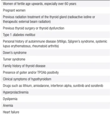
Unfortunately, many of these clinical features are nonspeciic, especially in elderly patients (39-41) (D), nor are frequent in SH. Therefore, some risk factors, listed in table 2, might raise a suspicion and prompt an evaluation for hypothyroidism.
Once the clinical aspects have been evaluated, a bio-chemical assessment will follow in order to diagnose or rule out hypothyroidism. If OH is suspected, TSH and free T4 should be determined (22,31) (D).
Cop
yright
© ABE&M t
odos os dir
eit
os r
eser
vados
.
relationship between them; TSH + TPOAb and TSH + free T4. The combination TSH + estimated free T4 is ideal, because TSH alone cannot detect patients with central hypothyroidism, and may lead to mis-diagnosis when the thyroid state is unstable at the beginning of the treatment with LT4. Moreover, this last approach has the advantage of permitting the cli-nical validation of the relationship of TSH to free T4 to detect method interference or clinical discrepan-cies in less common conditions. However, it has to be considered that free T4 levels do not result from a direct measurement of free hormone; they are an estimation of free T4 levels. Therefore, assessing free T4 has its own technical limitations, yielding false results, especially when there are alterations of bin-ding protein. In the context of nonthyroidal disease (NTI), when albumin is abnormal, affecting free T4 analog based immunoassays, or when the patient re-ceives drugs that displace T4 from the TBG, such as phenytoin, carbamazepine, or furosemide, TSH + total T4 should be determined.
It must also be considered that in certain, situations such as at the onset of hypothyroidism and during treatment of hyperthyroidism, discordance may occur between the plasma thyroid hormone concentrations and the TSH levels.
If a high TSH has been conirmed (either with or without low estimated free T4 or total T4), TPOAb measurement is a useful tool to establish that au-toimmunity is the cause of hypothyroidism.
The initial pair TSH + TPOAb, on the other hand, has gained some popularity. Among the different reasons are the recognition that autoimmunity is as-sociated with early elevations of TSH, improvements in the methods for determining autoantibodies that precede by years the development of autoimmune thyroid disease (AITD), and the weakness in current free T4 dosage. The knowledge that TPOAb prece-des the development of clinical diagnosis of AITD by years, suggests that their presence in apparently healthy individuals should not be overlooked. (43) (C).
Finally, if the patient belongs to the risk group for SH (see Screening Section below), an initial TSH is required. If found elevated, 2-3 months later TSH + free T4 will be required to conirm the diagnosis. TPOAb assessment is also necessary to certify thyroid autoimmunity.
Recommendation 1
For the general population, we recommend using tradi-tional TSH normal range values (0.45-4.5 mU/L). Gra-de A.
Higher cutoff TSH levels must be considered for el-derly patients. Grade A.
Recommendation 2
Clinicians cannot rely exclusively on physical examina-tion to conirm or rule out hypothyroidism. Patients with physical signs such as deeper voice (LR + = 7.1), hoarser voice (LR + = 5.2), puffy eyes (LR + = 4.0), bradycardia (LR + = 3.88), constipation (LR + = 3.6), coldness (LR + = 3.5), delayed ankle relex (LR + = 3.4), poor memory (LR + = 2.6), coarse skin (LR + = 2.3), puffy face (LR + = 1.15), pretibial edema (LR + = 1.13) and slow move-ments (LR+=1) are suspected of hypothyroidism and re-quire a diagnostic workup that includes thyroid hormone assays. Grade B.
If the patient has clinical indings or a high probabi-lity of overt hypothyroidism, the measurement of both TSH and free T4 are required. However, one must take into account that clinical indings of hypothyroidism are very unspeciic. Grade D.
TPOAb determination is also recommended to con-irm the presence of thyroid autoimmunity in primary hypothyroidism. Grade C.
Recommendation 3
To rule out SH when the patient belongs to any of the risk groups (described below in Screening Section Table 3), an initial TSH determination is suggested. If elevated, a re-peat test adding free T4 should be performed 2-3 months later to conirm the diagnosis. TPOAb determination is also suggested to deine the presence of thyroid autoimmu-nity. Grade D.
WHO SHOULD BE SCREENED FOR
HYPOTHYROIDISM?
Cop
yright
© ABE&M t
odos os dir
eit
os r
eser
vados
.
Until the beneit of treatment is conirmed, general population screening is not advisable for the detection of SH (45) (B).
The recommendations for screening differ substan-tially among professional societies and expert panels. However, case finding among high risk groups has been advocated, especially in pregnant women and in women who want to become pregnant.
Management strategies
Guidelines for early detection and treatment (Table 3)
Several guidelines for subclinical thyroid dysfunction have been published and they all have different scree-ning recommendations. The American College of Phy-sicians (in 1990 and 1998) recommended screening women older than 50 years of age for unsuspected but symptomatic thyroid disease. The goal of routine tes-ting was to ind overt, but overlooked, thyroid dysfunc-tion, not SH (46) (D). Similarly, in 1999, the American Association of Clinical Endocrinologists (AACE) re-commended screening asymptomatic women over the age of 60 (47) (D).
In 2000, the American Thyroid Association (ATA), using a consensus process to develop guidelines, re-commended screening for all patients over 35 years of age every 5 years (more frequently if the patient was at increased risk) (48) (D). In 2003, the Institute of Medicine (IOM) and in 2004, the U.S. Preventive Services Task Force (USPSTF) examined the issue of screening for thyroid dysfunction among asymptomatic persons and the general population or in speciic high- -risk groups respectively, and both concluded that there is insuficient evidence to recommend periodic, routine screening for thyroid dysfunction using serum TSH le-vels (49) (B). The conclusions about the evidence were that the risk-beneit ratio of screening asymptomatic adults for thyroid disease could not be determined.
In 2004, a panel sponsored by AACE (47) (D), ATA (48) (D) and the Endocrine Society evaluated data regarding the management of subclinical thyroid dysfunction (24) (D). The panel used a systematic re-view of the evidence to arrive at its recommendations, and found insuficient evidence to support population -based screening and recommended against popula-tion-based screening for thyroid disease, though it did advocate aggressive case-inding in those considered high-risk, including pregnant women and women over 60 (34) (A).
Besides the aforementioned group of women over 60 and pregnant women, the high-risk group for detec-tion of SH also includes the following situadetec-tions: indi-viduals with previous radiation treatment of the thyroid gland (radioactive iodine or therapeutic external beam radiation), previous thyroid surgery or thyroid dysfunc-tion (32) (A), Type 1 diabetes mellitus, personal history of autoimmune disease, or a family history of thyroid disease, presence of goiter, TPOAb positivity, clinical symptoms of hypothyroidism, hyperprolactinemia, dys-lipemia, or use of certain drugs such as lithium, amio-darone and interferon alpha (24) (D).
What is the importance of family history of
autoimmunity or other organ-speciic autoimmune diseases?
Autoimmune disorders can be divided into organ-spe-ciic and non-organ-speorgan-spe-ciic (or systemic) disorders. Autoimmune thyroid diseases (AITD), and Type 1 diabetes mellitus (T1DM) are considered organ-spe-ciic diseases. On the other hand, connective tissue diseases such as systemic lupus erythematosus (SLE) and primary Sjögren’s syndrome (pSS) are characteri-zed by nonspeciic systemic inlammation.
Table 3. Clinical conditions to be considered for thyroid testing
Women of fertile age upwards, especially over 60 years
Pregnant women
Previous radiation treatment of the thyroid gland (radioactive iodine or therapeutic external beam radiation)
Previous thyroid surgery or thyroid dysfunction
Type 1 diabetes mellitus
Personal history of autoimmune disease (Vitiligo, Sjögren’s syndrome, systemic lupus erythematosus, rheumatoid arthritis)
Down’s syndrome
Turner syndrome
Family history of thyroid disease
Presence of goiter and/or TPOAb positivity
Clinical symptoms of hypothyroidism
Drugs such as lithium, amiodarone, interferon alpha, sunitinib and sorafenib
Hyperprolactinemia
Dyslipemia
Anemia
Cop
yright
© ABE&M t
odos os dir
eit
os r
eser
vados
.
AITD
The presence of AITD among patients with systemic autoimmune diseases has been well recognized. Mo-reover, other autoantibodies associated with systemic autoimmune diseases have been detected in patients with AITD. Antinuclear antibodies (ANA) are detected in 1/3 of patients with AITD, a higher prevalence in comparison with healthy controls (50) (B).
The autoantibody proile in AITD and the dis-tribution of thyroid-related autoantibodies in other autoimmune disorders have been also evaluated (51) (B). Thyroid related autoantibodies were signiicantly elevated in all autoimmune diseases analyzed (DM1, primary biliary cirrhosis, autoimmune hepatitis, SLE, pSS and myasthenia gravis), suggesting a close rela-tionship between AITD and other immune-mediated diseases.
The etiology of AITD is multifactorial. Genetics play an important role in the development of AITD. Many patients have family members also affected by this con-dition. The inheritance of AITD is polygenetic and only a few of the susceptibility genes have been identiied to date (52,53) (B,B). A large prospective cohort study of subjects at risk of developing AITD (healthy fema-le relatives of AITD patients) was conducted. Only irst or second-degree female relatives of patients with do-cumented AITD were included. With a follow up of 5 years, of the 803 subjects seen at baseline, 29 (3.6%) were found to be hypothyroid, 10 of whom had overt disease (1.3%). Risk factors for hypothyroidism were ad-vanced age, and the strongest factor was the presence of TPOAb. Of the 759 participants with normal thyroid function, in 183 (24%), TPOAb was also positive. In this group, the presence of TPOAb was associated with age and pregnancy and correlated signiicantly with TSH levels. The authors detected only age and TPOAb as factors that increased the risk for hypothyroidism. Des-pite some limitations, this study strongly supports the importance of genetic factors in the development of au-toimmune thyroid disease in view of the high prevalence at baseline of autoimmune thyroiditis in this large group of subjects with a positive family history (54) (A).
Diabetes Type 1
The combination of AITD and Type 1 diabetes is the most frequent among the autoimmune diseases and it is included as part of the polyglandular autoimmune syndrome (55) (B). Perros and cols. (56) (B)
repor-ted thyroid dysfunction in up to 31.4% of adult Type 1 diabetic females. In children with Type 1 diabetes, 20% were found to be TPOAb positive and about 3-8% developed hypothyroidism (57) (B). Moreover, pos-tpartum thyroiditis is threefold higher (up to 25%) in women with Type 1 diabetes (58) (A). With regards to screening, a retrospective study (59) (B) in a large population of diabetic patients showed that incident hypothyroidism was associated with baseline TSH con-centration greater than 2.2 mU/L, so this cutoff TSH level has been proposed for selective annual thyroid screening in diabetics.
Primary Sjögren’s syndrome (pSS)
The irst descriptions of the prevalence of pSS and AITD in the same subject (60-62) (C,B,B), showed that both conditions were associated in as few as 10-21% of the cases, leading to the conclusion that AITD was rather uncommon in pSS. These results are at va-riance with other reports (63,64) (D,B) where AITD occurred in 37-50% of the patients. However, the ab-sence of controls is a limitation that questions the vali-dity of such signiicant results.
There are, however, a few studies with a comparison between case and controls and with good follow-up that conirm that thyroid disorders are more common in pSS than in controls (65-67) (B,B,C).
Rheumatoid arthritis (RA) and systemic lupus erythematosus (SLE)
Cop
yright
© ABE&M t
odos os dir
eit
os r
eser
vados
.
Vitiligo
Thyroid functional disorders and AITD have been re-ported in association with vitiligo. TPOAb was detec-ted in 18.1% of patients affecdetec-ted by vitiligo, while it was 7.3% in the control group. When analyzing subgroups, the difference in the frequency of TPOAb remained sig-niicant only in females and in patients in the age range of 18-25. A periodic follow-up of vitiligo patients for detecting thyroid diseases is further emphasized, espe-cially in young women with increased levels of TPOAb (71) (C).
Hegedus and cols. found 6% hypothyroidism and 25.8% with thyroid autoantibodies in patients with vitiligo (72) (B). More recently, a 40% prevalence of thyroid disease was described in vitiligo patients. The risk is exacerbated in patients with thyroid autoimmu-nity, emphasizing the need for regular screening of viti-ligo patients for AITD (73) (B).
What is the importance of genetic diseases such as Down’s syndrome and Turner’s syndrome?
Down’s syndrome (DS)
It is well recognized that thyroid dysfunction occurs more frequently in DS than in the general population. Prevalence igures for hypothyroidism in DS will vary not only according to the age of the population tested, but also to the type and extent of thyroid dysfunction measured.
A community study of patients with DS between 5-21 years found elevated TSH concentrations in 7% of participants (74) (B). Similar results were reported in a survey of 160 adults with DS with prevalences of decompensated and compensated hypothyroidism of 8.1% and 11.9%, respectively (75) (B).
More recently, a total of 50 DS patients were re-cruited and compared with a control group of 212 healthy adults. SH and TSH alterations were more frequent in the DS group (p = 0.008). TPOAb was positive in 26%. The prevalence of thyroid dysfunc-tion was 18% in DS patients. Thyroid funcdysfunc-tion testing should be performed periodically and treatment must be considered to prevent further neurocognitive alte-ration (76) (B).
Clinical diagnosis is dificult in DS. The hypothyroid features can be masked by the phenotypic appearance, and symptoms such as weight gain, poor growth, and loss of access to affect might be attributed to the syn-drome itself. Given these dificulties and the increased
prevalence of hypothyroidism in DS, regular screening has been recommended (75,77) (B,B).
With regards to the evolution of thyroid function in adults with DS, limited long-term follow-up data is available. One study investigated annual thyroid func-tion tests in 200 adults with DS over a 15-year period and found that the 15-year incidence for clinical hypo-thyroidism remains low. The authors recommended that the routine screening for adults with DS who are euthyroid can be reduced to every 5 years rather than every 1-2 years, as has been previously recommended (75) (B).
Turner syndrome (TS)
Individuals with (TS) are prone to develop autoimmu-ne conditions such as celiac disease (CD), AITD and T1DM. The described prevalence of hypothyroidism in girls with TS ranges from 0% to 40%. Some authors related the prevalence of hypothyroidism in 24% and positive antibodies in 42% in a group of patients with a mean period of observation of 8.4 ± 4.4 years (78) (B). Similar results were found by Fukuda and cols. (79) (B) who described that more than half of Japanese wo-men with TS in adulthood had thyroid autoantibodies. In women with TS, monitoring of thyroid hormone is important to detect hypothyroidism earlier and start adequate replacement therapy.
In 2009, Mortensen and cols. (80) (A) examined the TS of various karyotypes for autoantibodies and corresponding diseases in a prospective cross-sectional study of the Danish population. TPOAb was present in 45% of patients, of whom 33% were hypothyroid. TS girls and women had a high predisposition to hypo-thyroidism and celiac disease.
Likewise, a Brazilian study (81) (B) evaluated 71 girls with TS in order to determine the prevalence of AITD among them, and to look for an association with age and karyotype. They found 15.5%, 23.9% and 33.8% with hypothyroidism, positive antithyroid antibodies and goiter, respectively. There were no signiicant dif-ferences in thyroid indings among patients with a 45,X karyotype, mosaics, and structural rearrangements. Half of the patients exhibited one or more abnormalities, which demonstrates the importance of careful evalua-tion of thyroid funcevalua-tion in all girls with TS.
autoanti-Cop
yright
© ABE&M t
odos os dir
eit
os r
eser
vados
.
body and the clinical impact of thyroid dysfunction. In a review of 75 unselected TS patients (age range 3-30 years) for thyroid disease and thyroid autoantibodies, 10 of 75 (13.3%) TS patients had AITD. The prevalen-ce of AITD increased signiicantly from the irst (15%) to the third (30%) decade of life. Clinical AITD was diagnosed in 46% of TS patients with TPOAb and/or TgAb. They concluded that TS patients are at higher than average risk of developing AITD not only in ado-lescence and adulthood, but also in childhood. Hypo-thyroidism, mainly subclinical, is the most frequent thyroid dysfunction (82) (B).
Other situations where screening should be considered
Anemia
Although the deicit of thyroid hormones can lead to anemia, on the other hand, patients with certain types of anemia can be more susceptible to hypothyroidism and beneit from screening for thyroid dysfunction. b-thalassemia major can be complicated with hypo-thyroidism due to regular transfusion therapy that leads to iron overload of the thyroid gland. A study in a pe-diatric cohort of 50 b-thalassemia major patients sho-wed that the prevalence of overt hypothyroidism was 8.4% at baseline and increased to 13.9% after 12 ye-ars of follow-up. In 25% of the patients, the degree of thyroid dysfunction worsened with varying de grees of severity. Five (28%) of 17 patients with normal thyroid function at the beginning were classiied as having sub-clinical hypothyroidism at the end of the study (83) (B).
Pernicious anemia, probably due to a common au-toimmune origin, is another type of anemia related to primary hypothyroidism that has to be considered for thyroid screening (84) (B).
Another situation that might be considered for screening would be elderly patients with anemia. In a study of 316 patients aged 65 and older, hypothyroi-dism was found at a signiicantly higher incidence in those with anemia in comparison with those without anemia (20 vs. 9.9%, p = 0.01) (85) (B).
Hyperprolactinemia
The association between hyperprolactinemia and hy-pothyroidism is well known. In a prospective Brazilian study of 1,234 patients with various etiologies of
hyper-prolactinemia, 6.3% presented primary hypothyroidism (86) (B).
Lithium
Lithium is frequently used in the treatment of affective disorders, mainly bipolar disorder. This drug can impair thyroid hormone metabolism by various mechanisms. Subclinical and clinical hypothyroidism due to lithium are usually associated with circulating TPOAb but may occur in their absence (87) (D).
Bocchetta and cols studied a total of 116 patients on lithium treatment and followed them up for 2 years to determine the course and the clinical relevance of thyroid abnormalities. They found that the risk of deve-loping hypothyroidism was higher in women, especially in the presence of TPOAb (88) (B).
In a study that included 164 elderly patients with affective disorder, the prevalence of hypothyroidism (subclinical and clinical) was 35.4% among the lithium patients, women having a prevalence as high as 41.3% and men, 12.6%. In the non-lithium patients, however, the prevalence was 7.1%; similar to that in the general population (89) (B).
Interferon-a
Patients treated with interferon-a (IFNa) for Hepa-titis C are at risk of hypothyroidism. In a prospective study where distinct types of IFNa2 were administe-red for 48 weeks, 16.7% of previously euthyroid pa-tients developed abnormal TSH values during therapy, including 24 with TSH below 0.1 mU/liter, 69 with TSH over 5.5 mU/liter, and 76 with biphasic thyroi-ditis (90) (B).
Tyrosine kinase inhibitors
An increased incidence of hypothyroidism (20-40%) during treatment with sunitinib and sorafenib in metas-tatic renal cell carcinoma has been previously reported (91) (B).
Amiodarone
hypo-Cop
yright
© ABE&M t
odos os dir
eit
os r
eser
vados
.
thyroidism was found to be only 6% (94) (B). The mechanisms that could play a role in the development of amiodarone-induced hypothyroidism are the persis-tent Wolff-Chaikoff Effect (autoregulatory effect that blocks the synthesis and release of thyroid hormones from the gland), the preexistence of thyroid antibodies, higher baseline levels of thyroid-stimulating hormone, a positive family history of thyroid disease and residen-ce in iodine-suficient areas. Patients at high risk for amiodarone-induced hypothyroidism (such as women with preexisting thyroid antibodies) should be followed closely. Although thyroid dysfunction can develop at any time, in predisposed patients, it is usually an early phenomenon (92) (D).
Initial baseline thyroid function tests should be done in order to exclude underlying gland dysfunction that may predispose the patient to hyper- or hypothyroidism after amiodarone therapy is started. Serum levels of TSH and peripheral thyroid hormones may be assessed 3 months after amiodarone is started. Values obtained at this stage (normal TSH levels, slightly increased or normal thyroxine levels, and low normal triiodothyro-nine levels) may be considered reference values for sub-sequent comparisons. After the irst 3 months, periodic monitoring of serum TSH levels alone may sufice as a screening test (95) (B).
In order to determine the frequency of screening subclinical thyroid disorders, Pazin-Filho and cols. (93) (A) evaluated 121 patients under long term treatment with amiodarone. Patients were followed for a median period of 3.2 years. Thyroid dysfunction was detected in 59 (48.7%) of the 121 patients, amiodarone-induced hypothyroidism in 50 (41.3%) and hyperthyroidism in 9 (7.5%). The incidence rate for amiodarone-induced thyroid disease was 39.3 (95% CI 9.2-61.9) cases per 1,000 patients/year during the irst 6 months of fol-low-up. This data supports the need for screening at 6-month intervals.
Dyslipemia
Hypothyroidism is a recognized cause of secondary dyslipidemia and in any person who presents with ele-vated LDL cholesterol (> 160 mg/dL), it has been recommended to test for hypothyroidism (96) (D). A recent prospective study in Japan reported that the prevalence of hypothyroidism was 4.3% in patients with hypercholesterolemia (1.4% with primary overt hypothyroidism, 2.3% with subclinical
hypothyroi-dism, and 0.4% with central hypothyroidism) (97) (B). These values are relatively similar to those of the ge-neral population, nevertheless, the early detection of hypothyroidism and its treatment with levothyroxine would avoid unnecessary life-long use of antilipemic agents in these patients.
Depression
With regards to depression, although it has been re-lated to hypothyroidism, several studies have failed to ind a consistent relationship between these two entities (98-100) (A). On the other hand, in positive TPOAb subjects, depression was found to be more frequent. Prevalence of a lifetime of depression was higher in subjects with positive TPOAb (24.2%) in comparison to those without TPOAb (16.7%), with a relative risk of 1.4 (95% CI 1.0-2.1; p = 0.04 after adjustment for confounders) (101) (B). Therefore the need to rule out hypothyroidism in a depressive patient is still an unre-solved matter.
Heart failure
Hypothyroidism has detrimental effects on the cardio-vascular system (102) (D). Therefore, in patients with heart failure, it is important to detect it and eventu-ally treat it. With regards to SH, it has also been re-cognized as an important risk factor for HF in older adults. A meta-analysis of six prospective cohort studies for a total of 2,068 patients with SH, speciically with TSH > 10 mU/l, showed a higher risk of heart failure (103) (A). Therefore, heart failure patients would also be good candidates for whom hypothyroidism must be ruled out (104) (D).
Recommendation 4
Screening for hypothyroidism should be performed in wo-men of fertile age and upwards, especially older than 60 years. Grade A.
Cop
yright
© ABE&M t
odos os dir
eit
os r
eser
vados
.
Screening for hypothyroidism should be performed in the presence of goiter and clinical features of hypothyroi-dism. Grade D.
Screening for hypothyroidism should be performed in patients with a family history of AITD considering the high prevalence of this association. Grade A.
HOW SHOULD CASE FINDING BE DONE?
As the majority of cases of hypothyroidism and hyper-thyroidism encountered in general medical practice are caused by primary disease of the thyroid gland, TSH is ideal for biochemical testing.
Therefore, in the search for hypothyroidism, TSH should be the irst-line test in patients with stable thyroid condition and intact hypothalamus-pituitary axes, and is more sensitive than estimated free T4. An initial TSH measurement alone in ambulatory patients has been proposed, followed by free T4 to distinguish between clinical and SH, only if TSH is abnormal or if suspected abnormalities in its secre-tion (22) (D).
The appropriate time interval for retesting indivi-duals with subclinical thyroid dysfunction is unknown. A recent British guideline offered the following typical strategy for active surveillance: If screening is perfor-med, and a high serum TSH concentration and normal free T4 is found, repeat measurement 3-6 months later after excluding nonthyroidal illness and drug interfe-rence (31) (D), while other authors supported that a high TSH should be conirmed with a second measument performed 3 weeks apart, because laboratory re-ference ranges established from populations or adopted from guidelines tend to be wider than an intra-indivi-dual’s range, which only varies ± 0.5 mU/L over time, so that a change between measurements of 0.75 mU/L would be considered signiicant (26) (B).
Other guidelines based in determining precision for functional sensitivity of TSH over 6-8 weeks suggest that this would probably be a reasonable time between measurements (22) (D).
It should be pointed out that thyroid function should be evaluated based on TSH and total T4 (and not es-timated free T4) in the setting of hospitalized patients with severe nonthyroidal disease (NTI), when albumin is abnormal and affects free T4 analog based immunoas-says (IA), or when the patient receives medications that displace T4 from the TBG, such as phenytoin, carbama-zepine or furosemide. As previously mentioned, a
broa-der TSH reference range, about 0.05 to 10 mU/L is used to screen for hypothyroidism (22) (D).
If hypothyroidism has been conirmed, TPOAb mea-surement should be used to determine etiology. If SH has been detected and observation is decided upon, TSH can be repeated annually in TPOAb positive patients, if negative, it can be repeated every 3 years (31) (D). There is no consensus on initially determining TPOAb in conjunction with TSH for screening, except in some particular situations such as pregnancy (22) (D).
Recommendation 5
Case inding for hypothyroidism should be performed with TSH. If abnormal and conirmed after 2-3 months, free T4 should be done. Grade D.
If autoimmunity is suspected as the cause of hypo-thyroidism, TPOAb should be also determined. Grade B.
Although thyroid function testing is not routinely re-commended in hospitalized patients, if hypothyroidism has to be ruled out, an initial combination of TSH+T4+-TPOAb should be performed in order to make a differen-tial diagnosis with thyroid test alterations during non-thyroidal illness. Grade D.
What is the proportion of positive thyroid antibodies in the general population and what does it mean?
Estimates of TPOAb prevalence in the general popu-lation depend on the sensitivity and speciicity of the method employed. Sensitivity of TgAb methods may be improved using the detection limit (analytical sen-sitivity ) instead of manufacturer-recommended cutoffs (105) (D). It is necessary to assess whether or not the same occurs with TPOAb, without loss of speciicity.
A number of large-scale studies have established a high prevalence of TPO antibodies in normal euthyroid subjects. The NHANES survey of ±17,000 subjects wi-thout apparent thyroid disease, reported that TPOAb was detected in 12.6 % of subjects using a competitive immunoassay method. Furthermore, hypothyroidism was strongly associated with the presence of TPOAb (and not TgAb alone) (25) (B), with a clear female pre-ponderance (2:1) and with aging.
Cop
yright
© ABE&M t
odos os dir
eit
os r
eser
vados
.
clinical thyroid dysfunction over a timeframe of years or decades (107) (B).
Although the presence of TPOAb usually precedes the development of thyroid dysfunction, recent stu-dies suggest that a hypoechoic ultrasound pattern may precede a biochemical TPOAb abnormality (108,109) (A,B) The paradoxical absence of TPOAb in some pa-tients with unequivocal TSH abnormalities likely re-lects the suboptimal clinical and methodological sen-sitivity and/or speciicity of current TPOAb tests (25, 109) (B,B).
There are two classes of TPOAb and TgAb immu-noassay methods. Class 1 assays are characterized by a low detection limit and report undetectable Ab in normal subjects, suggesting that any Ab detected is pathologic. In contrast, Class 2 assays are characteri-zed by higher detection limits and report a “normal range”, consistent with the hypothesis that “natural” antibodies may circulate in normal individuals. These “normal” values may either represent non-speciic assay “noise” or have clinical signiicance (110) (B).
Overall, although the signiicance of positive Ab in healthy individuals remains unclear, their presence in apparently healthy individuals should not be neglected, as they may serve as a useful tool to screen for future AITD phenotype (43) (C).
When should anti-thyroid antibodies be measured?
TPOAb are the hallmark of AITD and are present in almost all patients with Hashimoto’s thyroiditis, with sensitivity and speciicity of 91.9% and 92.7% respecti-vely (+LR = 13.0; 95% CI 6.35-26.61). The probability of disease detection increases from 2% (pre-test disease prevalence) to 21% (111) (B), in two-thirds of patients with postpartum thyroiditis, with sensitivity and speci-icity of 86% and 94% respectively (+LR = 14.3; 95% CI 6.57-31.26), with increases from 22% (pre-test disease prevalence) to 80% (112,113) (A), and also in 75% of patients with Graves’ disease (114) with sensitivity and speciicity of 87% and 94% respectively (+LR = 14.5; 95% CI 6.65-31.6) (B). The antibodies are mainly pro-duced by a lymphocytic iniltrate in the thyroid gland, and only to a small extent by regional lymph nodes or in the bone marrow (115) (D).
Indeed, TPOAb levels relect the severity of the lymphocytic iniltration, regardless of the presence or absence of hypothyroidism. Several groups found a sig-niicant correlation between the degree of lymphocytic iniltration and the titer of microsomal antibodies. In
addition, an association exists between TPOAb titers and TSH levels in euthyroid subjects (54) (A). There-fore, TPOAb assay has been shown to be a sensitive and speciic marker for thyroid autoimmune disease, with clearly better accuracy than TgAb.
TPOAb predicts the development of overt hypo-thyroidism in patients with SH. Thyroid antibodies should be measured for the diagnosis of AITD and should be considered a risk factor for development of thyroid dysfunction in subjects with other autoimmu-ne disorders, and in patients using interferon, interleu-kin-2, lithium or amiodarone therapy (22) (D).
Although changes in autoantibody concentrations often relect a change in disease activity, serial thyroid autoantibody measurements are not recommended for monitoring treatment for AITD. As was previously des-cribed, TPOAb prevalence is increased in patients with nonthyroidal autoimmune diseases. Aging is also asso-ciated with an increased prevalence of TPOAb in both SH and overt hypothyroidism (25) (B).
Considering that isolated positivity of TgAb may occur in approximately 10% of patients without pro-ven thyroid disease, caution has been suggested in supporting simultaneous TPOAb and TgAb testing (25,106) (B,B). Isolated TgAb may also be obser-ved in some cases of TPOAb negative AITD; this generally occurs in the initial phase of AITD and in patients showing normal or slightly raised TSH, and in areas with high iodine intakes (116) (B). Conse-quently, in normal iodine intake areas, TgAb testing does not add relevant information for patient mana-gement (117) (D).
Recommendation 6
For diagnosis of AITD, we recommend measuring TPOAb. If positive, it does not need to be repeated. Grade B.
In the risk groups for AITD, TPOAb determination is recommended. Grade B.
WHEN SHOULD THYROID ULTRASONOGRAPHY BE
PERFORMED?
Cop
yright
© ABE&M t
odos os dir
eit
os r
eser
vados
.
treatment approach of patients with hypothyroidism has been investigated.
In a cross-sectional investigation of 4,649 randomly selected adult subjects, hypoechogenicity and irregular echo pattern at thyroid US were signiicantly associated with higher levels of serum TSH, even in subjects wi-thout overt thyroid disease, suggesting a possible use of thyroid US in detecting early and subclinical thyroid dysfunction (108). (A). In the same study, the Odds Ratios for being TPOAb positive in patients with mil-dly and markemil-dly decreased echogenicity were 5.0 (95% CI 3.6-7.1) and 33.7 (95% CI 5.5-97.9), respectively. Similarly, in another large multicenter study (109) (B) including 3,077 patients, the positive predictive value of reduced thyroid echogenicity as an indicator of autoimmune thyroiditis was 88.3%, and the nega-tive predicnega-tive value of this inding was 93%. In 451 ambulatory patients with unknown thyroid status, the negative predictive value of normal US for detection of euthyroidism in TPOAb negative subjects was 91%, and the positive predictive value of thyroid hypoecho-genicity for detection of autoimmune thyroiditis was 85% and 87% with overt or any degree of hypothyroi-dism, respectively (119) (B).
Thyroid US has been also applied to identify pa-tients with subclinical hypothyroidism who are prone to develop overt hypothyroidism. In a prospective study (120) (A) performed in 1184 consecutive ambulatory patients, thyroid function was normal in all patients with normal thyroid echogenicity, whereas hypothyroi-dism was found in 63.6% with hypoechogenicity. In this study, none of the patients with autoimmune thyroiditis and normal thyroid echogenicity became hypothyroid over an 18-month follow-up period. More recently, in a smaller prospective Latin American study among 117 women with subclinical hypothyroidism followed for a period of 3 years, the need for L-T4 was higher in the group with sonographic characteristics of autoimmu-ne thyroiditis (121) (A). L-T4 requirement was similar between TPOAb-positive and TPOAb-negative pa-tients as long as they exhibited US hypoechogenicity.
Altogether, these indings suggest that thyroid hy-poechogenicity pattern is indicative of autoimmune thyroiditis and could be useful to identify the etiology of hypothyroidism in patients with negative thyroid an-tibodies. In addition, thyroid US could also be useful in identifying patients with subclinical hypothyroidism with a higher risk to develop overt hypothyroidism.
A micronodular pattern on thyroid US is also highly diagnostic of Hashimoto’s thyroiditis (118) (B). Be-nign and malignant nodules can coexist, but whether thyroid US should be performed for all hypothyroid patients despite their thyroid palpation characteristics remains an unanswered question, because there is no cost-effective evidence for or against.
Recommendation 7
Routine thyroid US is not recommended in patients with either clinical or subclinical hypothyroidism. However, it should be considered for patients with negative thyroid antibodies to identify patients with autoimmune thyroi-ditis. Grade A.
Thyroid US can also be considered for those patients with subclinical hypothyroidism to help in the evaluation of the risk of progression to overt hypothyroidism. Grade A. The panel also recommends that thyroid US should be performed for patients with hypothyroidism and abnor-mal thyroid palpation. Grade D.
WHICH PATIENTS WITH SUBCLINICAL
HYPOTHYROIDISM SHOULD BE CONSIDERED FOR
TREATMENT WITH THYROID HORMONES?
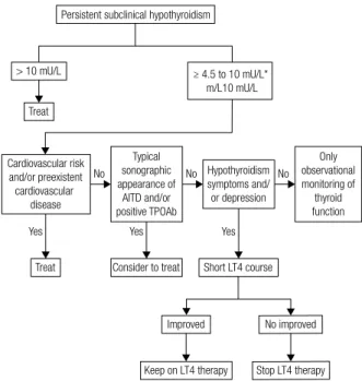
Despite the elevated prevalence of SH in the general population, treatment remains controversial in clinical practice since there are no adequately powered ran-domized clinical trials (RCT) to detect the beneit of levothyroxine replacement therapy. Thus, the choice between treating and not treating patients with SH will depend mainly on the best clinical judgment. Clinicians should bear in mind the potential risks to the individual’s health posed by not treating persis-tent SH. The likelihood of progression to overt hy-pothyroidism and the clinical signiicance of SH, in-cluding impact on quality of life, cognitive function, cardiovascular risk, and life expectancy have to be ca-refully considered.
What is the likelihood of progression to overt hypothyroidism?
worse-Cop
yright
© ABE&M t
odos os dir
eit
os r
eser
vados
.
ning of quality of life. After 9.2 years, 28% of patients developed overt hypothyroidism, with an annual pro-gression rate of SH to OH of 11.4% (32) (A). Ho-wever, before considering treatment, clinicians should verify if SH is persistent, since more than 50% of pa-tients with elevated TSH levels can exhibit normalized TSH in their second determination. The mean (SD) time between repeated TSH measurements was 18.93 ±13.11 months. The rate of progression from SH to OH during 5-year follow-up in patients in primary care was only 2.9% (122) (A).
Thyroid autoimmunity and TSH levels seem to in-luence the natural history of SH. In a 20 year follow-up of the Whickham survey, the annual progression rate of SH to overt hypothyroidism was 2.6% with elevated serum TSH levels (> 6 mU/L) and 4.3% with both elevated TSH and positive antithyroid antibodies (107) (B). In a 13 year longitudinal analysis by the Bussel-ton Health Study, female gender, and TSH cutoff of 2.5 mU/L, combined with positive thyroid antibo-dies were the strongest predictors for long-term risk of hypothyroidism. In women with positive thyroid anti-bodies (TPOAbs or TgAbs), the prevalence of hypo-thyroidism at follow-up was 12.0% (95% CI 3.0-21.0%) when baseline TSH was 2.5 mU/liter or less, 55.2% (95% CI 37.1-73.3%) for TSH between 2.5 and 4.0 mU/liter, and 85.7% (95% CI 74.1-97.3%) for TSH above 4.0 mU/liter. (123) (A) (Table 4).
In smaller prospective studies, the progression to overt hypothyroidism was also related to initial TSH levels and to thyroid autoimmunity. In a prospective study of SH patients who were followed over a mean period of 9.2 years, the annual incidence of overt hypo-thyroidism varied according to TSH stratiication, from 3.3% with TSH 6-12 mU/L to 11.4% with TSH > 12 mU/L. The cumulative incidence of overt hypothyroi-dism increased with higher TSH levels (0% with TSH 4.0-6.0 mU/L, and 55.3% with TSH > 6 mU/L), and with positive microsomal antibodies (58.5% vs. 23.2%; p = 0.03) (32) (A). Similarly, in another prospective study
with patients followed for a mean period of 31.7 mon-ths, TSH concentration was the only signiicant predic-tor for progression to overt hypothyroidism (124) (A). The incidence rate of overt hypothyroidism was 9.91% in the whole sample, and of 1.76%, 19.7% and 73.5% for patients with TSH levels of 5.0-9.9 mU/L, 10.0-14.9 mU/L, and 15.0-19.9 mU/L, respectively.
In Latin America, a study including women < 60 years of age with mild elevation of serum TSH (5-10 mU/L), showed that most patients did not progress to overt hypothyroidism in a 3-year follow-up period, however, the presence of TPOAb and hypoechographic ultrasound pattern increased the risk of progression to overt disease (121) (A). The role of thyroid US on the progression of SH was also investigated in a previous and larger study, in which none of the patients with autoimmune thyroidi-tis and normal thyroid echogenicity became hypothyroid over an 18-month follow-up period (120) (A).
In two studies of an elderly population performed in the United Kingdom (125) (A) and in the USA (126), (B), the persistence in SH (77% and 56%, respecti-vely) was associated with higher TSH and with positive thyroid antibodies. In the American study, TSH levels ≥ 10 mU/L were independently associated with progres-sion to overt hypothyroidism.
Recommendation 8
The progression to OH in patients with SH depends on the presence of thyroid antibodies and TSH baseline levels. Measurement of TPOAb and thyroid US in patients with SH is useful to help predict a higher risk of progression to overt hypothyroidism. Grade A.
ARE CLINICAL SYMPTOMS, DEPRESSION, AND
COGNITIVE DYSFUNCTION ASSOCIATED WITH
SUBCLINICAL HYPOTHYROIDISM?
The effects of overt hypothyroidism and its treatment on the quality of life and cognitive function are well Table 4. Sensitivity, speciicity, positive predictive value (PPV) of baseline serum TSH > 2.5 or > 4.0 mU/L for the presence of overt hypothyroidism (OH) and hypothyroidism (H) at follow-up (13 years)
TSH > 2.5 mU/L TSH > 4.0 mU/L
Sensitivity % Speciicity % PPV +LR (95% CI) Sensitivity % Speciicity % PPV +LR (95% CI)
OH 79 88 19 6.58 (3.84-11.30) 64 94 31 10.67 (4.84-23.49)
H 73 91 47 8.11 (4.30-15.30) 45 99 84 45.00 (6.32-320.17)