Rev Odontol UNESP. 2013 July-Aug; 42(4): 251-258 © 2013 - ISSN 1807-2577
ORIGINAL ARTICLE
In vitro evaluation of Biosilicate
dissolution on
dentin surface. A SEM analysis
Avaliação in vitro da dissolução do Biosilicato
sobre a superfície dentinária. Análise por meio da
microscopia eletrônica de varredura
Michele Carolina PINHEIRO
a, Beatriz Maria Valério LOPES
a, Rodrigo CAVASSIM
a,
Shelon Cristina Souza PINTO
a, José Eduardo Cezar SAMPAIO
aaFaculdade de Odontologia, UNESP – Univ Estadual Paulista, Araraquara, SP, Brasil
Resumo
Introdução: Biomateriais, tais como vidros bioativos e vidros cerâmicos têm sido propostos para o tratamento da hipersensibilidade dentinária. Objetivo: Avaliar por microscopia eletrônica de varredura (SEM), a dissolução de uma nova vitrocerâmica bioativa (Biosilicato, partículas de 1-20 µm) na superfície de amostras de dentina, com
diferentes métodos de aplicação e de meio de diluição diferente, usado para aplicação de Biosilicato.
Material e método: 280 amostras de dentina foram divididas aleatoriamente em quatro grupos: (1) Biosilicato mais flúor gel
aplicado com escova de Robinson, (2) Biosilicato mais flúor gel aplicado com microbrush, (3) Biosilicato mais
água destilada aplicado com escova deRobinson; (4) Biosilicato mais água destilada aplicado com microbrush.
Após o tratamento, as amostras foram imersas em saliva, em diferentes períodos (0, 30 e 15 minutos, 1, 2, 12 e 24 horas). Duas fotomicrografias foram obtidas a partir de cada amostra e foram analisadas por um examinador calibrado cego de acordo com um “Índice de Dissolução de Partículas”, criado para este estudo. Resultado: Os dados foram analisados usando o teste de Mann-Whitney, Kruskal-Wallis e Dunn. Não houve diferença estatística entre os graus de dissolução entre os quatro grupos em qualquer período. Conclusão: Biosilicato pode ser incorporado em
ambas às substâncias, sem diferenças no grau de dissolução das partículas em qualquer dos períodos de avaliação e a aplicação sobre da dentina pode ser realizada com os dois métodos avaliados.
Descritores: Materiais biocompatíveis; sensibilidade da dentina; flúor.
Abstract
Introduction: Biomaterials such as bioactive glasses and glass-ceramics have been proposed for the treatment of dentinal hypersensitivity. Objective: to evaluate by scanning electron microscopy (SEM), the dissolution of a novel bioactive glass-ceramic (Biosilicate 1-20 µm particles) on dentin surface samples, with different application
methods and different dilution medium used for applying Biosilicate.
Material and method: 280 dentin samples were randomly divided into four groups: (1) Biosilicate plus fluoride gel applied with Robinson brush;
(2) Biosilicate plus fluoride gel applied with microbrush; (3) Biosilicate plus distilled water applied with Robinson
brush; (4) Biosilicate plus distilled water applied with microbrush. After treatment, the samples were immersed
in saliva at different periods (0, 15 and 30 minutes, 1, 2, 12 and 24 hours). Two photomicrographs were obtained fromeach sample and were further analyzed by a blind calibrated examiner according to a “Particle Dissolution Index” created for this study. Result: The data were analyzed using the Mann-Whitney, Kruskal-Wallis and Dunn’s tests. There was no statistical difference among the degrees of dissolution between the 4 groups in any period.
Conclusion: Biosilicate can be incorporated in both substances without differences in the degree of dissolution
of the particles in any of the evaluated periods and the application of dentine can be performed with both methods evaluated.
INTRODUCTION
Cervical Dentin Hypersensitivity (DH) is a symptom frequently encountered in the population and its prevalence is estimated to range from 4 to 57%. his percentage tends to increase with a higher awareness of the population about the importance of maintaining healthy teeth in the oral cavity, allowing dental elements to be more susceptible to factors that could cause dentin exposures1.
DH is clinically deined as an acute, transient and well located pain which occurs in exposed dentin in response to tactile, thermal, evaporative, osmotic or chemical stimuli and which cannot be attributed to any other dental pathology. his pain does not occur spontaneously and does not persist ater the stimulus removal2. According to the hydrodynamic theory proposed
by Brännström, this pain occurs as a result of sensory nerve ibers activation in the pulp which is caused by the movement of luids within the dentinal tubules. hese sensory nerve ibers are activated by a hydrodynamic stimulus applied on the dentin exposed to the oral environment3.
Given the importance of dentin exposure and opened dentinal tubules, the relationship between conductivity of dentin and the hypersensitivity in the etiology of DH, it is certain that the treatment for such symptoms depends on the obliteration of dentinal tubules. hus, most researches on treatment for DH, focus on the capability of the available products to promote regression of the pain caused by exposure of these tubules by changing the diameter of the tubules, obliterating them or preventing the sensitization of the sensory nerve ibers4,5.
he proposed therapy includes periodontal surgery, restorative treatment, laser application and use of chemical substances for tubule obliteration. Numerous treatments are proposed but only those that eliminate the soreness and efectively prevent the recurrence of pain can be considered deinitive6.
Some studies search for a material that obliterates the dentinal tubules in an efective and lasting way. Some authors suggest the use of bioactive glass particles for the DH treatment7,8.
he elimination of DH is obtained with the dissolution of bioactive glass particles and subsequent precipitation of calcium and phosphate ions at the opening of the dentinal tubules in the form of amorphous calcium phosphate, which subsequently becomes a hydroxycarbonateapatite (HCA) layer8-10. he bioactive
glasses present in products for DH treatment are constituted of glassy particles with sharp edges. hese edges are produced from the grinding of glass material and may cause injury to the gingival margin during the application by friction11.
A novel particulate bioactive material free of glassy phase in an attempt has been developed for use in dentinal hypersensitivity. his material, commercially called Biosilicate was developed by
the Vitreous Materials Laboratory of the Federal University of São Carlos, Brazil and consists in a crystalline powder of bioactive quaternary P2O5-Na2O-CaO-SiO2 glass ceramic with the same advantages of the bioactive glasses12,13. Biosilicate consist in a
novel fully-crystallized glass-ceramic produced by modifying the structure and concentration of the components in the initial
bioglass through a thermal treatment, which results in formation of polycrystalline microstructure having crystals with controlled size and volume fraction11. he crystallization process results in
particles that can be safely added to any type of formulation to be used in the oral cavity, in addition, the particles can be more easily inserted into the dentinal tubules because there are no edges to delect away from the oriice13.
Previous studies evaluated through SEM the surface of dentin samples treated with Biosilicate mixed with water and
Biosilicate incorporated into a gel and observed obliteration of
dentinal tubules and the formation of HCA ater immersion in artiicial saliva11. Because it is a new product, was suggested by
laboratory responsible for creating the material, the evaluation of acidulated luoride as a vehicle for the incorporation of the biomaterial. he acidity of the luorine could accelerate the dissolution of Biosilicate and thereby enable a more rapid
formation of HCA.
he aim of this in vitro study was to evaluate the dissolution of Biosilicate particles in artiicial saliva using diferent application
methods (microbrush and Robinson brush) and diferent dilution mediums (luoride gel and distilled water).
MATERIAL AND METHOD
his study was approved by the Research Ethics Committee of Araraquara Dental School – UNESP – Brazil (protocol # 12/07). A total of 140 third molars were obtained from the Human Tooth Bank of the institution.
1.
Dentin sample preparation
Samples were prepared by making two parallel grooves on the most regular root surface of the teeth. Two grooves of approximately 0.5 mm in depth were made using a high speed cylindrical bur (4219, KG Sorensen, Barueri, SP, Brazil) under copious water irrigation. he irst groove was made horizontally at the cementoenamel junction and the second groove was made approximately 4 mm distant from the irst in the apical direction. he same bur was used to remove the surface layer of the root between the two grooves. Two samples were obtained from each tooth and all of them were stored in containers with saline solution (0.9% sodium chloride) at room temperature.
Two hundred and eighty dentin samples were obtained and randomly divided into four experimental groups (n = 70 for each group): 1) 0.6 g of Biosilicatepowder (Vitrovita, São Carlos,
Brazil), plus 1 mL of luoride gel (acidulated phosphate luoride 1.23%, DFL, Jacarepaguá, Brazil) plus distilled water applied with Robinson brush; 2) 0.6 g of Biosilicatepowder, plus 1 mL of
luoride gel plus distilled water (2 mL) applied with microbrush; 3) Biosilicate powder (0.6 g) plus distilled water (2 mL) applied
with Robinson brush; 4) Biosilicate powder plus distilled water
Prior to the application of Biosilicate, the samples were
etched with 35% phosphoric acid for 15 seconds and rinsed with 10 mL of distilled water, in order to remove the smear layer and open the dentinal tubules.
he Biosilicate was applied in the dentin samples for
15 seconds and remained on the samples for 5 minutes. Ater that, the samples were rinsed with 10 mL of artiicial saliva and were stored in plastic containers with 10 mL of artiicial saliva. he samples were placed in a chamber at constant temperature (37 °C) for 15 and 30 minutes, 1, 2, 12, and 24 hours.
2.
SEM Analysis
All samples were then mounted on metallic stubs (Senai, Sao Paulo, SP, Brazil) and dried overnight in a dehydration jar (Corning, Sao Paulo, SP, Brazil). Ater that, the samples were sputter-coated with a thin 25 nm layer of 99.99% pure gold.
Two photomicrographs were obtained from the center area of each sample with 1,500X and 3,500X magniications, using a scanning electron microscope operated at an accelerating voltage of 20 kV (Jeol T330 A, Jeol Ltd., Peabody, MA, USA). he photomicrographs were evaluated according to a Particle Dissolution Index (Figure 1). his index was created to assess, ater the scanning electron microscopy, if the particles were Biosilicate dissolved, partially-dissolved or non-dissolved
particles on the surface of the dentin samples.
hree evaluations at 15 day intervals were performed by a previously calibrated and experienced examiner. Good reproducibility was achieved with a weighted kappa score of 0.85.
3.
Statistical Analysis
he data were analyzed using statistical sotware (BioEstat Sotware, Belem/PA, Brazil). he level of signiicance was set at 5% for all tests. he data were explored for normality using the Shapiro-Wilk normality test. Once the data did not show normal distribution, the Mann-Whitney nonparametric test was used to determine the diference among the groups within each application method and within each dilution medium. Kruskal-Wallis and Dunn’s post hoc tests were used to determine
the diference among groups within each diferent saliva immersion period.
RESULT
Table 1 shows score comparison between the application methods for Biosilicate dilution in distilled water at diferent
saliva immersion periods. he results presented no statistically signiicant diferences between the application methods when the distilled water was used for dilution. Ater 2 hours in saliva immersion, there was a tendency of the microbrush application to be less eicient than Robinson Brush application.
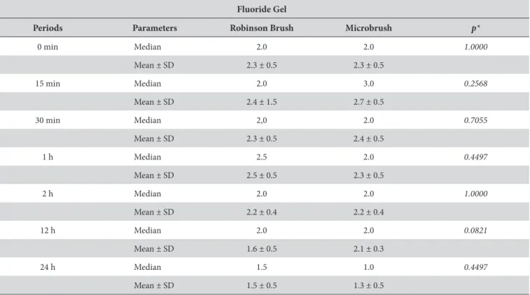
Table 2 shows comparison between the application methods for Biosilicate dilution in luoride gel at diferent
saliva immersion periods. he results presented no statistically signiicant diferences between the application methods when luoride gel was used for dilution at the evaluated periods.
Table 3 shows score comparison between the dilution mediums in relation to the use of Robinson brush at diferent saliva immersion periods. he results presented no statistically signiicant diferences between the application methods when the luoride gel was used for dilution at the evaluated periods. No statistically signiicant diferences were observed between the dilution mediums of the product when Robinson brush was used for application.
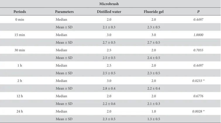
Table 4 shows the comparison between dilution mediums when microbrush is used for application at each saliva immersion period. Ater two hours of saliva immersion, there was a tendency of microbrush application to present lower particle dissolution in distilled water in comparison to luoride gel dissolution (p < 0.0233). Ater 24 hours of saliva immersion, the results kept showing lower particle dissolution in distilled water in comparison to luoride gel dissolution (p < 0,0028).
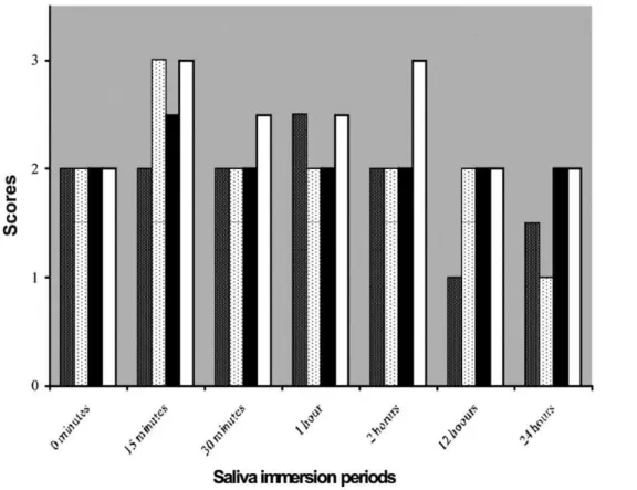
Figure 2 compares the saliva immersion periods according to the median of particle dissolution evaluation scores. here were no statistical signiicant diferences among the groups. here were statistically signiicant diferences between 15 minutes and 24 hours for the Microbrush plus luoride gel group and between the 2 and 24 hour subgroups for the Robinson brush plus luoride gel group.
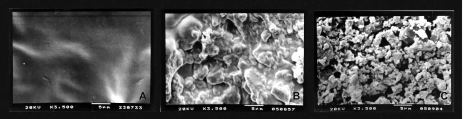
Figure 1. Evaluation criteria used in the Particle Dissolution Index. A) Score 1. Complete Biosilicate
particle dissolution with a homogeneous layer covering the opening of the dentinal tubules. B) Score 2. Partially particle dissolution with a non homogeneous layer and presenting traces of non-dissolved Biosilicate
particles. C) Score 3. No Biosilicate
Table 1. Score comparison between application methods for the dilution in distilled water at diferent saliva immersion periods (n = 10)
Distilled water
Periods Parameters Robinson Brush Microbrush p*
0 min Median 2.0 2.0 0,7055
Mean ± SD 2.0 ± 0.0 2.1 ± 0.3
15 min Median 2.5 3.0 0.4497
Mean ± SD 2.5 ± 0.5 2.7 ± 0.5
30 min Median 2.0 2.5 0.1306
Mean ± SD 2.1 ± 0.3 2.5 ± 0.5
1 h Median 2.0 2.5 0.1306
Mean ± SD 2.1 ± 0.3 2.5 ± 0.5
2 h Median 2.0 3.0 0.0588
Mean ± SD 2.3 ± 0.5 2.8 ± 0.4
12 h Median 2.0 2.0 0.1212
Mean ± SD 1.7 ± 0.5 2.2 ± 0.6
24 h Median 2.0 2.0 0.1620
Mean ± SD 1.9 ± 0.3 2.3 ± 0.5
*Mann-Whitney test; p < 0.05
Table 2. Score comparison between application methods for dilution in luoride gel at diferent saliva immersion periods (n = 10)
Fluoride Gel
Periods Parameters Robinson Brush Microbrush p*
0 min Median 2.0 2.0 1.0000
Mean ± SD 2.3 ± 0.5 2.3 ± 0.5
15 min Median 2.0 3.0 0.2568
Mean ± SD 2.4 ± 1.5 2.7 ± 0.5
30 min Median 2,0 2.0 0.7055
Mean ± SD 2.3 ± 0.5 2.4 ± 0.5
1 h Median 2.5 2.0 0.4497
Mean ± SD 2.5 ± 0.5 2.3 ± 0.5
2 h Median 2.0 2.0 1.0000
Mean ± SD 2.2 ± 0.4 2.2 ± 0.4
12 h Median 2.0 2.0 0.0821
Mean ± SD 1.6 ± 0.5 2.1 ± 0.3
24 h Median 1.5 1.0 0.4497
Mean ± SD 1.5 ± 0.5 1.3 ± 0.5
*Mann-Whitney test; p < 0.05.
DISCUSSION
Biosilicate is a novel bioactive vitroceramic developed in the
Laboratory of Vitreous Materials of São Carlos Federal University. Some studies have shown the efectiveness of this new material to form HCA when it is in contact with body luids12. hus, this
material can be used as bone substitute in areas such as dentistry and medicine12. his property permits the use of Biosilicate in
the DH treatment, once HCA has a structure similar to dentin surface. Hydroxycarbonateapatite binds to dentin, bringing a permanent solution to avoid pain12,14,15. However, the best form to
his study evaluated two mediums for adding Biosilicate
(luoride gel and distilled water), two application methods of Biosilicate on dentin surface (Robinson brush and microbrush),
and the necessary period to complete Biosilicate dissolution
on dentin surface. Non-dissolved particles of Biosilicate cover
the dentin surface; however, this layer may possibly be removed easily with toothbrushing, diet, or even with patient salivation.
he method used in this study allowed a morphological evaluation of the Biosilicate layer formed on dentin. In order
to evaluate the photomicrographs, an index was created. he
Table 4. Score comparison between dilution mediums when microbrush was used for application at diferent saliva immersion periods (n = 10)
Microbrush
Periods Parameters Distilled water Fluoride gel P
0 min Median 2.0 2.0 0.4497
Mean ± SD 2.1 ± 0.3 2.3 ± 0.5
15 min Median 3.0 3.0 1.0000
Mean ± SD 2.7 ± 0.5 2.7 ± 0.5
30 min Median 2.5 2.0 0.7055
Mean ± SD 2.5 ± 0.5 2.4 ± 0.5
1 h Median 2.5 2.0 0.4497
Mean ± SD 2.5 ± 0.5 2.3 ± 0.5
2 h Median 3.0 2.0 0.0233 *
Mean ± SD 2.8 ± 0.4 2.2 ± 0.4
12 h Median 2.0 2.0 0.6776
Mean ± SD 2.2 ± 0.6 2.1 ± 0.3
24 h Median 2.0 1.0 0.0028 *
Mean ± SD 2.3 ± 0.5 1.3 ± 0.5
*Mann-Whitney test; p < 0.05.
Table 3. Score comparison between dilution mediums when Robinson brush was used for application at diferent saliva immersion periods (n = 10)
Robinson Brush
Periods Parameters Distilled water Fluoride gel p*
0 min Median 2.0 2.0 0.2568
Mean ± SD 2.0 ± 0.0 2.3 ± 0.5
15 min Median 2.5 2.0 0.7055
Mean ± SD 2.5 ± 0.5 2.4 ± 1.5
30 min Median 2.0 2.0 0.4497
Mean ± SD 2.1 ± 0.3 2.3 ± 0.5
1 h Median 2.0 2.5 0.1306
Mean ± SD 2.1 ± 0.3 2.5 ± 0.5
2 h Median 2.0 2.0 0.7055
Mean ± SD 2.3 ± 0.5 2.2 ± 0.4
12 h Median 2.0 2.0 0.7055
Mean ± SD 1.7 ± 0.5 1.6 ± 0.5
24 h Median 2.0 1.5 0.1306
Mean ± SD 1.9 ± 0.3 1.5 ± 0.5
Particle Dissolution Index is able to verify the dissolution degree of Biosilicate particles. It can evaluate the samples with
non-dissolved Biosilicate particles, samples with partially-dissolved
Biosilicate particles, and samples with completely-dissolved
Biosilicateparticles.
According to the manufacturer (Vitrovita, São Carlos, Brazil), Biosilicate reaction starts immediately ater contact with luids,
so it’s important that the product reviews are mixed at the time of application.
Distilled water and luoride gel showed similar results in particle dissolution on dentin surface (Tables 1 and 2), and this mixture can be applied either with Robinson brush (Table 3) or with microbrush (Table 4). he results suggested that both substances can be used in order to dilute Biosilicate and that
both application methods can be performed.
Although the results for both substances are similar, the mixture of Biosilicate with luoride gel is more consistent and
it facilitates the application on dentin surface. his fact may be the reason why the best results were achieved with Biosilicate
plus luoride gel (Figure 2), and Biosilicate plus distilled water
showed no statistically signiicant diferences. Perhaps, the interaction between Biosilicate and luoride gel may have
facilitated the dissolution of Biosilicate due to luoride gel
acidity. Nevertheless, this elucidation was not the objective of the present study.
In clinical practice, both microbrush and Robinson brush can be used to apply the substances on dentin surface. Trying to evaluate if there is any diference between these two application
methods, the present study did not ind a statistically signiicant diference between them (Tables 1 and 2). hese results suggest using the microbrush in clinical practice instead of Robinson brush because the friction of the Biosilicate hard particle with
a Robinson brush on dentin surface can cause abrasion on the gingival margin and patient discomfort.
Pashley et al.16 (1986) showed that even the act of rubbing an
instrument on dentin may promote the obliteration of dentinal tubules through the formation of smear layer. his fact can explain why both Robinson brush and microbrush were able to form smear layer and obliterate the dentinal tubules. he formation of smear layer produced by rubbing Robinson brush or the microbrush associated with the abrasive power of the Biosilicate was probably responsible for a 2 median score in the
subgroups that were not immersed in saliva (Figure 2). Initially, it was expected to observe no dissolved particles (score 3) in these subgroups, as the surface with smear layer is visually similar to the surface with dissolved particles or partially dissolved particles in photomicrographs. herefore, possibly the layer formed on the dentin surface in the absence of saliva is diferent from the layer formed on the dentin surface ater 12 and 24 hours of immersing samples in saliva12.
Although smear layer can obliterate the dentinal tubules as a barrier that prevents the dentinal luid displacement, it can be easily removed by an acid diet, toothbrushing or the saliva soluble efect17-19. Probably, in the 15 minute subgroups (Figure 2), the
smear layer may have been removed by the saliva soluble efect. In the other subgroups, the smear layer may have been replaced by
hydroxycarbonateapatite, forming a more resistant layer to acids. In most groups, the worst scores were found at the 15 minute period. Another possibility for the presence of complete and partially-dissolved particles in samples not immersed in saliva is that the dissolution of Biosilicate particles may have started in
contact with the luoride and distilled water (time 0), similar to what occurs with other bioglasses when in contact with any luid containing water20,21 .
Considering the particle dissolution, there were no statistically signiicant diferences among the groups during the analyzed periods (Figure 2), because the HCA layer is formed according to a process that occurs ater 12 hours11. his fact may explain the
diferences between 1 and 24 hour subgroups into the Biosilicate
and luoride gel group applied with Robinson brush, and between 15 minutes and 24 hour subgroups into Biosilicate plus luoride
gel group applied with microbrush (Figure 2).
According to the evaluation of the obtained data, it was possible to observe that all application methods were eicient in dentinal tubules obliteration by particle dissolution (Tables 3 and 4). Considering the diferent dilution mediums, luoride gel or distilled water, there were no statistically signiicant diferences
in relation to obliteration of dentinal tubules (Tables 1 and 2) and among the groups at each saliva immersion period. However, the best results were observed when the Biosilicate was diluted
with luoride gel and applied with microbrush ater 24 hours of saliva immersion (Figure 2). Nevertheless, studies about dentin permeability and acid challenges must be performed in order to evaluate the permanence of the layer formed on dentin surface. Furthermore, clinical researches with Biosilicate must be
performed in order to test clinical eicacy.
CONCLUSION
According to the results obtained and within the limitations of the methodology, it can be concluded that there were no diferences between the evaluated application methods. In addition, no statistical diferences were observed according to the evaluated dissolution periods.
Both dilution mediums were able to initiate the dissolution of the glass-ceramic particles. Notwithstanding the lack of statistical signiicance between the dilution mediums, the Biosilicate plus
luoride gel ater 24 hours tended to produce samples with a more homogeneous layer covering the opening of the dentinal tubules.
REFERENCES
1. Orchardson R, Gillam DG. Managing dentin hypersensitivity. J Am Dent Assoc. 2006; 137: 990-8. PMid:16803826.
2. Canadian Advisory Board on Dentin Hypersensitivity. Consensus-based recommendations for diagnosis and management of dentin hypersensitivity. J Can Dent Assoc. 2003; 69: 221-6. PMid:12662460.
3. Brännström M, Aström A. he hydrodynamics of the dentine; its possible relationship to dentinal pain. Int Dent J. 1972; 22: 219 –27. PMid:4505631.
4. Bánóczy J. Dentine hypersensitivity – general practice considerations for successful management. Int Dent J. 2002; 95: 223-8.
5. Bartold PM. Dentinal hypersensitivity: a review. Aust Dent J. 2006; 51: 212-8. PMid:17037886. http://dx.doi.org/10.1111/j.1834-7819.2006. tb00431.x
6. Gillam GD, Tang JY, Mordan NJ, Newman HN. he efects of a novel Bioglass
dentifrice on dentine sensitivity: a scanning electron microscopy investigation. J Oral Rehabil. 2002; 29: 305-13. PMid:11966962. http://dx.doi.org/10.1046/j.1365-2842.2002.00824.x 7. Clinical trial designs for testing of products for dentin hypersensitivity: a review. J West Soc Periodontol. 1997; 45: 37- 46.
8. Lee BS, Chang CW, Chen WP, Lan WHL, Lin CP. In vitro study of dentin hypersensitivity treated by Nd: YAP laser and Bioglass. Dent Mater. 2005; 21: 511-9. PMid:15904693. http://dx.doi.org/10.1016/j.dental.2004.08.002
9. Gillam DG, Bulmam JS, Eijkman MAJ, Newman HN. Dentist’ perceptions of dentine hypersensitivity and knowledge of its treatment. J Oral Rehabil. 2002; 29: 219-25. PMid:11896837. http://dx.doi.org/10.1046/j.1365-2842.2002.00812.x
10. Tirapelli C, Panzeri H, Soares RG, Peitl O, Zanotto ED. A novel bioactive glass-ceramic for treating dentin hypersensitivity. Braz Oral Res.2010; 26: 381-7. http://dx.doi.org/10.1590/S1806-83242010000400002
11. Peitl O, Zanotto ED, Serbena FC, Hench LL. Compositional and microstructural design of highly bioactive P2O5–Na2O–CaO–SiO2 glass-ceramics. Acta Biomater. 2012, 8, 321-32 PMid:22032913. http://dx.doi.org/10.1016/j.actbio.2011.10.014
12. Moura J, Teixeira LN, Ravagnani C, Peitl O, Zanotto ED, Beloti MM, et al. Osteogenesis on a highly bioactive glass- ceramic ( Biosilicate). J Biomed Mater Res. 2007; 82: 545-57. PMid:17311315. http://dx.doi.org/10.1002/jbm.a.31165
13. Siqueira RL, Zanotto ED. Biosilicate
: historical of a highly bioactive brazilian glass-ceramic. Quím Nova. 2011; 34; (suppl 7): 1231-41 http://dx.doi.org/10.1590/S0100-40422011000700023
14. Massuda ED, Maldonato LL, de Lima-junior JT, Peitl O, Hyppolito MA, de Oliveira MAA. Biosilicate
Ototoxicity and Vestibulotoxicity evaluation in guinea-pigs. Braz J Otorhinolaryngol. 2009; 75: 665-8. PMid:19893933. http://dx.doi.org/10.1590/S1808-86942009000500009 15. Roriz VM, Rosa AL, Peitl O, Zanoto ED, Panzeri H, de Oliveira PT. Eicacy of a bioactive glass–ceramic (Biosilicates) in the maintenance
16. Pashley DH. Dentin permeability, dentin sensitivity, and treatment through tubule oclusion. J Endod. 1986; 12: 465-74. http://dx.doi. org/10.1016/S0099-2399(86)80201-1
17. Fogel HM, Pashley DH. Efect of periodontal root planning on dentin permeability. J Clin Periodontol. 1993; 20: 673-7. http://dx.doi. org/10.1111/j.1600-051X.1993.tb00714.x
18. Pashley DH, O’Meara JA, Kepler EE, Galloway SE, hompson SM, Stewart FP. Dentin permeability. Efects of desensitizing dentifrices in vitro. J Periodontol. 1984; 55: 522-5. PMid:6592326. http://dx.doi.org/10.1902/jop.1984.55.9.522
19. Zandim DL, Correia FO, Rossa Junior C, Sampaio JEC. In vitro evaluation of the efect of natural orange juices on dentin morphology. Braz Oral Res. 2008; 22: 176-183. PMid:18622489. http://dx.doi.org/10.1590/S1806-83242008000200014
20. Hench LL. he story of Bioglass
. J Mater Sci Mater Med. 2006; 17: 967-8. PMid:17122907. http://dx.doi.org/10.1007/s10856-006-0432-z 21. Hench LL, West, JK. Biological applications of bioactive glass. Life Chemistry Reports. 1996; 13: 187-241.
CONFLICTS OF INTERESTS
he authors declare no conlicts of interest.
CORRESPONDING AUTHOR
José Eduardo Cezar Sampaio
Departamento de Diagnóstico e Cirurgia, Faculdade de Odontologia, UNESP – Univ Estadual Paulista, Rua Humaitá, 1680, 14801-903 Araraquara - SP, Brasil
e-mail: [email protected]