Rev Odontol UNESP. 2013 July-Aug; 42(4): 273-282 © 2013 - ISSN 1807-2577
ORIGINAL ARTICLE
Inorganic elemental analysis and identiication of
residual monomers released from diferent glass
ionomer cements in cell culture medium
Análise elementar inorgânica e identiicação de monômeros residuais orgânicos
liberados por diferentes cimentos de ionômero de vidro
Marcia Hiromi TANAKA
a, Alberto Camilo ALÉCIO
b, Danilo Luiz FLUMIGNAN
b,
José Eduardo de OLIVEIRA
b, Elisa Maria Aparecida GIRO
aaFaculdade de Odontologia, UNESP – Univ Estadual Paulista, Araraquara, SP, Brasil bInstituto de Química, UNESP – Univ Estadual Paulista, Araraquara, SP, Brasil
Resumo
Introdução: Os cimentos de ionômero de vidro (CIVs) liberam elementos inorgânicos e monômeros orgânicos residuais que têm o potencial de causar efeitos deletérios sobre as células pulpares. Objetivo: Identiicar e quantiicar os elementos inorgânicos presentes em diferentes CIVs, bem como os componentes liberados por estes materiais em meio de cultura celular. Material e método: Espécimes cilindricos de dois CIVs modiicados por resina para base/forramento (Vitrebond e Fuji Lining LC), dois CIVs modiicados por resina restauradores (Vitremer e Fuji II LC) e dois CIVs convencionais restauradores (Ketac Fil Plus e Ketac Molar Easymix) foram preparados e analisados por Espectrometria de Fluorescência de Raios X por Energia Dispersiva (EDXRF). Em seguida, extratos de 24h desses materiais foram obtidos e analisados por EDXRF e por Cromatograia Gasosa/Espectrometria de Massa (CG/EM).
Resultado: Os elementos inorgânicos identiicados em maior porcentagem nos CIVs Vitrebond, Fuji Lining LC, Vitremer, Fuji II LC e Ketac Fil Plus foram estrôncio, silício e alumínio, enquanto o zinco foi detectado apenas no Vitrebond. O Ketac Molar Easymix apresentou maior porcentagem dos elementos lantânio, cálcio, alumínio e silício. Estrôncio foi detectado nos extratos de todos os materiais, exceto no Ketac Molar Easymix; cálcio estava presente no extrato do Ketac Fil Plus; zinco apenas no Vitrebond; e silício no extrato do Fuji II LC . O HEMA foi identiicado nos extratos de todos os CIVs modiicados por resina, e o iodobenzeno, somente no Vitrebond. Conclusão: Entre os CIVs estudados, o Vitrebond é o que libera mais componentes com potencial citotóxico.
Descritores: Cimentos de ionômeros de vidro; compostos inorgânicos.
Abstract
Introduction: Glass ionomer cements (GICs) release inorganic elements and organic residual monomers with the potential for deleterious efects on pulp cells. Objective: To identify and quantify inorganic elements present in diferent GICs and released components from these materials in cell culture medium. Material and method: Samples of two resin-modiied GICs for base/liner (Vitrebond and Fuji Lining LC), two resin-modiied restorative GICs (Vitremer and Fuji II LC) and two conventional restorative GICs (Ketac Fil Plus and Ketac Molar Easymix) were prepared and analyzed by Energy-Dispersive X-Ray Fluorescence Spectrometry (EDXRF). Extracts of these materials were obtained by immersion of each sample in separate containers of DMEM for 24 h (total surface-liquid ratio = 45.7 mm2/mL). he extracts were analyzed by EDXRF and Gas Chromatography-Mass Spectrometry (GC-MS). Result: Higher percentages of strontium, silicon and aluminum were identiied in Vitrebond, Vitremer, Fuji Lining LC, Fuji II LC, and Ketac Fil Plus, while zinc was detected only in Vitrebond. Ketac Molar Easymix presented a greater atomic composition of lanthanum, calcium, aluminum and silicon. Strontium was detected in the extracts from all materials except Ketac Molar Easymix; calcium was present in extracts from Ketac Fil Plus; zinc only in Vitrebond; and silicon in Fuji II LC extract. he analysis by GC-MS detected 2-hydroxyethyl-methacrylate (HEMA) in the extracts from all resin-modiied GICs, and iodine benzene was detected only in the Vitrebond extract.
Conclusion: Of the GICs sampled, Vitrebond released the highest number of components with cytotoxic potential.
INTRODUCTION
Due to their anticariogenic properties, glass ionomer cements (GICs) play an important role in preventing secondary caries, which is the most frequent cause for replacement of restorations1,2. Based on their chemical compositions, these materials can be classiied as (1) chemically cured GICs and (2) resin-modiied GICs. Chemically cured GICs consist of a powder made up of glass particles (calcium aluminum luoride silicate glass) and a liquid made up of polyacrylic acids3, and their setting reaction depends on the acid reaction with the glass particle surface ater mixing (acid-base reaction). Resin-modiied GICs incorporate organic resin monomers like 2-hydroxyethyl methacrylate (HEMA) and photosensitive polymerization initiators in addition to glass particles and polyacrylic acids4. In these materials, light activation causes an initial set that is followed by an acid-base setting reaction.
he amounts of luoride released by GICs indicate their anticariogenic potential5-7. However, several other inorganic elements or ions are released from GICs, and the type and amount of these depend both on the chemical composition of the glass used to manufacture the cement powder and the local pH conditions, with greater release occurring under acidic conditions8-10.
In resin-modiied GICs, organic residual monomers are also released, along with products of degradation of the photoinitiators. Together with the release of ions and/or inorganic elements, these constitute important factors that can inluence the cytotoxicity of these materials11-16.
Since the ions, residual monomers, and other components released from GICs have the potential for deleterious efects on pulp cells, it is important to evaluate the release of these components in cell culture medium with a pH near neutral before assessing their in vitro and in vivo cytotoxic efects. hus, this study aimed to identify the inorganic elements present in diferent GICs as well as to evaluate inorganic elements and organic residual monomers present in the extracts from these materials. he null hypothesis advanced was that the lixiviated components from the GIC are not dependent on the type of GIC.
MATERIAL AND METHOD
he commercially available glass ionomer cements used in this study as well as the main composition of the products, powder/liquid ratio by weight, and curing times are listed in Table 1. he experiments were carried out at 24 ± 1 °C room temperature. Moreover, each measurement was repeated three times to ensure reliable results.
1.
Identiication and Analysis of Inorganic Elements
Present in Glass Ionomer Cements
he study materials were handled at room temperature (24 ± 1 °C), according to the respective manufacturers’ instructions, and placed with the aid of a Centrix syringe (DFL Indústria e Comércio SA, Rio de Janeiro, Brazil) in a stainless steel matrix measuring 10 mm in diameter and 1 mm
thick. A plastic matrix strip and glass slide were placed on the surface of the material and pressed with a weight of 500 gf to promote the overlow of the material. Resin-modiied GICs were light-activated using an halogen light unit (Optilux 500, Kerr Company, Orange, USA) positioned about one millimeter from the surface of the specimen. he light intensity was monitored with a radiometer (average 450 ± 10 mW/cm2). Conventional GICs remained in the matrix for 10 minutes in the presence of ambient light to ensure the initial setting.
he specimens were removed from the matrix and placed in an incubator with 100% humidity at 37 °C for 60 minutes. Ater this period, the specimens were analyzed by Energy-Dispersive X-Ray Fluorescence Spectrometry (EDX800, Shimadzu, Tokyo, Japan) for simultaneous quantitative determination of atomic inorganic multi-elements (at. %) ranging from Sodium (Na) to Uranium (U). Before the analysis, the spectrometer was calibrated to a standard aluminum sample. he X-ray generator with a rhodium (Rh) tube was operated at a tube voltage of 50 kV, tube current of 20 mA, and collimator size of 10 mm diameter without primary ilters and with an air cooling method. he sample chamber was conditioned to a vacuum atmosphere and the detector was cooled to –174 °C by the LN2 method (liquid nitrogen).hree specimens of each material were analyzed under these conditions.
Next, the specimens were immersed in DMEM cell culture medium (Sigma Chemical CO., St. Louis, MO, USA) without fetal calf serum (total surface-liquid ratio = 45.7 mm2/mL) for 24 hours in separate containers. Finally, they were rinsed with distilled water and analyzed by EDXRF again.
2.
Identiication and Analysis of Inorganic Elements
Present in Extracts from the Glass Ionomer Cements
hree specimens of each material were placed in individual compartments of sterile acrylic plates with 12 wells (Costar Corp., Cambridge, MA, USA) containing 4.1 mL of DMEM culture medium (Sigma Chemical Co., St. Louis , MO, USA) without fetal calf serum (total surface-liquid ratio = 45.7 mm2/mL). he specimens were incubated for 24 hours at 100% humidity and 37 °C to obtain the extracts from the materials. he extracts were then analyzed by Energy-Dispersive X-Ray Fluorescence Spectrometry (EDX800, Shimadzu, Tokyo, Japan) for determination of atomic inorganic multi-elements (at. %). Before the analyses, the spectrometer was calibrated to a standard aluminum sample. he X-ray generator with a rhodium (Rh) tube was operated at a tube voltage of 50 kV, tube current of 20 mA, and a collimator size of 10 mm diameter without primary ilters and with an air cooling method. he sample chamber was conditioned to air atmosphere, and the detector was cooled to –174 °C by the LN2 method (liquid nitrogen).he DMEM pure culture medium was used as a negative control of the experiment.
3.
Identiication of 2-Hydroxyethyl-methacrylate (HEMA)
and Iodine Benzene in the Extracts from Glass Ionomer
Cements
serum for 24 hours in an incubator with 100% humidity at 37 °C to obtain the extracts (total surface-liquid ratio = 45.7 mm2/mL). hese extracts were analyzed by Gas Chromatography-Mass Spectrometry (GC-MS) to identify the organic residual monomers released from GICs into the DMEM culture medium.
Analyses were conducted with a Shimadzu GC-MS automated gas chromatograph-mass spectrometer model GC-17A/QP-5050A coupled to a model AOC20i auto-sampler. It was manipulated with GCMS Solutions v.1.02 workstation sotware (Shimadzu, Kyoto, Japan). All analyses were obtained
using EI/MS, positive ion mode, full-scan acquisition mode (40 to 500 m/z), and “hard” energy ionization (70 eV).
We performed chromatographic separation on a fused-silica capillary nonpolar column SPB™–1 (30 m × 0.25 mm i.d. × 0.10 µm ilm thickness; Supelco Inc., Bellefonte, PA, USA) with dimethylpolysiloxane as the stationary phaseand helium as carrier gas at a constant low (1 mL min–1). Sample aliquots (1 µL) were injected in split mode (1:20) with solvent cut time (3 min). he injector and interface detector temperatures were maintained at 260 °C and 280 °C, respectively. he oven temperature was
Table 1. Commercially available glass ionomer cements used in the study
GICs (manufacturer) Composition (% weight) Classiication : Indication17 Powder/ Liquid ratio
MRT or initial curing time
Vitrebond
(3M, ESPE Dental Prod-ucts, St. Paul, MN, EUA)
Powder
glass powder (>95%) diphenyliodonium chloride (<2%)
Resin-modiied
GIC : lining/base 1.4/1 30 s
Liquid
copolymer of acrylic and itaconic acids (35-45%) 2-hydroxyethyl methacrylate - HEMA (20-30%)
water (30-40%)
Vitremer
(3M, ESPE Dental Prod-ucts, St. Paul, MN, EUA)
Powder
silane treated glass (90-100%)
potassium persulfate < 1 %
Resin-modiied
GIC : restoration 2.5/1 40 s
Liquid
copolymer of acrylic and itaconic acids (45-50%) 2-hydroxyethyl methacrylate - HEMA (15-20%)
water (25-30%)
Fuji Lining LC
(GC, Tokyo, Japão)
Powder
alumino-silicate glass (100%)
Resin-modiied
GIC : lining/base 1.4/1 30 s
Liquid
polyacrylic acid (65-70%)
2-hydroxyethyl methacrylate, HEMA (8-10%) Proprietary ingredient (5-15%)
Fuji II LC
(GC,Tokyo, Japão)
Powder
alumino-silicate glass –100%
Resin-modiied
GIC : restoration 3.0/1 40 s
Liquid
polyacrylic acid (20-22%)
2-hydroxyethyl methacrylate, HEMA (35-40%) proprietary ingredient (5-15%) 2,2,4 trimethyl hexamethylene dicarbonate (5-7%)
triethylene glycol dimethacrylate (4-6%)
Ketac Fil Plus
(3M, ESPE Dental Prod-ucts, St. Paul, MN, EUA)
Powder
glass powder (≈100%)
Convencional
GIC : restoration 3.2/1 7 min
Liquid
water (60-65%)
polyethylene polycarbonic acid (30-40%) tartaric acid (5-10%)
Ketac Molar Easymix
(3M, ESPE Dental Prod-ucts, St. Paul, MN, EUA)
Powder
glass powder (85-95%) polyacrylic acid - 5-15%
Convencional
GIC : restoration 2.9/1 5 min
Liquid
water (55-65%)
polyethylene polycarbonic acid (25-35%) tartaric acid (5-10%)
initially kept at 60 °C for 2 min then increased to 180 °C at 2 °C min–1. Finally, the temperature was raised to 240 °C at 10 °C min–1, and kept constant for 10 min, completing 78 min of total analysis.
Residual organic monomers 2-hydroxyethyl methacrylate (HEMA) and iodine benzene (IB) were identiied by comparing retention times and mass spectra of chromatograms of the extracts from GICs with chromatograms of authentic standard HEMA (Sigma Chemical Co., St. Louis, USA) and iodine benzene (Sigma Chemical Co., St. Louis, USA) injected under the same conditions.
RESULT
1.
Identiication and Analysis of Inorganic Elements
Present in Glass Ionomer Cements
he quantitative atomic inorganic multi-elemental composition (at.%) of the glass ionomer cements Vitrebond (VB), Vitremer (VT), Fuji Lining LC (FL), Fuji II LC (FII), Ketac Fil Plus (KP), and Ketac Molar Easymix (KM) determined by EDXRF are listed in Table 2.
Figure 1 shows EDXRF wide scans up to 40 keV binding energy for the glass ionomer cements. All glass ionomer cement samples exhibit signiicant signals of their main components with
varying intensities quantiied in Table 2. Moreover, as can be seen in Figure 1, all samples have very similar spectral proiles, except for Ketac Molar Easymix (KM).
he quantitative atomic inorganic multi-elementals (at.%) identiied at higher levels in the VB, VT, FL, FII and KP glass ionomer cements were strontium (Sr), silicon (Si) and aluminum (Al), while zinc (Zn) was only detected in VB. On the other hand, KM presented lanthanum (La), calcium (Ca), aluminum (Al), and silicon (Si) as its main atomic inorganic elements, and strontium (Sr) was not detected in this material. Moreover, ater 24 hours in contact with DMEM culture medium, the inorganic multi-elemental composition was unchanged in all glass ionomer cements. Only a small decrease or increase of main elements could be seen.
2.
Identiication and Analysis of Inorganic Elements
Present in DMEM Extracts from Glass Ionomer Cements
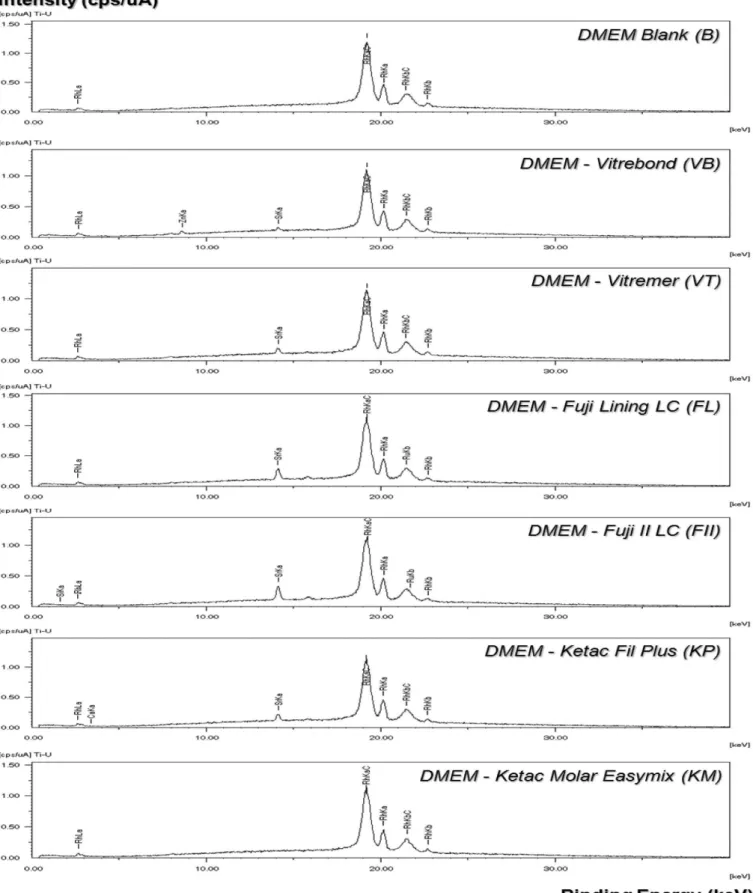
he quantitative atomic inorganic multi-elemental compositions (at.%) of all GICs extracts detected by EDXRF are listed in Table 3. Pure DMEM was used as an internal negative control. Figure 2 shows EDXRF wide scans up to 40 keV binding energy for extracts from all glass ionomer cements. As can be seen in Figure 2, all samples have very similar spectral proiles except for Ketac Molar Easymix (KM). he most intensive
Table 2. Main atomic inorganic multi-elemental composition (at.%) in glass ionomer cements identiied by Energy Dispersive X-Ray Fluorescence Spectrometry (EDXRF) at initial time and ater 24 hours of immersion in DMEM
Group Period Main Atomic Inorganic Elements in GIC
Zn Sr La Si Al P Ca Na Fe
VB
Initial 32.9(0.4) 32.8(0.2) - 20.8(0.4) 10.8(0.1) 1,0(0.7) 1,0(1.0) - 1,9(1.1)
24 hours 33.2(0.1) 33.8(0.5) - 19.9(0.3) 9.4(0.1) 1.6(0.2) 1.4(0.1) - 0.2(0.1)
VT
Initial - 52.8(1.6) - 27.5(0.2) 13.8(0.2) 2.4(0.1) - 1.6(0.08) 0.2(0.1)
24 hours 0.2(0,1) 54.8(2.3) - 25.7(0.2) 11.9(0.4) 2.3(1,7) 2.4(0.3) 3.0(0.1) 0.6(0.5)
FL
Initial 0.1(0.1) 59.5(0.5) - 23.2(0.6) 12.6(0.3) 2.6(0.04) 0.2(0.1) - 0.9(0.9)
24 hours - 57.4(1.8) - 25.5(0.4) 10.3(0.2) 3.6(0.4) 1.5(0.1) 2.1(1,2) 0.8(1.0)
FII
Initial - 56.8(1.7) - 29.4(1.1) 12.3(0.4) 0.9(0.3) 0.2(0.3) - 0.2(0.01)
24 hours - 56.8(0.8) - 28.8(0.3) 10.1(0.3) 2.4(0.1) 1.7(0.1) -
-KP
Initial - 46.9(0.2) 7.0(0.1) 24.2(0.3) 14.5(0.3) 3.2(0.09) 0.2(0.03) 2.6(0.4)
-24 hours - 47.6(1.9) 7.1(0.3) 23.0(0.5) 12.5(0.6) 3.0(0.1) 2.1(0.5) 5.0(1.7)
-KM Initial
-- 30.8(2.7) 17.2(0.8) 20.0(0.7) 2.4(0.2) 26.2(1.5) 3.4(2.0)
-24 hours - - 29.5(0.2) 14.7(0.1) 16.6(0.6) 2.9(0.2) 28.5(1.0) 7.4(1.5)
Table 3. Main atomic inorganic multi-elemental composition (at.%) identiied in 24-hour extracts of glass ionomer cements by Energy Dispersive X-Ray Fluorescence Spectrometry (EDXRF)
Main Inorganic Elements*
Glass Ionomer Cements Extracts
blank VB VT FL FII KP KM
Zn - 86.5 - - - -
-Sr - 13.5 100.0 100.0 71.2 52.1
-Si - - - - 28.8 -
-Ca - - - 47.9
-* mean of three samples.
Figure 2. Spectral proile of the extracts from glass ionomer cements in cellular culture medium analyzed by Energy Dispersive X-Ray Fluorescence Spectrometry (EDXRF).
signals, binding energy from 18 to 23 keV, came from the X-ray generator with a rhodium (Rh) tube. Strontium was detected in all glass ionomer cement extracts except Ketac Molar Easymix (KM). Zinc (Zn) was only identiied in Vitrebond (VB) extract; silicon (Si) in Fuji II LC (FII) extract, and calcium (Ca) in Ketac Fil Plus (KP) extract.
3.
Identiication of 2-Hydroxyethyl-methacrylate (HEMA)
and Iodine Benzene (IB) in Extracts from Glass Ionomer
Cements
mass spectra for each of the standards injected in the same chromatographic conditions as the extracts from the cements (Figure 3).
he chromatographic proiles obtained by GC/MS of DMEM culture medium containing organic residual monomers released from resin-modiied glass ionomers Vitrebond, Vitremer, Fuji Lining, and Fuji II LC, along with the chromatographic proiles of standards HEMA and iodine benzene, are illustrated in Figure 4.
HEMA was detected in all the extracts from resin-modiied GICs we examined, and iodine benzene was identiied only in the Vitrebond chromatogram (Figure 4). he chromatograms of extracts from resin-modiied GICs showed several peaks with diferent retention times, suggesting the release of other monomers and/or decomposition products of initiators of polymerization, but these were not identiied in this study.
DISCUSSION
he type of glass ionomer cement (conventional or resin-modiied cement/liner or restorative) and the amount of released components may play an important role in the biological behavior of these materials12,13,16. Some ions and/or inorganic elements, such as luoride, calcium, aluminum, silicon, strontium, and zinc, among others, may be released during the cure reaction or by solubilization of the glass ionomer cements in humid conditions15,18-21. However, according to Stanislawski et al.22
(2000), the concentration of ions and/or inorganic elements released, with the exception of zinc, is insuicient to induce toxic efects in cells. In some GICs, cytotoxic efects are attributed to the release of small amounts of aluminum, iron, or copper, which can cause oxidative stress on cells in culture23 by depletion of glutathione, generation of reactive oxygen species,11,22 and other molecular mechanisms that can lead to apoptosis or cell death24.
he nature of the ions and/or inorganic elements released depends on the chemical composition of the material. herefore, in this study, before analyzing the extracts of GICs, we examined specimens of each material using Energy-Dispersive X-Ray Fluorescence Spectrometry (EDXRF). his analysis showed the percentage by mass of inorganic chemical elements present in each material (Table 2). We also analyzed extracts from the materials in cell culture medium (DMEM) using EDXRF. In the 24-hour extracts, we identiied several inorganic elements (Table 3) that generally corresponded to those present in highest percentages in the materials (Table 2). We found strontium in extracts from all materials except Ketac Molar Easymix; calcium in extracts from Ketac Fil Plus; zinc only in Vitrebond; and silicon in Fuji II LC. As the methodology only allows the analysis of inorganic elements ranging from sodium (Na) to uranium (U), luoride ion was not availed in samples. Other chemical elements were likely present at levels below those detectable by our methodology and were not identiied. Diferences may occur between various studies due to methodology. Forss8 (1993) used the molybdenum blue method for detection of silicon and atomic absorption spectrometry
Figure 4. Authentic standard chromatograms of HEMA and iodine benzene (IB) and chromatograms of the diferent GIC extracts injected under the same conditions (rt = retention time in minutes).
for detection of sodium, calcium, strontium and aluminum to analyze extracts from Vitrebond and Fuji Lining LC in distilled water. he author found that sodium, silicon and strontium were present in 24-hour extracts from the two materials, while aluminum was only detected in the extract from Vitrebond and calcium was not detected.
he resin-modiied glass ionomers can induce varying degrees of cytotoxicity, probably due to diferences in their
causing its solubilization26. hus, it has a highly cytotoxic efect even at low concentrations27. Moreover, HEMA inhibits the intracellular phosphorylation of tyrosine28 and cell growth29, and is associated with glutathione depletion and the production of reactive oxygen species that are determinants of apoptosis29.
Toxic monomers that difuse through dentin, reaching the underlying cells, are represented by residual molecules not converted into polymers during the polymerization process and molecules released by hydrolytic degradation of the polymer over time. he concentration at which these monomers reach pulp space can be considered potentially toxic, resulting in a signiicant reduction in cell viability and evident morphological cell changes14,27,30,31. In this study, since the samples were placed in the culture medium 60 minutes ater its preparation and stayed in contact with it for 24 hours, the HEMA detected in GC/MS represents the percentage of unconverted monomer.
In addition to HEMA, other monomers with the potential to induce cytotoxic efects and cell apoptosis have been identiied in aqueous extracts from resin materials32,33. while only HEMA and iodine benzene were analyzed in this study, the chromatograms
of some extracts showed several peaks with diferent retention times, suggesting the release of other monomers and/or decomposition products of initiators of polymerization. hese can be easily identiied by GC/MS, according to Geurtsen et al.11,32 (1998, 1999).
herefore, further studies are needed to determine the quantity of all substances released and their role in the cytotoxicity of some glass ionomer cements. Furthermore, although the methodology of the present study only allowed the analysis of some released components from the GICs, it suggests that Vitrebond represents the most potentially cytotoxic material, since along with the release of HEMA, which occurs in other resin-modiied GICs, this material also releases iodine benzene and zinc, a highly cytotoxic ion.
ACKNOWLEDGEMENTS
his study was supported in part by the Brazilian research agency CNPq (grant306029/2004-9).
REFERENCES
1. Tyas MJ. Placement and replacement of restorations by selected practioners. Aust Dent J. 2005; 50: 81-9. PMid:16050086. http://dx.doi. org/10.1111/j.1834-7819.2005.tb00345.x
2. Mjör IA. Clinical diagnosis of recurrent caries. J Am Dent Assoc. 2005; 136: 1426-33. PMid:16255468.
3. Wilson AD, Kent BE. A new translucent cement for dentistry: the glass-ionomer cement. Br Dent J. 1972; 132(4): 133-5. http://dx.doi. org/10.1038/sj.bdj.4802810
4. Wilson AD. Resin-modiied glass-ionomer cements. Int J Prosthodont. 1990; 3(5): 215-9.
5. Smales RJ, Gao W. In vitro caries inhibition at enamel margins of glass ionomer restauratives developed for the ART approach. J Dent. 2000; 28(4): 249-56. http://dx.doi.org/10.1016/S0300-5712(99)00071-8
6. Hayacibara MF, Ambrosano GMB, Cury JA. Simultaneous release of luoride and aluminium from dental materials in various immersiom media. Oper Dent. 2004; 29(1): 16-22. PMid:14753327.
7. Exterkate RA, Damen JJ, Ten Cate JM. Efect of luoride-releasing illing materials on underlying dentinal lesions in vitro. Caries Res. 2005; 39(6): 509-13. PMid:16251797. http://dx.doi.org/10.1159/000088188
8. Forss H. Release of luoride and other elements from resin-modiied glass-ionomers in neutral and acidic conditions. J Dent Res. 1993; 72: 1257-62. PMid:8360372. http://dx.doi.org/10.1177/00220345930720081601
9. Czarnecka B, Limanowska-Shaw H, Nicholson JW. Bufering and ion-release by a glass-ionomer cement under near-neutral and acidic conditions. Biomaterials. 2002; 23: 2783-8. http://dx.doi.org/10.1016/S0142-9612(02)00014-5
10. Hazar-Yoruc B, Bavbek AB, Özcan M. he erosion kinetics of conventional and resin-modiied glass-ionomer luting cements in acidic bufer solutions. Dent Mater J. 2012; 31(6):1068-74. PMid:23207217. http://dx.doi.org/10.4012/dmj.2012-115
11. Geurtsen W, Spahl W, Leyhausen G. Residual monomer/additive release and variability in cytotoxicity of light-curing glass-ionomer cements and compomers. J Dent Res. 1998; 77(12): 2012-9. PMid:9839790. http://dx.doi.org/10.1177/00220345980770121001
12. Stanislawski L, Daniau X, Lauti A, Goldberg M. Factors responsible for pulp cell cytotoxicity induced by resin-modiied glass ionomer cements. J Biomed Mater Res. 1999; 49(3): 277-88. http://dx.doi.org/10.1002/(SICI)1097-4636(1999)48:3<277::AID-JBM11>3.0.CO;2-T 13. Costa CAS, Hebling J, Garcia-Godoy F, Hanks CT. In vitro citotoxicity of ive glass-ionomer cements. Biomaterials. 2003; 24(21): 3853-8.
http://dx.doi.org/10.1016/S0142-9612(03)00253-9
14. Nicholson JW, Czarnecka B. he biocompatibility of resin-modiied glass-ionomer cements for dentistry. Dent Mater. 2008 Dec; 24(12):1702-8. PMid:18539324. http://dx.doi.org/10.1016/j.dental.2008.04.005
15. Kanjevac T, Milovanovic M, Volarevic V, Lukic ML, Arsenijevic N, Markovic D, et al. Cytotoxic efects of glass ionomer cements on human dental pulp stem cells correlate with luoride release. Med Chem. 2012 Jan; 8(1):40-5. PMid:22420549. http://dx.doi. org/10.2174/157340612799278351
17. McLean JW, Nicholson JW, Wilson AD. Proposed nomenclature for glass-ionomer dental cements and related materials. Quintessence Int. 1994; 25(9): 587-9. PMid:7568709.
18. Kan KC, Messer LB, Messer HH. Variability in cytotoxicity and luoride release of resin-modiied glass-ionomer cements. J Dent Res. 1997; 76(8): 1502-7. PMid:9240387. http://dx.doi.org/10.1177/00220345970760081301
19. Lönnroth EC, Dahl JE. Cytotoxicity of dental glass ionomers evaluated using dimethylthiazol diphenyltetrazolium and neutral red tests. Acta Odontol Scand. 2001; 59: 34-9. http://dx.doi.org/10.1080/000163501300035760
20. Kawai K, Takaoka T. Fluoride, hydrogen ion and HEMA release from light-cured GIC restoratives. Am J Dent. 2002; 15: 149-52. PMid:12469750.
21. Upadhyay S, Rao A, Shenoy R. Comparison of the amount of luoride release from nanoilled resin modiied glass ionomer, conventional and resin modiied glass ionomer cements. J Dent (Tehran). 2013 Mar; 10(2):134-40.
22. Stanislawski L, Soheili-Majd E, Perianin A, Goldberg M. Dental restorative biomaterials induce glutathione depletion in cultured human gingival ibroblast: protective efect of N-acetyl cysteine. J Biomed Mater Res. 2000; 51: 469-74. http://dx.doi.org/10.1002/1097-4636(20000905)51:3<469::AID-JBM22>3.0.CO;2-B
23. Soheili Majd E, Goldberg M, Stanislawski L. In vitro efects of ascorbate and Trolox on the biocompatibility of dental restorative materials. Biomaterials. 2003; 24: 3-9. http://dx.doi.org/10.1016/S0142-9612(02)00221-1
24. Goldberg M. In vitro and in vivo studies on the toxicity of dental resin components: a review. Clin Oral Invest. 2008; 12: 1-8. PMid:18040729. http://dx.doi.org/10.1007/s00784-007-0162-8
25. Felton DA, Cos CF, Odom M, Kanoy BE. Pulpal response to chemically cured and experimental light-cured glass ionomer cavity liners. J Prosthet Dent. 1991; 65: 704-12. http://dx.doi.org/10.1016/0022-3913(91)90210-N
26. Fujisawa S, Kadoma Y, Komoda Y. 1H and 13C NMR studies of the interaction of eugenol, phenol, and triethyleneglycol dimethacrylate with phospholipids liposomes as a model system for odontoblast membranes. J Dent Res. 1988; 67: 1438-41. PMid:3183163. http://dx.doi. org/10.1177/00220345880670111501
27. Bouillaguet S, Wataha JC, Hanks CT, Ciucchi B, Holz J. In vitro cytotoxicity and permeability of HEMA. J Endod. 1996; 22(5): 244-8. http://dx.doi.org/10.1016/S0099-2399(06)80141-X
28. Kaga M, Noda M, Ferracane JL, Nakamura W, Oguchi H, Sano H. he in vitro cytotoxicity of eluates from dentin bonding resins and their efect on tyrosine phosphorylation of L929 cells. Dent Mater. 2001; 17: 333-9. http://dx.doi.org/10.1016/S0109-5641(00)00091-9 29. Chang HH, Guo MK, Kasten FH, Chang MC, Huang GF, Wang YL, et al. Stimulation of glutathione depletion, ROS production and cell
cycle arrest of dental pulp cells and gingival epithelial cells by HEMA. Biomaterials. 2005; 26: 745-53. PMid:15350779. http://dx.doi. org/10.1016/j.biomaterials.2004.03.021
30. Gerzina TM, Hume WR. Difusion of monomers from bonding resin-resin composite combinations through dentine in vitro. J Dent. 1996; 24(1-2): 125-8. http://dx.doi.org/10.1016/0300-5712(95)00036-4
31. Hamid A, Hume WR. he efect of dentine thickness on difusion of resin monomers in vitro. J Oral Rehabil. 1997; 24: 20-5. http://dx.doi. org/10.1046/j.1365-2842.1997.00490.x
32. Geurtsen W, Spahl W, Muller K, Leyhausen G. Aqueous extracts from dentin adhesives contain cytotoxic chemicals. J Biomed Mater Res. 1999; 48: 772-7. http://dx.doi.org/10.1002/(SICI)1097-4636(1999)48:6<772::AID-JBM2>3.0.CO;2-X
33. Becher R, Kopperud HM, Al RH, Samuelsen JT, Morisbak E, Dahlman HJ, et al. Pattern of cell death ater in vitro exposure to GDMA, TEGDMA, HEMA and two compomer extracts. Dent Mater. 2006; 22: 630-40. PMid:16223522. http://dx.doi.org/10.1016/j. dental.2005.05.013
CONFLICTS OF INTERESTS
he authors declare no conlicts of interest.
CORRESPONDING AUTHOR
Elisa Maria Aparecida Giro
Departamento de Clínica Infantil, Faculdade de Odontologia, UNESP – Univ Estadual Paulista Rua Humaitá, 1680, 14801-903 Araraquara - SP, Brasil
e-mail: [email protected]