○ ○ ○ ○ ○ ○ ○ ○ ○ ○ ○ ○ ○ABST RAC T○ ○ ○ ○ ○ ○ ○ ○ ○ ○ ○ ○ ○ ○ ○ ○IN T RO D U C T IO N○ ○ ○ ○ ○ ○ ○ ○ ○ ○
Szabó & Gellen1 were the first to report a relationship between accumulated fetal nuchal fluid and fetal abnormalities. T hey observed 105 normal karyotype fetuses and found more than 3 mm of nuchal fluid accumulated in 7 fetuses with trisomy 21 and in one normal karyotype fetus.
In 1992, Nicolaides et al.2 introduced the new term “nuchal translucency (NT )” which was defined as the thickness of the translucent space between the skin and the soft tissue over-lying the fetus cervical spine, measured in millimeters and tenths of a millimeter via ul-trasound. T his study was performed in a high-risk population sample, at 10 weeks to 14 weeks pregnant. T he NT measures considered abnormal were 3 mm and above and with 64% sensitivity for trisomy 21.
T he etiology for this nuchal fluid accu-mulation has still not been defined and vari-ous theories offer an explanation. T he most cited are: deficient and transitory lymphatic drainage of the cervical region due to disor-ders in the lymphatic connections,3,4 exces-sive perfusion of the protective mechanism of the central nervous system as a result of the rapid growth of the initial placenta which consequently increases the circulatory vol-ume5 and cardiac alterations - mainly the nar-rowing of the aortic isthmus and conse-quently increasing the vascular flow of the fetal cervical region.6,7
Many authors8-17 have published studies showing that an increase in the nuchal thick-ness measured in the first and at the begin-ning of the second trimester of pregnancy is
associated with greater prevalence of fetal aneu-ploidy. T he sensitivity of these publications with different cutoff points varies from 20% to 93.5%.
Some publications consider NT ³ 2.5 mm18,19 as positive screening values, whereas the great majority use a fixed value of -NT ³ 3 mm.2,10,15,20,21,22
T he Harris Birthright Research Centre for Fetal Medicine has coordinated the largest study to assess NT accuracy. It was conducted at 22 ultrasound centers in England on 96,127 women who were 10 weeks to 14 weeks preg-nant. T he risk for trisomy 21 was calculated by multiplying the NT probability ratio by the prevalence of this trisomy at different mater-nal and gestatiomater-nal ages. T he test was positive for 5% of the population which included 77% of the trisomy 21 cases.23 T his study consid-ered the patient’s age as well as the gestational age and used a software program to calculate the risk for trisomy 21.
T he aim of this study was to define the best fixed cutoff point for nuchal translucency, with the assistance of the receiver operator characteristic curve (ROC curve),24 and the accuracy of this cutoff for all fetal aneuploidy screening and for trisomy of chromosome 21 in a South American population.
○ ○ ○ ○ ○ ○ ○ ○ ○ ○ ○ ○ ○ ○M ET H O D S○ ○ ○ ○ ○ ○
T he study was submitted for assessment to the Research Ethics Committee of the State University of Campinas (UNICAMP) and was approved.
T his was a diagnostic validation study. T he sample size was estimated using Schäfer’s24
O
ri
gi
n
a
l
A
rt
ic
• W alter Pinto Júnior
• Renato Luís Silveira Ximenes
• Heverton Pettersen
• M arcos Faria
ultrasound marker for fetal
chromosomal abnormalities
Faculty of Medical Sciences, Universidade Estadual de Campinas,
Campinas, Brazil
CO N TEX T: The literature sho ws an asso ciatio n between several ultraso und markers and chro mo so me abno r-mality. Amo ng these, measurement o f nuchal trans-lucency has been indicated as a screening metho d fo r aneuplo idy. The triso my o f chro mo so me 2 1 has been mo st evaluated.
OBJECTIVE: To define the best fixed cuto ff po int fo r nuchal translucency, with the assistance o f the RO C curve, and its accuracy in screening all fetal aneuplo idy and triso my 2 1 in a So uth American po pulatio n.
TYPE O F STUDY: Validatio n o f a diag no stic test.
SETTIN G: This study was carried o ut at the State Univer-sity o f Campinas, Campinas, Braz il.
PARTICIPAN TS: 2 3 0 patients examined by ultraso und at two tertiary-level private centers, at 1 0 to 1 4 weeks o f g estatio n.
DIAGN O STIC TEST: The participants co nsisted o f all tho se p a tie nts w ho ha d und e rg o ne ultra so und imag ing at 1 0 to 1 4 weeks o f g estatio n to measure nuchal translucency and who had had the fetal o r neo natal karyo type identified.
M AIN M EASUREM EN TS: Maternal ag e, g estatio nal a g e , nuc ha l tra nsluc e nc y me a sure me nt, fe ta l o r neo natal karyo type.
RESULTS: Prevalence o f chro mo so mal defects – 1 0 %; mean ag e – 3 5 .8 years; mean g estatio nal ag e – 1 2 weeks and 2 days; nuchal translucency (N T) thick-ness – 2 .1 8 mm. The best balance between sensitiv-ity and specificsensitiv-ity were values that were equal to o r hig her than 2 .5 mm fo r o verall chro mo so mal ab-no rmalities as well as fo r the iso lated triso my 2 1 . The sensitivity fo r o verall chro mo so mal abno rmali-ties and triso my 2 1 were 6 9 .5 % and 7 5 %, respec-tively, and the po sitive likeliho o d ratio s were 5 .5 and 5 .0 , respectively.
CON CLUSION : The measurement o f nuchal translucency was fo und to be fairly accurate as an ultraso und marker fo r fetal abno rmalities and measurements equal to o r hig her than 2 .5 mm were the best fixed cuto ff po ints.
Table 1. Distribution according to karyotype fetus
N orma l Affected
N % N %
Tota l 2 0 7 9 0 2 3 1 0
Table 2. T he Crown-Rump Length (CRL)
N Average (mm) *CI (95%) M inimum (mm) M ax imum (mm)
Tota l 2 3 0 5 9 .7 0 5 8 .3 0 to 6 1 .0 8 3 9 .0 8 6 .0
* co nfidence interval.
Table 3. Nuchal translucency values
N Average (mm) *CI (95%) M inimum (mm) M ax imum (mm)
Tota l 2 3 0 2 .1 8 1 .9 6 to 2 .3 9 0 .9 0 1 4 .0 0
* co nfidence interval.
method – a specific method for validation stud-ies that uses the ROC curve assuming a 70% sensitivity and 90% specificity.25 A minimum of 217 participants were necessary.
T he study included patients who had un-dergone ultrasound imaging at tertiary level private centers and who, according to the crown-rump length (CRL),26 were at the stage of between 10 and 14 weeks gestation, with a single gestation and live fetus. Nuchal trans-lucency was measured in all the cases and later,
Figure 1. Ultrasound image of the caliper method used for measuring nuchal translucency.
fetal karyotyping was carried out for indica-tions that excluded abnormal NT.
Nuchal translucency was defined as the thickness of the translucent space between the skin and the soft tissues overlying the fetus cer-vical spine, measured in millimeters and tenths of a millimeter by ultrasound and following the criteria set by the Fetal Medicine Founda-tion 27 (Figure 1).
Five physicians certified by the London or Brazilian Fetal Medicine Foundation measured the CRL and NT using the Sequoia® , Aspen 128 X P 10 - Acuson® and Toshiba® SH 140 equipment. T he ultrasound examination was transvaginal or abdominal.
Chorionic villus biopsy, amniocentesis, blood or placenta provided fetal cells for fetal karyotyping.
A univariate descriptive analysis was
con-ducted and the sensitivity, specificity and the nuchal translucency ratio were calculated for normal or altered karyotype (the gold standard). T he ROC curve for the nuchal translu-cency values was drawn in order obtain the best cutoff point for this measure. Logistic re-gression analysis was used to find out whether the translucency value was a predictor for fe-tal abnormalities. All the data used in this study were obtained from the files of patients who had previously undergone nuchal translucency measurement and fetal karyotyping.
○ ○ ○ ○ ○ ○ ○ ○ ○ ○ ○ ○ ○ ○ ○R ESU LT S○ ○ ○ ○ ○
T he study included 230 patients. T here was a 10% prevalence of fetal abnormalities (Table 1). T he patients’ ages ranged from 21 years to 45 years – the mean age was 35.8 years. T he minimum crown-rump length of the fe-tus (CRL) was 39 mm (which corresponds to a 10-week gestational age) and its maximum value was 86 mm (14 weeks gestational age). T he mean CRL was 59.7 mm (12 weeks and 2 days gestational age) (Table 2). T he mean value of nuchal translucency (NT ) was 2.18 mm – the minimum value was 0.9 mm and the maximum value was 14 mm (Table 3).
A higher frequency of normal karyotype fetuses was observed when the NT values were low and when the NT values were higher; the frequency of aneuploidy was also higher (P < 0.01) (Table 4). T he most frequent chromo-somal abnormality was trisomy 21 followed by trisomy 18 (Table 5).
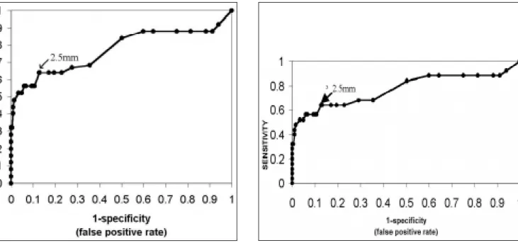
T he RO C curve (Figure 2) was drawn based on the sensitivity and specificity values and identified NT ³ 2.5 mm as a good bal-ance between sensitivity and specificity for screening aneuploidy. T he measurement of NT ³ 2.5 mm helped to correctly classify 16 fe-tuses out of 23 fefe-tuses with chromosomal al-terations and 181 fetus out of 207 normal karyotype fetuses (Table 6). T he ROC curve also demonstrates that an NT measurement ³ 2.5 mm gives a good balance between sensi-tivity and specificity for screening trisomy 21 (Figure 3). Using the cut-off point of NT ³ 2.5 mm (Table 7), 9 out of 12 fetuses with trisomy 21 (75% sensitivity) and 185 out of 218 normal fetuses (84.9% specificity, 5.0 like-lihood ratio) were correctly classified.
○ ○ ○ ○ ○ ○ ○ ○ ○ ○ ○ ○ ○D ISC U SSIO N○ ○ ○ ○ ○ ○ ○
T he mean age of the women in this study was 35.8 years. T his age group showed a high risk for aneuploidy and, as such, this fact may have positively influenced the accuracy of the test. T here can be two explanations for the 10% aneuploidy prevalence – the women’s age and a concentration of high risk cases at the two clinics, which were Fetal Medicine refer-ral centers.
T he tendency tests used showed that the NT measurement for normal fetuses was lower than that for fetuses that had chromosomal ab-normalities. T his was in keeping with the data in the literature10,17,19,28-32 and reinforced the use of the NT measure for screening aneuploidy.
T he ROC curve helped define the best cut-off point for the NT measurement at ³ 2.5 mm18,19for all aneuploidy. Some other studies have also obtained the same NT value, but a greater number of research studies have fixed the value as NT ³ 3 mm.2,10,15,20-22
When the NT measurement ³ 2.5 mm, all the aneuploidy showed a 69.5% sensitivity, 87.4% specificity and 5.5 likelihood ratio. T hese results coincided with the 65% sensi-tivity obtained by Hafner et al.19b in a 0.4% aneuploidy population and also with the re-sults of Pandya et al.18 - 75% sensitivity and 0.2% aneuploidy.
T he most frequent chromosomal altera-tion was trisomy 21 (12 cases) followed by tri-somy 18 (5 cases), and in 3 cases the chromo-some X monosomy, one of which was a case of mosaicism. Trisomy 21 is also the most fre-quent occurrence in the literature. In neonates, its frequency of identification is tenfold when compared to trisomy 18,33 and as in this study, three times more frequent in 9 to 14 week gestations.34 In the literature, the frequency of the X monosomy is 1.5% of all recognized gestations, although only 1% of these survive beyond the 28th gestational week. T he propor-tions of trisomy 2 1 , trisomy 1 8 and X monosomy were similar to those found by Snijders et al.23 in 1998.
T he largest collaborative study published to date on nuchal translucency consisted of 96,127 patients, used sequential risk, and its accuracy for trisomy 21 is better than that found in this study (82.2%). However, soft-ware is needed to classify the positively and negatively screened cases. Probably the reason for the high accuracy of this large collabora-tive study by Snijders is that it took into ac-count the risk related to gestational age, ma-ternal age and the nuchal translucency meas-ure when calculating sequential risk.
T he criteria for sample selection had the aim of reducing the verification bias36 by
in-Table 7. Fetuses with trisomy 21 and those without, according to the chosen cutoff point
Present (n) Absent (n)
TN ³ 2 .5 9 3 3
TN < 2 .5 3 1 8 5
Tota l 1 2 2 1 8
Sensitivity = 7 5 .0 %; Specificity = 8 4 .9 %; Po sitive likeliho o d ratio 5 .0 .
Table 6. Altered fetal karyotype and normal karyotype - proportion at the chosen cutoff point
Altered N ormal
TN ³ 2 .5 1 6 2 6
TN < 2 .5 7 1 8 1
Tota l 2 3 2 0 7
Sensitivity = 6 9 .5 %; Specificity = 8 7 .4 %; Po sitive likeliho o d ratio = 5 .5 .
Table 5. Maternal age, gestational age (GI), nuchal translucency (NT ) and fetal karyotype with aneuploidies
Case number M aternal age (years) GA (w eek s) N T (mm) Karyotype
1 2 6 1 1 weeks 1 .0 4 7 ,XY,+2 1
2 2 6 1 1 weeks and 4 days 1 .7 6 9 ,XXY
3 3 4 1 4 weeks 1 .8 9 2 ,XXYY
4 4 1 1 3 weeks and 4 days 1 .8 4 6 ,XX / 4 5 ,X0
5 3 6 1 2 weeks and 5 days 1 .8 4 7 ,XY,+2 1
6 3 7 1 2 weeks and 5 days 1 .8 4 7 ,XY,+1 8
7 4 2 1 2 weeks 2 .0 4 7 ,XY,+2 1
8 3 4 1 2 weeks and 4 days 2 .5 4 7 ,XY,+2 1
9 4 1 1 1 weeks and 6 days 2 .5 4 7 ,XX,+1 8
1 0 4 2 1 2 weeks and 4 days 3 .0 4 7 ,XY,+1 8
1 1 3 8 1 1 weeks and 3 days 3 .8 4 7 ,XX,+2 1
1 2 4 1 1 2 weeks 4 .5 4 7 ,XX,+7
1 3 4 1 1 1 weeks and 6 days 4 .8 4 7 ,XY,+2 1
1 4 3 8 1 3 weeks and 5 days 5 .1 4 7 ,XY,+2 1
1 5 3 0 1 1 weeks and 2 days 5 .3 4 7 ,XY,+2 1
1 6 3 8 1 1 weeks and 1 day 6 .9 4 5 ,X0
1 7 4 2 1 2 weeks 7 .2 4 7 ,XX+2 1
1 8 2 5 1 3 weeks and 5 days 7 .7 4 7 ,XX,+2 1
1 9 3 8 1 2 weeks and 3 days 8 .8 4 7 ,XX,+2 1
2 0 3 7 1 1 weeks and 2 days 9 .4 4 7 ,XX+1 8
2 1 4 1 1 3 weeks and 5 days 1 0 .0 4 7 ,XY,+2 1
2 2 2 6 1 3 weeks and 6 days 1 2 .0 4 5 ,X0
2 3 4 3 1 3 weeks and 5 days 1 4 .0 4 7 ,XY,+1 8
Table 4. Fetal karyotype according to nuchal translucency
N ormal Affected
Translucency (mm) n % n %
< 1 3 1 .5 0
-1 .0ú ¾1 .5 5 0 2 4 .1 1 4 .3
1 .5ú ¾2 9 4 4 5 .5 5 2 1 .8
2 .0ú ¾2 .5 3 4 1 6 .5 1 4 .3
2 .5ú ¾3 .0 1 4 6 .7 2 8 .7
3 .0ú ¾4 .0 9 4 .3 2 8 .7
4 .0ú ¾5 .0 1 0 .5 2 8 .7
5 .0ú ¾1 0 .0 2 0 .9 7 3 0 .5
1 0 o r mo re 0 - 3 1 3 .0
Tota l 2 0 7 1 0 0 .0 2 3 1 0 0 .0
Co chran-Armitag e tendency test: P < 0 .0 1 .
cluding in the study only those cases where the indication had not been an increased NT but the karyotype as a definite diagnosis. However, the number of cases where the indication for fetal karyotype was unknown was high. Maybe the fetal karyotype was studied in these cases because the NT measure was high.
Studies published with large samples have used NT as an indication of fetal karyotype and karyotype for high risk patients only (ad-vanced maternal age, previous malformations), whereas the assessment of the perinatal phe-notype has been reserved for only low risk cases and low NT.2,10,15,18-23
are present.37 T herefore, the neonate pheno-type assessment may have underestimated the number of fetuses, at 10 to 14 weeks, with chromosomal abnormalities that were nor-mally aborted later, and overestimated the sen-sitivity test described by Pajkrt et al,17 leading to biased verification as highlighted by Begg & McNeil.36
In this study, an increase in fetal nuchal translucency, at 10 to 14 weeks’ gestation, showed a relationship with increased chromo-somal alterations and was therefore useful in screening overall or individual chromosomal abnormalities. T he accuracy was best for tri-somy 21.
An important fact to be kept in mind is that the sample size for this study was calcu-lated for overall chromosomal abnormalities and that an individualized analysis of the results for trisomy 21 would possibly have a statistical power inferior to that initially obtained.
T he fact that this study analyzed a sample with a high risk for aneuploidy should be un-derscored, as it raised the accuracy of the screen-ing tests. Consequently, this accuracy cannot
be generalized for populations where the initial risk related to age and obstetric variables is smaller. It has been found that in neonates, polyploidy is extremely rare and that 30% of the fetuses with trisomy 21, 80% of those with trisomy 18, and nearly 99% of those with X monosomy are spontaneously aborted or evolve to death by 40 weeks of gestation.35,38 If these rates were applied to this study and the maxi-mum loss rate for each aneuploidy was accepted, the NT screening would have identified 8 cases of fetal trisomy, 21 live births, 1 case of trisomy 18 and no cases of polyploidy or monosomy X. In countries where abortion in legally al-lowed when chromosomal abnormalities are identified, research on screening tests is focused on trisomy 21 because of its low lethality.
In Brazil, the law does not provide for preg-nancy interruption when chromosomal abnor-malities are identified. However, a considerable number of these fetuses have had cardiac mal-formations or other defects in addition to aneu-ploidy, which can turn the gestational progno-sis poor. T he recognition of these fetuses with aneuploidy allows more specific examinations,
1. Szabo J, Gellen J. Nuchal fluid accumulation in trisomy 21 de-tected by vaginosonography in first trimester. Lancet 1990;3:1133. 2. Nicolaides KH, Azar G, Byrne, D, Mansur C, Marks K. Fetal nuchal translucency: ultrasound screening for chromosomal de-fects in first trimester of pregnancy. Br Med J 1992;304:867-9.
○ ○ ○ ○ ○ ○ ○ ○ ○ ○ ○ ○ ○ ○ ○ ○ ○ ○ ○ ○ ○ ○ ○ ○ ○ ○ ○ ○ ○ ○ ○ ○ ○ ○ ○ ○ ○ ○ ○ ○ ○ ○ ○ ○ ○ ○ ○ ○ ○ ○ ○ ○ ○ ○ ○ ○ ○ ○ R EFER EN C ES○ ○ ○ ○ ○ ○ ○ ○
3. Greco P, Loverro G, Vimercati A, Marzulo A, Caruso G, Selvaggi L. Pathological significance of first-trimester fetal nuchal oedema. Prenat Diagn 1996;16:503-09.
4. Kaisenberg CS, Nicolaides KH, Brand-Saberi B. Lymphatic ves-sel hypoplasia in fetuses with Turner syndrome. Human Reprod
1999;3:823-6.
5. Moscoso G. Fetal nuchal translucency: a need to understand the physiological basis. Ultrasound Obstet Gynecol 1995;5:381-3. 6. Pandya PP, Johnson SP, Malligianis P, Nicolaides KH. First-trimester
fetal nuchal translucency and screening for chromosomal
abnormali-Figure 2. Balance between NT sensitivity and specificity to screen all chromosomal abnormalities at different cutoff points of Receiver Operator Characteristic Curve (ROC).
such as fetal echocardiography and morphologi-cal ultrasound, that will assist in the risk classi-fication of these fetuses, thereby allowing pre-natal programs and delivery in tertiary services to be prepared to receive them. From a psycho-logical point of view, screening and posterior diagnosis gives the couple the possibility of knowing the risks in the gestation and being ready for unfavorable situations, and it also helps to make them aware of the risks they face regarding future gestations.
In this study, the measurement of nuchal translucency demonstrated a high level of ac-curacy for a population that had a high preva-lence of aneuploidy, and therefore it can be in-dicated in the same kind of circumstances.
○ ○ ○ ○ ○ ○ ○ ○ ○ ○ ○ ○C O N C LU SIO N○ ○ ○ ○ ○ ○ ○ ○
T he measurement of nuchal translucency in a South American population showed a high degree of accuracy in screening overall chromo-somal abnormalities and even higher accuracy for trisomy 21. T he best cutoff point obtained for nuchal translucency was values ³ 2.5 mm.
Figure 3. Balance between NT sensitivity and specificity for trisomy 21 at different cutoff points of Receiver Operator Characteristic Curve (ROC).
CONTEXTO: A literatura mostra uma associação entre diversos marcadores ultra-sonográficos e riscos de cromossomopatias. D entre os marcadores ultra-sonográficos, a medida da translucência nucal têm sido apontada como um método de rastreamento de aneuploidias, sendo a trissomia do cromossomo 21 a mais investigada.
OBJETIVO: Definir, com auxilio da curva ROC, o melhor ponto de corte fixo da translucência nucal e sua acurácia no rastreamento das aneuploidias fetais como um todo, e para a trissomia do cromossomo 21 numa população sul americana.
T IPO DE EST U DO : Validação de teste diagnóstico.
LOCAL: O estudo foi realizado na Universidade Estadual de Campinas, Campinas, Brasil.
PARTICIPANTES: 230 pacientes que realizaram ultra-sonografia em dois centros privados de nível terciário, entre 10 e 14 semanas de gestação.
PROCEDIMENTOS: Foram incluídas as pacientes que realizaram ultra-sonografia, entre 10 e 14 semanas de gestação, onde se mediu a translucência nucal, e a pesquisa do cariótipo fetal ou do recém-nascido.
○ ○ ○ ○ ○ ○ ○ ○ ○ ○ ○ ○ ○ ○ ○ ○ ○ ○ ○ ○ ○ ○ ○ ○ ○ ○ ○ ○ ○ ○ ○ ○ ○ ○ ○ ○ ○R ESU M O○ ○ ○ ○ ○ ○
VARIÁVEIS ESTUDADAS: Idade materna, idade gestacional, medida da translucência nucal e resultado do cariótipo fetal ou do recém-nascido.
RESULTADOS: A prevalência de cromossomopatias foi de 10%. A idade média das pacientes foi de 35,8 anos. A idade gestacional média foi de 12 semanas e dois dias. A medida da translucência nucal (TN) apresentou uma média de 2,18 mm. O ponto de corte fixo, com melhor equilíbrio entre sensibilidade e especificidade, foram os valores iguais ou maiores que 2,5 mm para todas as cromossomopatias e, também, para a trissomia do cromossomo 2 1 isoladamente. A sensibilidade do teste foi de 69,5% e 75% para todas as cromossomopatias e para a trissomia do cromossomo 21 respectivamente, com uma razão de verossimilhança positiva de 5,5 e 5,0 para todas as cromossomopatias e para a trissomia do cromossomo 21 respectivamente.
CONCLUSÕES: A medida translucência nucal mostrou boa acurácia como marcador ultra-sonográfico de cromossomopatias fetais, sendo o valor de 2,5 mm o melhor ponto de corte fixo.
PALAVRAS-CHAVE: Aneuplodia. Ultra-sonografia. Cromossomos. Feto. Diagnóstico pré-natal.
Gregório Lorenzo Acá cio, M D, M Sc. Faculty o f Medical Sc ie nc e s a nd a ffilia te d to Unive rsid a d e d e Ta ub a té , Universidade Estadual de Campinas, Campinas, Braz il.
Rica rdo Ba rini, M D, PhD. Faculty o f Medical Sciences and a ffilia te d to DTG , C A ISM , Unive rsid a d e Esta d ua l d e Campinas, Campinas, Braz il.
W a lter Pinto Júnior M D, PhD. Titular Pro fesso r, Depart-ment o f G enetics, Faculty o f Medical Sciences and affiliated to C e me sp c linic fo r he re d ita ry d ise a se s, Unive rsid a d e Estadual de Campinas, Campinas, Braz il.
Rena to Luís Silveira X imenes, M D. Affiliated toCentrus -Center fo r Feta l Medic ine a nd Ultra so und in Ca mpina s, Campinas, Braz il.
Heverton Pettersen, M D. Affiliated to G ennus - N ucleus fo r Fetal Medicine in Belo Ho riz o nte, Belo Ho riz o nte, Braz il.
M a rcos Fa ria , M D. Affiliated to G ennus - N ucleus fo r Fetal Medicine in Belo Ho riz o nte, Belo Ho riz o nte, Braz il.
Sources of funding: The principal autho r was spo nso red fo r 3 mo nths by CAPES.
Conflict of interest: N o t declared
La st received: 0 1 Aug ust 2 0 0 0
Accepted: 0 4 September 2 0 0 0
Address for correspondence:
G reg ó rio Lo renz o Acácio
Rua G ino Bio ndi, 5 1 7 - Jardim Primavera Taubaté/ SP – Brasil - CEP 1 2 0 3 1 -2 2 0 e-mail: g lacacio @ uo l.co m.br
CO PYRIG HT© 2 0 0 1 , Asso ciação Paulista de Medicina
○ ○ ○Pu b l i sh i n g i n f o r m a t i o n○ ○ ○ ○ ○ ○ ○ ○ ○ ○ ○ ○ ○ ○ ○ ○ ○ ties. In Jurkovic D, Jauiaux ??, editors. Ultrasound and Early Preg-nancy. London and New York: Parthenon Publishing; 1996:81-94.
7. Snijders RJM, Nicolaides KH. Assessment of risks. In:_ Ultra-sound markers for fetal chromosomal defects. New York and Lon-don: Parthenon publishing group; 1996a:63-120.
8. Bronshtein M, Rottem S, Yoffe N, Blumenfeld Z. First trimester and early second-trimester diagnosis of nuchal cystic hygroma by transvaginal sonography: Diverse prognosis of the septate from the non-septate lesion. Am J Obstet Gynecol 1989;161:78-82. 9. Johnson MP, Johnson A, Holzgreve W, et al. First-trimester
sim-ple hygroma: cause and outcome. Am J O bstet Gynecol 1993;168:156-61.
10. Nicolaides KH, Brizot ML, Snijders RJM. Fetal nuchal translu-cency: ultrasound screening for fetal trisomy in the first trimester of pregnancy Br J Obstet Gynaecol 1994;101:782-6. 11. Brambati B, Cislaghi C, Tului L, et al. First-trimester Down’s
syn-drome screening using nuchal translucency: a prospective study in patients undergoing chorionic villus sampling. Ultrasound Obstet Gynecol 1995;5:9-14.
12. Comas C, Martinez JM, Ojuel J, et al. First-trimester nuchal edema as a marker of aneuploidy. Ultrasound Obstet Gynecol 1995;5:26-9. 13. Szabo J, Gellen J, Szemere G. First-trimester ultrasound screen-ing for fetal aneuploidy in women over 35 and under 35 years of age. Ultrasound Obstet Gynecol 1995;3:161-3.
14. Kornman LH, Morssink LP, Beekhuis JR, De Wolf BT HM, Heringa MP, Mantingh A. Nuchal translucency cannot be used as a screening test for chromosomal abnormalities in the first trimes-ter of pregnancy in a routine ultrasound practice. Prenat Diagn 1996;16:797-805.
15. Faria M, Quintino S, Pettersen H. Rastreamento ultra-sonográfico de anomalias cromossômicas através da medida da translucência nucal - Análise de 231 fetos. Rev Bras Ginec Obstet 1997;19:19-30. 16. Taipale P, Hilesma A, Salonen R, Ylostalo P. Increased nuchal
trans-lucency as a marker for the chromosomal defects. N Engl J Med 1997;23:1654-8.
17. Pajkrt E, Mol BWJ, Van Lith JMM, Bleker OP, Bilardo CM. Screening for Down’s syndrome by fetal nuchal translucency measurement in a high-risk population. Ultrasound Obstet Gynecol 1998b;12:156-62. 18. Pandya PP, Goldberg H, Walton B, et al. T he implementation of
first-trimester scanning at 10-13 weeks’ gestation and the meas-urement of fetal nuchal translucency thickness in two maternity units. Ultrasound Obstet Gynecol 1995c;5:20-5.
19. Hafner E, Schuchter K, Liebhart E, Philipp K. Results of routine fetal nuchal translucency measurement at weeks 10-13 in 4233 unselected pregnant women. Prenat Diagn 1998;18:29-34. 20. Bewley S, Roberts LJ, Mackinson AM, Rodeck CH. First
trimes-ter fetal nuchal translucency: Problems with the general popula-tion 2. Br J Obstet Gynaecol 1995;102:386-8.
21. Cha’ban FK, Van Splunder P, Los FJ, Wladimiroff JW. Fetal out-come in nuchal translucency with emphasis on normal fetal karyo-type. Prenat Diagn 1996;16:537-41.
22. Reynders CS, Pauker SP, Benacerraf BR. First trimester isolated fetal nuchal lucency: significance and outcome. J Ultrasound Med 1997;16:101-05.
23. Snijders RJM, Noble P, Sebire N, Souka A, Nicolaides KH. UK multicentre project on assessment of risk of trisomy 21 by mater-nal age and fetal nuchal-translucency thickness at 10-14 weeks of gestation. Fetal medicine foundation first-trimester screening group. Lancet 1998;352:337-8.
24. Schäfer H. Efficient confidence bounds for ROC curves. Statis-tics in medicine 1994;13:1551-61.
25. Pandya PP, Snijders RJM, Johnson SP, Brizot ML. Screening for fetal trisomy by maternal age and fetal nuchal translucency thick-ness at 10 to 14 weeks of gestation. Br J O bstet Gynaecol 1995b;102:957-62.
26. Robinson HP, Fleming JE. A critical evaluation of sonar crown-rump length measurements. Br J Obstet Gynaecol 1975;82:702-10.
27. Nicolaides KH, Sebire NJ, Snijders RJM. Nuchal translucency and chromosomal defects. In: The 11-14 weeks scan: the
diag-nosis of fetal abnormalities. 1st ed.New York and London:
Par-thenon Publishing; 1999:03-63.
28. Hafner E, Schuchter K, Philipp K. Screening for chromosomal abnormalities in an unselected population by fetal nuchal translu-cency. Ultrasound Obstet Gynecol 1995;330-3.
29. Braithwaite JM, Morris RW, Economides DL. Nuchal translu-cency measurements: frequency distribution and changes with gestation in a general population. B r J O bstet G ynaecol 1996;103:1201-04.
30. Snijders RJM, Nicolaides KH. First-trimester fetal nuchal trans-lucency. In: Ultrasound markers for fetal chromosomal defects. New York and London: Parthenon Publishing; 1996b:121-56. 31. Pajkrt E, De Graff IM, Mol BWJ, Van Lith JMM, Bleker OP,
Bilardo CM. Weekly nuchal translucency measurements in nor-mal fetuses. Obstet Gynecol 1998a;91:208-11.
32. Schuchter K, Wald N, Hackshaw AK, Hafner E, Liebhart E. T he distribution of nuchal translucency at 10-13 weeks of pregnancy. Prenat Diagn 1998;18:281-6.
33. T hompson MW, McInnes RR, Willard HF. Citogenética clínica: princípios gerais e anormalidades autossômicas. In: T hompson & T hompson. Genética médica. 5th ed. Rio de Janeiro: Guanabara Koogan; 1993:138-68.
34. Snijders RJM, Holzgreve W, Cuckle H, Nicolaides KH. Maternal age-specific risks for trisomy at 9-14 weeks’ gestation. Prenat. Diagn 1994;14:543-52.
35. Hook EB. Prevalence of chromosome abnormalities during hu-man gestation and implications for study of environmental mutagens. Lancet 1981;6:169-72.
36. Begg BC, McNeil BJ. Assessment of radiological tests: Control of bias and other design considerations. Radiology 1988;167:565–9. 37. Snijders RJM, Sundberg K, Holzgreve W, Henry G, Nicolaides KH. Maternal age and gestation-specific risk for trisomy 21. Ul-trasound Obstet Gynecol 1999;13:167-70.