Invasive ductal carcinoma: relationship between pathological
characteristics and the presence of axillary metastasis in 220 cases
Carcinoma ductal invasor: relação de características anatomopatológicas com a
presença de metástases axilares em 220 casos
RannieRe GuRGel FuRtadode aquino1,3; Paulo HenRique dióGenes Vasques, tCBC-Ce1; diane isaBelle MaGno CaValCante2; ayane
laynede sousa oliVeiRa3; BRuno Masato KitaGawade oliVeiRa4; luiz GonzaGa PoRto PinHeiRo, eCBC-Ce5.
INTRODUCTION
B
reast cancer is the most common malignant neoplasm in the world and still accounts for a number of unfavorable outcomes that make it the second leading cause of cancer death in women1.The variety of outcomes observed in clinical practice demonstrates that the biological behavior of this disease can still be uncertain and often does not depend on the type of treatment offered to the patient, which indicates that the mechanisms involved in this process are not fully known.
Morphological, genetic and enzymatic methods allow us to evaluate its degree of aggressiveness and are routinely used satisfactorily2,3.
Anatomopathological analysis, due to its simplicity, speed and low-cost, is still widely used for its reliability
even in the face of current molecular and genetic approaches4.
The tumor histological grade (HG) is one of the most important anatomopathological features. The Nottingham classification system, which is a modification of the Scarff-Bloom-Richardson (SBR)5,6
one, is the most commonly used classification system to determine HG in breast cancer7. In 1991, its prognostic
value was demonstrated for the first time5, and since
then several studies have validated it, which has made it a recommended classification system worldwide8,9.
In breast cancer, HG indicates the degree of tumor tissue differentiation, established through a semi-quantitative evaluation of the tumor’s morphological characteristics: tubular grade (TG), pleomorphism or nuclear grade (NG), and Number of mitoses per large increase field – mitotic index (MI)5.
1 - Federal University of Ceará (UFC), Post-graduation Program in Surgery, Fortaleza, Ceara State, Brazil. 2 - Federal University of Ceará (UFC), Depart-ment of Pathology and Legal Medicine, Fortaleza, Ceara State, Brazil. 3 - University of Fortaleza (UNIFOR), Faculty of Medicine, Fortaleza, Ceara State, Brazil. 4 - Federal University of Ceara (UFC), Faculty of Medicine, Fortaleza, Ceara State, Brazil. 5 - Federal University of Ceará (UFC), Department of Surgery, Fortaleza, Ceara State, Brazil.
A B S T R A C T
Objective: to analyze the relation of anatomopathological features and axillary involvement in cases of invasive ductal carcinoma. Methods: this is a cross-sectional study of 220 breast cancer patients submitted to radical mastectomy or quadrantectomy with axilar emptying, from the Mastology Service of the Assis Chateaubriand Maternity School, Ceará, Brazil. We submitted the tumors to histological processing and determined the histological (HG), tubular (TG) and nuclear (NG) grades, and the mitotic index (MI) by the classification of Scarff-Bloom-Richadson, verified the presence of angiolymphatic invasion (AI) and measured the largest tumor diameter (TD). We then correlated these variables with the presence of axillary metastases. Results: the mean patients’age was 56.81 years ± 13.28. Tumor size ranged from 0.13 to 22 cm, with an average of 2.23cm ± 2.79. HG3, TG3 and NG3 prevailed, respectively 107 (48.6%), 160 (72.7%) and 107 (48.6%). Mitotic indexes 1, 2 and 3 presented a homogeneous distribution, respectively 82 (37.2%), 68 (31%) and 70 (31.8%). We observed no relation between the HG, TG and NG with the occurrence of axillary metastases (p=0.07, p=0.22 and p=0.21, respectively). Mitotic indices 2 and 3 were related with the occurrence of axillary metastases (p=0.03). Tumors larger than 2cm and cases that presented angiolymphatic invasion had a higher index of axillary metastases (p=0.0003 and p<0.0001). Conclusion: elevated mitotic indexes, tumors with a diameter greater than 2cm and the presence of angiolymphatic invasion were individuallyassociatedwith the occurrence of axillary metastases.
Aquino Invasive ductal carcinoma: relationship between pathological characteristics and the presence of axillary metastasis in 220 cases 164
Another aspect analyzed is the tumor size. Currently, it has a high prognostic value, since it is highly related to disease aggressiveness. Tumors with larger diameters are associated with axillary lymph node involvement, lower disease free interval, and mortality. Small tumors are invariably related to a better prognosis both for overall survival and disease-free time10,11.
Regarding the presence of angiolymphatic invasion, it is known that the involvement of peritumoral blood and lymphatic vessels is necessary for metastasis to occur in most cases. In cases of breast cancer, these findings are directly associated with axillary lymph node involvement and worse prognoses12-14.
The axillary status reflects the involvement or not of the axillary lymph nodes by neoplastic cells. It is one of the most important prognostic factors of invasive carcinoma, since patients with disease-free armpits display better prognosis, both for overall and for disease-free survival. Survival at ten years is associated with the number of lymph nodes involved15.
Due to its indisputable clinical applicability and the different outcomes still observed in our clinical practice, this study aimed to verify the relationship between the tumor anatomopathological characteristics and the axillary metastatic involvement, since this is an excellent predictor of aggression and prognosis.
METHODS
This is a cross-sectional study based on the histopathological analysis of surgical specimens of patients with breast cancer of the invasive ductal histological subtype who underwent surgical treatment by mastectomy or quadrantectomy and investigation of the presence of axillary metastases by sentinel lymph node biopsy or axillary emptying. The patients were treated at the Mastology Service of the Assis Chateaubriand School Maternity from January 2005 to December 2014, and we carried out the research at the Department of Pathology and Legal Medicine – DPML –, Faculty of Medicine of the Federal University of Ceará (UFC), between February and May 2015.
We selected female patients with confirmed histological diagnosis of invasive ductal carcinoma, regardless of age, who did not undergo neoadjuvant chemotherapy. We excluded cases that did not have sufficient material in good condition for histological processing and evaluation or did not meet the selection criteria.
The following variables were analyzed: Nuclear Grade (NG), Tubular Grade (TG), Mitotic Index (MI) and Histological Grade (HG) by the Scarf-Bloom-Richardson (SBR) method modified by Elston and Ellis5, which considers the sum of the tubular,
nuclear and mitotic index scores (Table 1). We also studied the presence of angiolymphatic invasion,
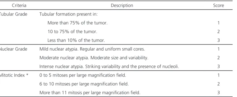
Table 1. Scores for assessment of tubular and nuclear grades, and mitotic index.
Criteria Description Score
Tubular Grade Tubular formation present in:
More than 75% of the tumor. 1
10 to 75% of the tumor. 2
Less than 10% of the tumor. 3
Nuclear Grade Mild nuclear atypia. Regular and uniform small cores. 1
Moderate nuclear atypia. Moderate size and variability. 2
Intense nuclear atypia. Striking variability and the presence of nucleoli. 3
Mitotic Index * 0 to 5 mitoses per large magnification field. 1
6 to 10 mitoses per large magnification field. 2
More than 11 mitosis per large magnification field. 3
metastasis to axillary lymph nodes and tumor diameter. We made the histological sections 5μm-thick, processed them from the paraffin-embedded tumor tissue stained with Hematoxylin and Eosin (HE) and observed them under light microscopy (Nikon CX40 model microscope).
We considered: HG 1- Well differentiated (sum of 3 to 5 scores); HG 2- Moderately differentiated (sum of 6 to 7 scores); HG 3- Poorly differentiated (sum of 8 to 9 scores).
We confirmed angiolymphatic invasion (AI) by histopathological examination and considered it present when tumor cells invaded the lumen of blood vessels and/or peritumoral lymphatic vessels.
We assessed axillary metastases (Mx) by the histopathological study of lymph nodes harvested during surgery. We considered the cases positive for axillary metastasis when we found the presence of breast tissue neoplastic cells infiltrating the lymph nodes.
We measured tumor diameter (TD) in centi-meters from the specimen sent to anatomopatholo-gical study. For this study, we considered the largest diameter measured in the primary tumor.
We performed statistical analysis using the SPSS® software version 20.0, using the chi-square test, in order to show the correlation of each anatomopathological variable with the presence or absence of axillary metastases, considering p<0.05 as statistically significant.
This study was approved by the Ethics in Research Committee of the Pro-Rectory of Research of the Federal University of Ceará, via Plataforma Brasil, under the number 651,657.
RESULTS
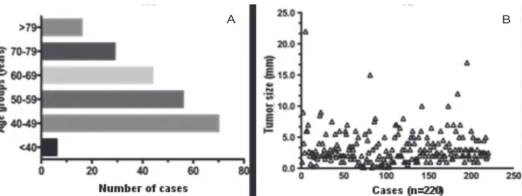
We studied 220 cases of invasive ductal carcinoma. The mean patients’age was 56.81 years ± 13.28. The minimum age was 35 years and the maximum, 93, and the patients less than 50 years old predominated (Figure 1A). Tumor size ranged from 0.13 to 22 cm, with a mean of 2.23cm ± 2.79; we also recorded isolated cases of extensive tumors (Figure 1B).
When analyzing the degree of differentiation of the tumors regarding the histological grade and its variables, we observed a predominance of poorly differentiated tumors. HG3, TG3 and NG3 prevailed in this series, respectively 107 (48.6%), 160 (72.7%) and 107 (48.6%). Mitotic indexes 1, 2 and 3 presented a homogeneous distribution in this study, respectively 82 (37.2%), 68 (31%) and 70 (31.8%) (Table 2).
We classified tumors according to their largest diameter and distributed them in two groups: less than or equal to 2cm and greater than 2cm. Tumor
≤2.0 cm were more prevalent (142 – 65.5%). The involvement of peritumoral blood and/or lymphatic vessels was present in most of the cases studied, being represented by 65% of the sample (n=144) (Table 2).
Aquino Invasive ductal carcinoma: relationship between pathological characteristics and the presence of axillary metastasis in 220 cases 166
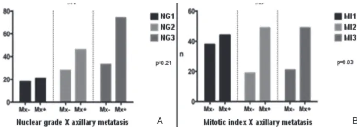
When relating the histological grades and their variables with the presence of axillary metastases, we observed that the variables that prevailed in the cases with metastatic disease were those that indicated less tissue differentiation: HG3, TG3 and NG3. However, when analyzing the relation with the occurrence of axillary metastases, we found no statistical significance, with p=0.07,
p=0.22 and p=0.21, respectively (Figures 2A, 2B and 3A).
When analyzing the number of mitoses per microscopic field, we observed that the highest mitotic indexes was significantly related (p=0.03) with the occurrence of axillary metastases (Figure 3B).
Tumor diameter and angiolymphatic invasion were also statistically correlated with
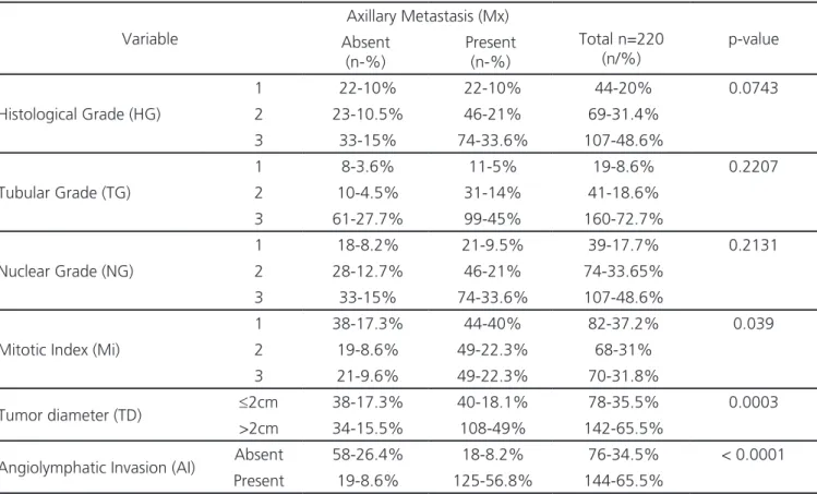
Table 2. Distribution of frequencies of anatomopathological variables according to the occurrence of axillary metastases.
Variable
Axillary Metastasis (Mx)
Total n=220 (n/%)
p-value Absent
(n-%)
Present (n-%)
Histological Grade (HG)
1 22-10% 22-10% 44-20% 0.0743
2 23-10.5% 46-21% 69-31.4%
3 33-15% 74-33.6% 107-48.6%
Tubular Grade (TG)
1 8-3.6% 11-5% 19-8.6% 0.2207
2 10-4.5% 31-14% 41-18.6%
3 61-27.7% 99-45% 160-72.7%
Nuclear Grade (NG)
1 18-8.2% 21-9.5% 39-17.7% 0.2131
2 28-12.7% 46-21% 74-33.65%
3 33-15% 74-33.6% 107-48.6%
Mitotic Index (Mi)
1 38-17.3% 44-40% 82-37.2% 0.039
2 19-8.6% 49-22.3% 68-31%
3 21-9.6% 49-22.3% 70-31.8%
Tumor diameter (TD) ≤2cm 38-17.3% 40-18.1% 78-35.5% 0.0003
>2cm 34-15.5% 108-49% 142-65.5%
Angiolymphatic Invasion (AI) Absent 58-26.4% 18-8.2% 76-34.5% < 0.0001
Present 19-8.6% 125-56.8% 144-65.5%
axillary involvement in the present study. The group of cases that presented tumors >2.0cm in their largest diameter and cases in which angiolymphatic invasion was present presented more axillary metastases, with p=0.0003 and p<0.0001, respectively (Figure 4).
DISCUSSION
Breast cancer has several outcomes in spite of the treatments used, and the presence of axillary metastases is a strong indicator of poor prognosis15.
Therefore, it is important in the clinical practice to better know the tumor characteristics that can predict aggressiveness and axillary involvement. The morphological aspects are directly related to tumors evolution and, in this context, the main determinant is the degree of cellular differentiation5,16,17.
Hammond et al.18 and Fitzgibbons et al.19
considered HG as the first-choice morphological feature to predict prognosis in cases of breast cancer. Leong et al.20 evidenced HG as a good predictor of
prognosis. However, they stated that low-grade tumors might also present unfavorable outcomes, with axillary metastases in cases with a long survival time.
Nevertheless, Younes et al.21 studied cases
of breast cancer without axillary involvement and could not establish a relationship between HG and the patients’prognosis. Lopes et al.22, analyzing the
relationship between HG and distant recurrence in five years, also did not find statistical significance. Ladekarl23
considers that there are flaws in the reproducibility of HG that compromise its clinical applicability as a prognostic factor. In this study, we could not establish a significant correlation between HG and axillary involvement either.
Figure 3. Grouped distribution according to the metastatic involvement of axillary lymph nodes (n = 220). A) Nuclear Grade; B) Mitotic index.
Aquino Invasive ductal carcinoma: relationship between pathological characteristics and the presence of axillary metastasis in 220 cases 168
We did not find a significant relationship between NG and the occurrence of axillary metastases when considered in isolation. This finding is in agreement with those of Lopes et al.22, who, studying the relation of the NG with
the metastatic relapse in five years, did not observe statistical correlation. Martinez-Arribas et al.24 found
a direct relation of NG3 with worse prognosis when associated with immunohistochemical parameters, such as absence of hormonal receptors and high Cerb-B2 and Ki67.
MI is a variable of the histological grade established by counting the number of mitoses per high magnification field. Lopes et al.22 showed that a
mitotic count greater than ten per field was associated with axillary metastases and with systemic relapse in five years. In the study by Medri et al.25, the high
number of mitoses was associated with a greater probability of distant metastases in cases of previously negative axilla breast cancer.
Caly et al.26, Baak et al.27 and van Diest et
al.28 analyzed MI as a prognostic factor and considered
it as effective as HG, tumor size and NG in cases of breast cancer. In our series, the results agree with the literature presented here, since tumors with high MI statistically related with the metastatic involvement of axillary lymph nodes.
Barbosa et al.29 observed that tumor size
has a significant correlation with axillary lymph node involvement. A meta-analysis involving 11 studies30
concluded that tumor size >2cm and angiolymphatic invasion indisputably increase the probability of axillary lymph node involvement. The literature data presented here corroborate the results of our study, which demonstrated that the present AI was significantly associated with axillary metastases and tumor diameter >2cm.
High mitotic indexes, tumors with a diameter >2.0cm and the presence of angiolymphatic invasion were individually associated with the occurrence of axillary metastases in the cases studied and should therefore continue to be part of the initial evaluation of breast cancer and direct the conducts despite current molecular and genetic classifications.
REFERENCES
1. Ban KA, Godellas CV. Epidemiology of breast can-cer. Surg Oncol Clin North Am. 2014;23(3):409-22. 2. Vich P, Brusint B, Alvarez-Hernández C,
Cuadra-do-Rouco C, Diaz-García N, Redondo-Margüello E. Update of breast cancer in primary care (I/V). Se-mergen. 2014;40(6):326-33.
3. Shah R, Rosso K, Nathanson SD. Pathogenesis, pre-vention, diagnosis and treatment of breast cancer.
World J Clin Oncol. 2014;5(3):283-98.
4. Aquino RGF, Pinheiro LGP, Ferreira MVP, Cavalcante DIM, Oliveira ALS, Gomes NN, et al. Ductal carcino-ma of the breast: morphological aspects according to the age. J Bras Patol Med Lab. 2015;51(4):252-7. 5. Elston CW, Ellis IO. Pathological prognostic factors in
breast cancer. I. The value of histological grade in bre-ast cancer: experience from a large study with long--term follow-up. Histopathology. 1991;19(5):403-10. 6. Zhang R, Chen HJ, Wei B, Zhang HY, Pang ZG, Zhu
Objetivo: analisar a relação das características anatomopatológicas com o comprometimento axilar em casos de carcinoma ductal invasor. Métodos: estudo transversal de 220 pacientes com câncer de mama, submetidas à mastectomia radical ou quadrantectomia com esvaziamento axilar, oriundos do Serviço de Mastologia da Maternidade Escola Assis Chateaubriand, Ceará, Brasil. Os tumores foram submetidos a processamento histológico e, em seguida, foram determinados os graus histológico (GH), tubular (GT), nuclear (GN), índice mitótico (IM) pela classificação de Scarff-Bloom-Richadson, verificada a presença de invasão angiolinfática (IA) e mensurado o maior diâmetro do tumor (DT). Tais variáveis foram correlacionadas com a presença de metástases axilares. Resultados: a média de idade das pacientes foi 56,81 anos ± 13,28. O tamanho do tumor variou de 0,13 a 22 cm, com média de 2,23cm ± 2,79. Os GH3, GT3 e GN3 prevaleceram: n=107 (48,6%), n=160 (72,7%) e n=107 (48,6%), respectivamente. Os índices mitóticos 1, 2 e 3 apresentaram distribuição homogênea: n=82 (37,2%), n=68 (31%) e n=70 (31,8%), respectivamente. Não foi evidenciada relação do GH, GT e GN com a ocorrência de metástases axilares (p=0,07; p=0,22 e p=0,21). Índices mitóticos 2 e 3 apresentaram relação com a o ocorrência de metástases axilares (p=0,03). Tumores maiores do que 2cm e casos com invasão angiolinfática apresentaram maior índice de metástases axilares (p=0,0003 e p<0,0001). Conclusão: índices mitóticos elevados, tumores com diâmetro maior do que 2cm e presença de invasão angiolinfática apresentaram isoladamente relação com a ocorrência de metástases axilares.
Descritores: Neoplasias da Mama. Metástase Linfática. Patologia Cirúrgica. Gradação de Tumores.
H, Zhang Z, et al. Reproducibility of the Nottingham modification of the Scarff-Bloom-Richardson his-tological grading system and the complementary value of Ki-67 to this system. Chin Med J (Engl). 2010;123(15):1976-82.
7. Rakha EA, Reis-Filho JS, Baehner F, Dabbs DJ, De-cker T, Eusebi V, et al. Breast cancer prognostic classification in the molecular era: the role of histo-logical grade. Breast Cancer Res. 2010;12(4): 207. 8. Blamey RW, Hornmark-Stenstam B, Ball G,
Blichert--Toft M, Cataliotti L, Fourquet A, et al. ONCOPOOL – a European database for 16,944 cases of breast cancer. Eur J Cancer. 2010;46(1):56-71.
9. Rakha EA, El-Sayed ME, Lee AH, Elston CW, Grainge MJ, Hodi Z, et al. Prognostic significance of Nottin-gham histologic grade in invasive breast carcinoma. J Clin Oncol. 2008;26(19):3153-8.
10. Nikolic-Vukosavljevic D, Kanjer K, Markicevic M, Todorovic-Rakovic N, Vukotic D, Neskovic-Kons-tantinovic Z. Natural course of node-negative bre-ast cancer: high risk-related subgroups. J Exp Clin Cancer Res. 2003;22(4):543-9.
11. Elston CW, Ellis IO. Pathological prognostic fac-tors in breast cancer. I. The value of histological grade in breast cancer: experience from a large study with long-term follow-up. Histopathology. 2002;41(3A):154-61.
12. Cao Y, Paner GP, Rajan PB. Sentinel node sta-tus and tumor characteristics: a study of 234 in-vasive breast carcinomas. Arch Pathol Lab Med. 2005;129(1):82-4.
13. Chen YW, Lai YC, Hsu CC, Chuang YW, Hou MF. Value of nodal drainage patterns and tumor loca-tion from lymphoscintigraphic mapping in detec-ting axillary sentinel lymph node status in breast cancer: experience at Kaohsiung Medical University Hospital. Kaohsiung J Med Sci. 2005;21(6):251-7. 14. Lopes CM, Montemor Netto MR, Mansani FP,
Stival RSM, Cassapula MR, Oliveira TFB. Clinical, histomorphological, and therapeutic progmostic factors in patients with triple-negative invasive breast câncer. 2015;51(6):397-406.
15. Follana P, Barrière J, Chamorey E, Largillier R, Da-done B, Mari V, et al. Prognostic factors in 401 elderly women with metastatic breast cancer.
On-cology. 2014;86(3):143-51.
16. Fisher ER, Redmond C, Fisher B. Histologic gra-ding of breast cancer. Pathol Annu. 1980;15(Pt 1):239-51.
17. Arihiro K. Pathological approach to evolving prog-nostic and predictive factors of breast cancer. Bre-ast Cancer. 2004;11(4):343-9.
18. Hammond ME, Fitzgibbons PL, Compton CC, Grignon DJ, Page DL, Fielding LP, et al. College of American Pathologists Conference XXXV: solid tumor prognostic factors-which, how and so what? Summary document and recommendations for implementation. Cancer Committee and Conference Participants. Arch Pathol Lab Med. 2000;124(7):958-65.
19. Fitzgibbons PL, Page DL, Weaver D, Thor AD, All-red DC, Clark GM, et al. Prognostic factors in bre-ast cancer. College of American Pathologists Con-sensus Statement 1999. Arch Pathol Lab Med. 2000;124(7):966-78.
20. Leong AC, Hanby AM, Potts HW, Tan DS, Skilton D, Ryder K, et al. Cell cycle proteins do not predict outcome in grade I infiltrating ductal carcinoma of the breast. Int J Cancer. 2000;89(1):26-31. 21. Younes M, Laucirica R. Lack of prognostic
sig-nificance of histological grade in node-negati-ve invasinode-negati-ve breast carcinoma. Clin Cancer Res. 1997;3(4):601-4.
22. Lopes LAF, Ferraro O, Linhares JJ, Lopes RGC, Baracat FF. Valor prognóstico do grau histológi-co (GH), grau nuclear (GN) e índice mitótihistológi-co (IM) para pacientes com carcinoma da mama estádios II e III com linfonodos axilares comprometidos. Rev bras cancerol. 2006;52(3):245-51.
23. Ladekarl M. Objective malignancy grading: a re-view emphasizing unbiased stereology applied to breast tumors. APMIS Suppl. 1998;79:1-34. 24. Martínez-Arribas F, Martín-Garabato E, Lafuente P,
Tejerina A, Lucas R, Sánchez J, et al. Proliferation measurement in breast cancer by two different methods. Anticancer Res. 2006;26(1A):199-202. 25. Medri L, Volpi A, Nanni O, Vecci AM, Mangia A,
Aquino Invasive ductal carcinoma: relationship between pathological characteristics and the presence of axillary metastasis in 220 cases 170
26. Caly M, Genin P, Ghuzlan AA, Elie C, Fréneaux P, Klijanienko J, et al. Analysis of correlation be-tween mitotic index, MIB1 score and S-phase fraction as proliferation markers in invasive breast carcinoma. Methodological aspects and prognos-tic value in a series of 257 cases. Anprognos-ticancer Res. 2004;24(5B):3283-8.
27. Baak JP. The relative prognostic significance of nucleolar morphometry in invasive ductal breast cancer. Histopathology. 1985;9(4):437-44.
28. van Diest PJ, Baak JP, Matze-Cok P, Wisse-Brekel-mans EC, van Galen CM, Kurver PH, et al. Repro-ducibility of mitosis counting in 2,469 breast can-cer specimens: results from the Multicenter Mor-phometric Mammary Carcinoma Project. Hum Pathol. 1992;23(6):603-7.
29. Barbosa EM, Francisco AARF, Araujo Neto JT, Al-ves EMF, Tavares MGM, Góes JCS. Fatores clínico--patológicos de predição do acometimento axilar
em pacientes com metástases de câncer de mama no linfonodo sentinela. Rev Bras Ginecol Obstet. 2010;32(3):144-9.
30. Degnim AC, Griffith KA, Sabel MS, Hayes DF, Cimmino VM, Diehl KM, et al. Clinicopatholo-gic features of metastasis in nonsentinel lym-ph nodes of breast carcinoma patients. Cancer. 2003;98(11):2307-15.
Received in: 13/09/2016
Accepted for publication: 11/10/2016 Conflict of interest: none.
Source of funding: Coordination of Improvement of Higher Level Personnel – CAPES.
Mailing address:
Ranniere Gurgel Furtado de Aquino E-mail: [email protected]