Use of blends of bioabsorbable
poly(L-lactic
acid)/poly(hydroxybutyrate-co-hydroxyvalerate) as surfaces for
Vero cell culture
1Departamento de Biologia Celular, Instituto de Biologia,
Universidade Estadual de Campinas, Campinas, SP, Brasil
2Instituto de Ciências Biológicas, Centro Regional Universitário de Espírito Santo
do Pinhal, Espírito Santo do Pinhal, SP, Brasil
3Departamento de Engenharia de Materiais, Faculdade de Engenharia Mecânica,
UNICAMP, Campinas, SP, Brasil
4Departamento de Ciências Fisiológicas, Faculdade de Ciências Biológicas,
Pontifícia Universidade Católica de São Paulo, Sorocaba, SP, Brasil A.R. Santos Jr.1,2,
B.M.P. Ferreira3,
E.A.R. Duek4,
H. Dolder1
and M.L.F. Wada1
Abstract
Vero cells, a cell line established from the kidney of the African green monkey (Cercopithecus aethiops), were cultured in F-10 Ham medi-um supplemented with 10% fetal calf sermedi-um at 37°C on membranes of poly(L-lactic acid) (PLLA), poly(hydroxybutyrate-co-hydroxyvalerate) (PHBV) and their blends in different proportions (100/0, 60/40, 50/50, 40/60, and 0/100). The present study evaluated morphology of cells grown on different polymeric substrates after 24 h of culture by scanning electron microscopy. Cell adhesion was also analyzed after 2 h of inoculation. For cell growth evaluation, the cells were main-tained in culture for 48, 120, 240, and 360 h. For cytochemical study, the cells were cultured for 120 or 240 h, fixed, processed for histologi-cal analysis, and stained with Toluidine blue, pH 4.0, and Xylidine ponceau, pH 2.5. Our results showed that cell adhesion was better when 60/40 and 50/50 blends were used although cells were able to grow and proliferate on all blends tested. When using PLLA/PHBV (50/50) slightly flattened cells were observed on porous and smooth areas. PLLA/PHBV (40/60) blends presented flattened cells on smooth areas. PLLA/PHBV (0/100), which presented no pores, also sup-ported spreading cells interconnected by thin filaments. Histological sections showed that cells grew as a confluent monolayer on different substrates. Cytochemical analysis showed basophilic cells, indicating a large amount of RNA and proteins. Hence, we detected changes in cell morphology induced by alterations in blend proportions. This suggests that the cells changed their differentiation pattern when on various PLLA/PHBV blend surfaces.
Correspondence
A.R. Santos Jr.
Departamento de Biologia Celular Instituto de Biologia, UNICAMP Caixa Postal 6109
13084-971 Campinas, SP Brasil
Fax: +55-19-3788-6111 E-mail: [email protected] Research supported by CNPq, FINEP-PRONEX, and FAPESP (No. 97/14275-7).
Received January 10, 2004 Accepted May 20, 2005
Key words
•Poly(L-lactic acid) •
Poly(hydroxybutyrate-co-hydroxyvalerate)
Introduction
A research area that has recently received increased attention is tissue engineering, in which functional tissue is restored from na-tive or synthetic sources by using engineer-ing principles. Biomaterials play an impor-tant role working as scaffolds to guide tissue regeneration, releasing drugs and growth factors to stimulate tissue response, or creat-ing a new functional structure when dam-aged tissue does not regenerate (1-3). In this context, a clear understanding of cell re-sponses to the biomaterial used is needed. This information would permit the use of biomaterials and cell culture techniques to create tissue-like structures that simulate the mechanical and physiological characteris-tics of tissues in vivo. These devices could be also produced for healing manipulation and to stimulate natural tissue regeneration.
In many cases, depending on the regen-erative capacity of damaged tissues, the use of biodegradable materials is clinically rec-ommended. In such situations, there is only a need for the temporary presence of bioma-terials to provide support to the damaged area, or substitute it, so as to direct the growth and restoration of tissues (4). A wide variety of bioabsorbable polymers have been used in biological systems, and polyesters derived from α-hydroxy acids are those most frequently employed (5).
Bioabsorbable polymers slowly degrade after implantation. This feature may be im-portant for many tissue regeneration appli-cations, since the polymer will disappear as functional tissue restoration occurs. Poly(L-lactic acid) (PLLA) is a biodegradable poly-ester that has been used experimentally as a support for cell culture or experimental treat-ment of some damaged tissues mainly due to its high biocompatibility. Applications of this polymer have been studied in our labo-ratory (6-8). Poly(hydroxybutyrate-co-hy-droxyvalerate) (PHBV) is also a biodegrad-able polyester produced by microorganisms.
The biocompatibility of PHBV has been studied (9) and the material has also been investigated for the improvement of blood vessel restoration (10).
Cell culture is a very important tool for the study of biomaterials. It provides a quick and reliable pre-selection method so as to choose the best sample that will later be tested in animals. Testing in animals is very expensive and hinders viewing the direct effects of biomaterials on the stimulation of important parameters such as cell prolifera-tion, growth and differentiation. Thus, our objective in the present study was to evalu-ate the biological interactivity of blends of PLLA/PHBV, with respect to their capacity to modify cell adhesion and growth in vitro.
Material and Methods
Preparation of PLLA/PHBV blends
etha-nol and the sterility of the samples was tested. The samples were washed three times in F-10 Ham medium (Sigma, St. Louis, MO, USA) and then incubated in the same culture medi-um for 24 h at 37ºC before cell inoculation.
Cell culture
Vero cells, a cell line established from the kidney of the African green monkey ( Cerco-pithecus aethiops), were obtained from the Adolfo Lutz Institute, São Paulo, SP, Brazil. Cells were cultured in F-10 Ham medium (Sigma) supplemented with 10% fetal calf serum (FCS, Nutricell Nutrientes Celulares, Campinas, SP, Brazil) at 37ºC. Vero cells are recommended for studies of cytotoxicity and for cell-substrate interactions with biomateri-als (11,12). The culture medium was replaced whenever had the acidification of it. Before inoculation, the cells were maintained in cul-ture flasks of 25 or 50 ml with a surface area of 25 or 50 cm2, respectively (Corning,
Cam-bridge, MA, USA).
Cell adhesion assay
The method of Mosmann (13) with some modifications, was used for the cell adhe-sion assay. Briefly, the sterilized PLLA/ PHBV blends (N = 6) were placed in a 96-well plate with 32 mm2 of surface area
(Corn-ing) with 100 µl of culture medium and kept at 37°C for 24 h. After incubation, 1.0 x 10⁶
cells/ml in 100 µl F-10 Ham medium supple-mented with 10% FCS were added to the wells containing the substrates. The cells were cultured for 2 h in F-10 Ham supple-mented with 10% FCS at 37ºC, washed twice with 0.1 M phosphate-buffered saline (PBS), pH 7.4, at 37ºC and incubated with 100 µl F-10 Ham medium.
The assay mixture (10 µl per well) contain-ing 5 mg/ml of 3-(4,5-dimethylthiazol-2-yl)-2,5-diphenyl-tetrazolium bromide (MTT, Sigma) was added to each well and incubated for 4 h at 37ºC. After 4 h, 100 µl isopropanol
acid (Isofar Ind., Jacaré, RJ, Brazil) was added to each well and, after 3 h, was quantified spectrophotometrically based on absorbance at 540 nm, using a Bio-Rad Model 550 micro-plate reader (Bio-Rad Laboratories Inc., Her-cules, CA, USA). The MTT is an incolor salt of tetrazolium that, when oxidized by mito-chondria, form a dark composed detected by spectrophotometry. The measurements are proportional to cell number (13). Cells on the culture plate itself (polypropylene) without other substrates and silicone membranes were used as positive and negative controls, respec-tively. We also read the absorbance of all experimental conditions (different PLLA/ PHBV blends, negative or positive controls) without cells for control of the MTT reac-tion. The statistical significance (P < 0.05) was assessed using one-way analysis of vari-ance (ANOVA) followed by Tukey HSD multiple comparison test.
Cell growth on PLLA/PHBV blends
For the cell proliferation assay we used a modification of the method described by Murakami et al. (14). Briefly, the PLLA/PHBV blends (N = 9) were added to a 96-well plate with F-10 Ham medium for 24 h at 37ºC. After incubation, 1.0 x 10³ cells/ml in 200 µl F-10
(PLLA/PHBV blends, negative/positive con-trols) in a cell-free condition for dye staining control.
Scanning electron microscopy
The three-dimensional blends of differ-ent compositions were fractured by immer-sion in liquid nitrogen, covered with gold using a sputter coater (Baltec SCD 050, Fursteintum Liechtenstein, Germany) and observed with a JEOL JXA-840 (Jeol Co., Tokyo, Japan) scanning electron microscope. For the analysis of cell morphology, 1.0 x 10⁵ cells/ml were inoculated into different
PLLA/PHBV blends in 24-well culture plates (with 2 cm2 of surface area; Corning) in F-10
Ham medium with 10% FCS. Cells cultured on a glass coverslip under the same culture conditions were used as control. After 24 h, the samples were fixed in 3% glutaralde-hyde (Sigma) in 0.1 M phosphate buffer, pH 7.2, for 45 min at 4ºC, and postfixed with 1% OsO4 (Sigma) for 2 h at 4ºC. The specimens
were then dehydrated in graded ethanol
se-ries, critical point dried (Balzers CDT 030, Balzers Inc., Elgin, IL, USA) and coated with gold in a sputter coater. The coated specimens were observed and photographed with a JEOL JSM-5800 scanning electron microscope.
Cytochemical analysis
For cytochemical study, 1.0 x 10⁵ cells/
ml were cultured on membranes prepared with the different PLLA/PHBV blends in F-10 Ham medium supplemented with F-10% FCS. After 48, 120, and 240 h in culture, the samples were fixed with Karnovsky fixative (4% paraformaldehyde/2.5% glutaraldehyde in 0.2 M phosphate buffer, pH 7.2), dehy-drated in graded ethanol series at room tem-perature, cleared in xylene, and embedded in paraplast. Sections of 5 µm were obtained with a microtome (Reicher-Jung, New York, NY, USA) with a steel knife (Leica Instru-ments, Nussloch, Germany) and stained with Toluidine blue (TB), pH 4.0, a basophilic dye, for the detection of DNA, RNA and
glycosaminoglycans, with Alcian blue, pH 2.5, a basophilic dye, for the detection of glycosaminoglycans, and with Xylidine ponceau (XP), pH 2.5, an anionic dye, for the identification of cationic proteins (15-17). Staining time was 15 min for TB and 20 min for XP, both at room temperature. The coverslips obtained were photographed un-der an inverted IX-50 Olympus microscope (Tokyo, Japan) and figures were prepared using Adobe Photoshop software.
Results
PLLA/PHBV substrates
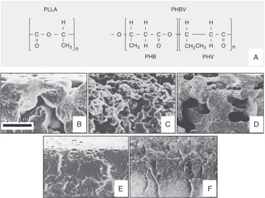
The molecular structure and electron mi-crographs of PLLA, PHBV and the PLLA/ PHBV blends used in the present experi-ment were shown in Figure 1.
Cell adhesion
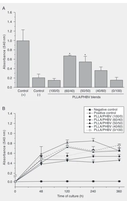
All membranes presented a low capacity to stimulate cell adhesion compared with the positive control used (P < 0.05). The PLLA/ PHBV blends (100/0, 40/60 and 0/100) showed a cell adhesion capacity similar to that of the negative control. Only the 60/40 and 50/50 PLLA/PHBV blends proved to be more receptive to cell interaction (P < 0.05). These results are illustrated in Figure 2A.
Growth curve on different PLLA/PHBV blends
All samples showed a higher mitotic cell rate than the negative control. It is interest-ing to note that the pure polymers used (100/ 0 and 0/100) showed a similar proliferative rate, whereas 60/40 and 40/60 blends showed a lower proliferative index. In all cases, cells grew slower than the positive control. How-ever, in long-term cultures there was a de-crease in cell number for the positive con-trol, suggesting the senescence of the cell layer. This behavior was not observed in the different PLLA/PHBV blends (Figure 2B).
Figure 2. A, Adhesion of Vero cells cultured to different PLLA/PHBV blends. All samples studied displayed the capacity to stimulate less adhesion than the positive control. The (100/0), (40/60) and (0/100) blends were not different from the negative control. *P < 0.05 compared to negative control (Tukey multiple comparison test). B, Growth curve of Vero cells cultured on different PLLA/PHBV blends. In all samples, the cells grew slower than the positive control. PLLA = poly(L-lactic acid); PHBV = poly(hydroxybutyrate-co-hydroxyvalerate).
Scanning electron microscopy
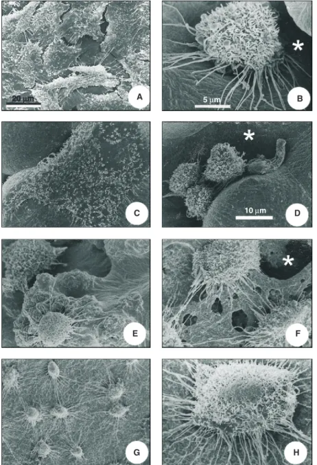
microscopy of fractured blend surfaces. PLLA/PHBV (100/0) membranes were po-rous, with a rounded structure (Figure 1B). The decreased concentration of PLLA in the blends, such as 60/40 and 50/50 samples, results in alterations of porosity (Figure 1C and D, respectively). PLLA/PHBV (40/60) shows that the blend acquired a dense struc-ture, with the presence of cavities on the surface (Figure 1E). PLLA/PHBV (0/100) was completely dense as verified by its frac-ture (Figure 1F). In the scanning electron microscopy of cells cultured on the glass coverslip we could see the formation of a non-confluent cell layer. The cells showed irregular morphology, with a flattened and elongated shape (Figure 3A). In the PLLA/ PHBV (100/0) blends we found cells grow-ing on porous or non-porous areas. In the pores, the cells were round, with microvilli on their surface. They also showed numer-ous long and thin filaments budding from the basal portion of the cell (Figure 3B). On the other hand, on the same membranes, the cells that grew on non-porous areas were flattened on the substrate surface (Figure 3C). In the PLLA/PHBV (60/40) blends we found cells with a growth pattern very simi-lar to that of the former samples, although the pores seen in blends were more irregular (Figure 3D). In PLLA/PHBV (50/50), in contrast to the previous samples, surfaces were irregular, with cells producing many thin filaments (Figure 3E). The PLLA/PHBV (40/60) blends displayed flattened cells, which were shrunk along their edges (Figure 3F). In PLLA/PHBV (0/100) samples, which do not present pores, we found flat or round cells with many interconnecting thin fila-ments (Figure 3G,H).
Cytochemical analysis
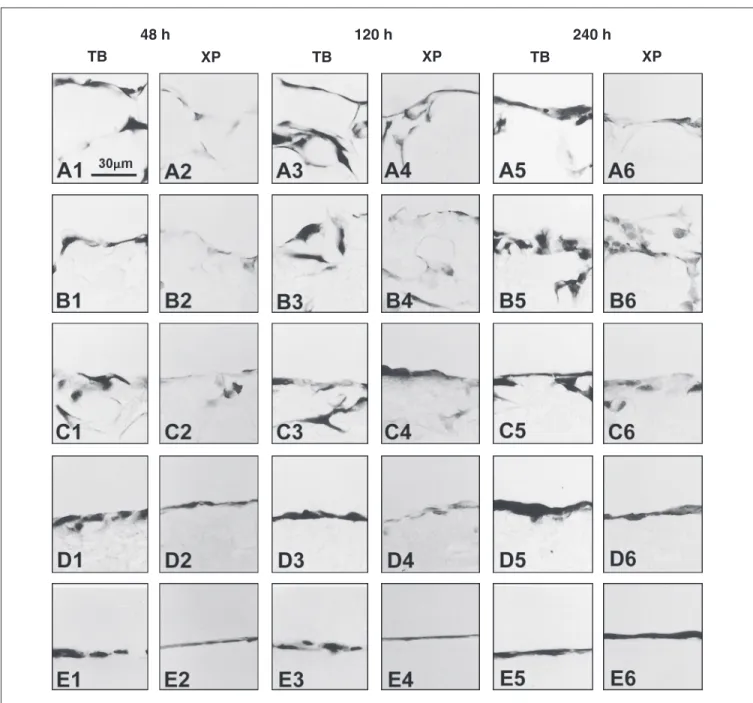
During the culture period cells were able to reach confluence on the substrate and to migrate into different PLLA/PHBV blends through the pores on the blend surface. In all Figure 3. Scanning electron microscopy of Vero cells cultured on the polymers for 24 h. A,
samples, at different time, we detected cells strongly stained by TB or XP, which are basophilic and acidophilic dyes, respective-ly. We did not find cytochemical alterations
induced by the different PLLA/PHBV mem-branes on which the cells were cultured (Fig-ure 4). All samples were negative for Alcian blue at pH 2.5 (data not shown).
Discussion
In this study, we observed variations in cell morphology induced by the physico-chemical characteristics of the membranes upon which they were cultured. In homopoly-mers, PLLA/PHBV (100/0) or (0/100), rounded or flattened cells were observed. In porous areas of PLLA/PHBV (100/0) or (60/ 40) cells were flat, possibly because of the effects of surface tension. The physicochemi-cal properties of different materials could modulate cell morphology and behavior. Sig-naling from polymers that drive the cell growth pattern is complex and may originate from dipole and electric charge interaction forces, hydrogen bonds, electrostatic forces, hydrophilicity/hydrophobicity or surface free energy, and roughness and rigidity, surface tension, and substrate topography (18-20). The morphology of Vero cells on pure PLLA scaffolds observed here was similar to previ-ous results obtained by our group (7,8,21), which showed that round or spreading cells attached on PLLA surface by thin cytoplas-matic filaments. Each new form of produc-tion of biomaterial can bring variaproduc-tions in its structure and porosity. All these characteris-tics can be sensed by the cells that react to them by changing its morphology. The producibility of the cell morphological re-sults indicates a good standardization of poly-mer production, a desirable characteristic to development of biomaterials for clinical uti-lization.
The cell morphology of PLLA/PHBV had not been previously described. In a re-cent study on long-term cultures, Köse et al. (22), reported that osteoblastic cells cultured on PHBV scaffolds had a spread or polygo-nal morphology with extended filopodia simi-lar to that observed for some blends in the present study. Thus, the signaling from PHBV that drives osteoblast growth may be similar to that observed here for fibroblastic cells.
The occurrence of pores on a biomaterial
surface can also modulate the cell prolifera-tion rate. Pores represent an increased area for cell growth and division. In vivo,the pore structure is a desirable characteristic that could create a three-dimensional environ-ment for tissue growth, increasing the tissue-polymer interaction. In vitro, PLLA devices have been used to support the growth and proliferation of endothelial cells without stimulating platelet activation (23). It is in-teresting to note that in our proliferation assay we observed a similar multiplication rate on homopolymers, PLLA/PHBV (100/ 0 or 0/100), compared with the PLLA/PHBV 50/50 blend. It was recently reported that mouse NIH/3T3 fibroblasts cultured on PHBV showed a satisfactory adhesion and proliferation rate and that this property was increased with a physicochemical modifica-tion of the PHBV surface that increased the hydrophilicity of the polymer (24). In an-other report PHBV sustained cell prolifera-tion rates similar to that observed in collagen sponges for at least 35 days of culture (25). Thus, our results obtained for PLLA/PHBV blends agree with those reported for pure PHBV and indicate that our blends are good scaffolds for cell culture.
addition, we observed Vero cell multilayers growing on dry collagen I sponges (29) or on collagen I gels in short- (30) or long-term (31) culture studies.
Cytochemistry has not shown alterations in cell behavior induced by different PLLA/ PHBV blend proportions. In all samples, we found basophilic cells stained by TB that, in pH 4.0, can stain nucleic acids and gly-cosaminoglycans. Although Vero cells can synthesize glycosaminoglycans (32), they produce these molecules in a soluble form in the culture medium. Thus, the basophilic cytoplasm suggests that cells are able to active protein synthesis. The XP, that can stain proteic groups, confirms these results. A similar cytochemical pattern was found in Vero cells cultured on other biomaterials such as poly-HEMA hydrogels (27,28) or collagen I (31). These results agree with the description that different cells are able to produce extracellular matrix on PHBV
poly-mers (33,34).
Due to an intense search for better bio-materials over the last years, clinical ap-plications of reabsorbable polymers have been increasing. Several studies have been carried out to search for biomaterials that could help in the repair of articular cartilage and bones (3,35). In a recent study, PHBV was implanted in the tibia of rabbits as an antibiotic carrier, with no significant symp-toms of chronic inflammation or toxicity observed (36). The results described in the present study show promising results for the biological use of PLLA/PHBV. Usually blends present advantageous physical and mechanical properties if compared to pure polymers. Our results demonstrate that PLLA/PHBV have the desirable character-istics for use in bioimplants. By changing the blend proportion, apparently the cells respond by changing their differentiation pattern.
References
1. Langer R & Vacanti JP (1993). Tissue engineering. Science, 260: 920-926.
2. Hench LL (1998). Biomaterials: a forecast for the future. Biomateri-als, 19: 1419-1423.
3. An YH, Woolf SK & Friedman RJ (2000). Pre-clinical in vivo evalua-tion of orthopaedic bioabsorbable devices. Biomaterials, 21: 2635-2652.
4. Hubbell JA (1995). Biomaterials in tissue engineering. Biotechnol-ogy, 13: 565-576.
5. Törmälä P, Pohjonen T & Rokkanen P (1998). Bioabsorbable poly-mers: materials technology and surgical applications. Proceedings of the Institution of Mechanical Engineers Part H, Journal of Engi-neering in Medicine, 212: 101-111.
6. Barbanti SH, Zavaglia CAC & Duek EAR (2002). Porous and dense poly(L-lactic acid) membranes: in vitro degradation. Acta Microsco-pica, 11: 85-89.
7. Zoppi RA, Contant S, Duek EAR et al. (1999). Porous poly(L-lactide) films obtained by immersion precipitation process: morphology, phase separation and culture of VERO cells. Polymer, 40: 3275-3289.
8. Santos Jr AR, Barbanti SH, Duek EAR et al. (2001). Growth and differentiation of Vero cells on poly(L-lactic acid) membranes of different pore diameters. Artificial Organs, 25: 7-13.
9. Baran ET, Özer N & Hasirci V (2002). Poly(3-hydroxybutirate-co-3hydroxyvalerate) nanocapsules as enzyme carriers for cancer therapy: an in vivo study. Journal of Microencapsulation, 19: 363-376.
10. Peng T, Gibula P, Yao KD et al. (1996). Role of polymer in improving the results of stenting in coronary arteries. Biomaterials, 17: 685-694.
11. International Standart (1992). Biological evaluation of medical de-vices - Part 5. Tests for cytotoxicity: in vitro methods. ISO 10993-5, 1992(E).
12. Kirkpatrick CJ (1992). Biological testing of materials and medical devices - A critical view of current and proposed methodologies for biocompatibility testing: cytotoxicity in vitro. Regulatory Affairs, 4: 13-32.
13. Mosmann T (1983). A rapid colorimetric assay of cellular growth and survival: application to proliferation and cytotoxicity assays. Journal ofImmunological Methods, 65: 55-63.
14. Murakami N, Fukuchi S, Takeuchi K et al. (1998). Antagonistic regulation of cell migration by epidermal growth factor and glucocor-ticoid in human gastric carcinoma cells. Journal of Cellular Physiolo-gy, 176: 127-137.
15. Lison L (1960). Histochemie et Cytochemie Animales - Principles et Methodes. Gauthier Villars, Paris, France.
16. Módis L (1991). Organization of the Extracellular Matrix: A Polariza-tion Microscopy Approach. CRC Press, Boca Raton, FL, USA. 17. Mello MLS (1997). Cytochemistry of DNA, RNA and nuclear
pro-teins. Brazilian Journal of Genetics, 20: 257-264.
18. Lee JH, Lee JW, Khang G et al. (1997). Interaction of cells on chargeable functional group gradient surfaces. Biomaterials, 18: 351-358.
are regulated by substrate flexibility. Proceedings of the National Academy of Sciences, USA, 94: 13661-13665.
20. Dalby MJ, Childs S, Riehle MO et al. (2003). Fibroblast reaction to island topography: changes in cytoskeleton and morphology with time. Biomaterials, 24: 927-935.
21. Lombello CB, Santos Jr AR, Malmonge SM et al. (2002). Adhesion and morphology of fibroblastic cells cultured on different polymeric biomaterials. Journal of Material Science. Materials in Medicine, 13: 867-874.
22. Köse GT, Kenar H, Hasirci N et al. (2003). Macroporous poly(3-hydroxybutyrate-co-3-hydroxyvalerate) matrices for bone tissue en-gineering. Biomaterials, 24: 1949-1958.
23. Hsu SH, Tseng HJ & Fang ZH (1999). Polyurethane blended with polylactides for improved cell adhesion and reduced platelet activa-tion. Artificial Organs, 23: 958-961.
24. Lee SJ, Lee YM, Khang G et al. (2002). Effect of poly(3-hydroxybu-tyrate-co-hydroxyvalerate) surface with different wettability on fibro-blast behavior. Macromolecular Research, 10: 150-157.
25. Rivard CH, Chaput C, Rhalmi S et al. (1996). Bio-absorbable syn-thetic polyesters and tissue regeneration. A study of three-dimen-sional proliferation of ovine chondrocytes and osteoblasts. Annales de Chirurgie, 50: 651-658.
26. Wald HL, Sarakinos G, Lyman MD et al. (1993). Cell seeding in porous transplantation devices. Biomaterials, 14: 270-278. 27. Lombello CB, Malmonge SM & Wada MLF (2000). Morphology of
fibroblastic cells cultured on poly(HEMA-co-AA) substrates. Cyto-bios, 101: 115-122.
28. Lombello CB, Malmonge SM & Wada MLF (2000). PolyHEMA and
poly-HEMA-poly(MMA-co-AA) as substrates for culturing Vero cells. Journal of Material Science. Materials in Medicine, 11: 541-546. 29. Wada MLF & Vidal BC (1991). Growth and differentiation of Vero
cells cultivated in three-dimensional type I collagen. Cytobios, 67: 101-109.
30. Santos Jr AR, Dolder H & Wada MLF (2003). Effects of fetal calf serum and dexamethasone in the differentiation of fibroblastic cells cultured on collagen I gel. Journal of SubmicroscopicCytology and Pathology, 35: 35-42.
31. Maria SS & Wada MLF (1997). Cytochemical analysis of Vero cells on type I collagen gels in long-term culture. In Vitro Cellular and Developmental Biology. Animal, 33: 748-750.
32. Stabellini G, Calastrini C, Scapoli L et al. (2002). The effect of polyamines and dialysate fluid on extracellular matrix synthesis in VERO cell cultures. Journal of Nephrology, 15: 539-546.
33. Deng Y, Lin X-S, Zheng Z et al. (2003). Poly(hydroxybutyrate-co-hydroxyhexanoate) promoted production of extracellular matrix of articular cartilage chondrocytes in vitro. Biomaterials, 24: 4273-4281.
34. Santos Jr AR, Ferreira BMP, Duek EAR et al. (2004). Differentiation pattern of Vero cells cultured on poly(l-lactic acid)/poly(hydroxybu-tyrate-co-hydroxyvalerate) blends. Artificial Organs, 28: 381-389. 35. Temenoff JS & Mikos AG (2000). Review: tissue engineering for
regeneration of articular cartilage. Biomaterials, 21: 431-440. 36. Gürsel I, Korkusuz F, Türesin F et al. (2001). In vivo application of