Vol.48, Special n.: pp. 71-77, June 2005
ISSN 1516-8913 Printed in Brazil
BRAZILIAN ARCHIVES OF
BIOLOGY AND TECHNOLOGY
A N I N T E R N A T I O N A L J O U R N A L
Influence of Culture Conditions on Vero Cell Propagation
on Non-Porous Microcarriers
Marta Cristina de Oliveira Souza
1, Marcos da Silva Freire
2and Leda dos Reis Castilho
1*1Programa de Engenharia Química
-COPPE; Universidade Federal do Rio de Janeiro; C. P. 68502; Ilha do
Fundão; leda@peq.coppe.ufrj.br; 21941-970; Rio de Janeiro - RJ - Brasil. 2Instituto de Tecnologia em
Imunobiológicos (Bio-Manguinhos) -FIOCRUZ; Av. Brasil, 4365; freire@bio.fiocruz.br; 21045-900; Rio de
Janeiro - RJ - Brasil
ABSTRACT
Animal cell cultures are widely employed for the production of viral vaccines and for recombinant protein expression. The cell line Vero is a continuous, adherent cell line, which has been recommended by the World Health Organization for the production of human vaccines. For the large-scale production of vaccines, microcarriers, which are microspheres that serve as support for the cells, are being increasingly used. The use of microcarriers in stirred bioreactors allows high cell densities and, consequently, high virus titres to be achieved. With the aim of selecting appropriate culture conditions for the cultivation of Vero cells at high cell densities, in this work the influence of several variables (agitation rate, ratio of inoculated cells to microcarrier mass and fetal bovine serum concentration) on cell growth on Cytodex 1 microcarriers was studied. Under the best conditions determined, a comparison with Vero cell cultivation on Cytodex 3 microcarriers was carried out.
Key words: Vero cells, microcarriers, Cytodex 1, Cytodex 3, high-cell-density cultivation
*
Author for correspondence
INTRODUCTION
Animal cell technology is largely employed in the manufacture of viral vaccines and recombinant therapeutic proteins (Doyle and Griffiths, 1998). Many of the cell lines employed are anchorage dependent, and their proliferation relies on their adhesion to surfaces (Cruz et al., 1999; Butler et al., 2000). For large-scale cultivation of adherent cells, systems using microcarriers are advantageous, since they provide a large surface area in a relatively reduced culture volume and allow cells to grow in a homogeneous environment, where parameters such as temperature, dissolved oxygen and pH can be
monitored and controlled (Kadouri, 1994; Spier and Kadouri, 1997).
Vero cell line propagation on microcarriers is being increasingly employed in the commercial production of vaccines such as rabies and poliomyelitis (Butler et al., 2000; Frazzati-Gallina et al., 2001). When using microcarriers, an approach to further increase cell density and virus production is to operate bioreactors in perfusion mode (Mendonça and Pereira, 1998; Mendonça and Pereira, 1995).
of several different materials, such as dextran, polystyrene, collagen, gelatin, cellulose and glass (Kadouri, 1994). The initial adhesion of cells onto microcarriers, which is essential for further cell proliferation, is influenced by different factors. One of them is the ratio of inoculated cells to microcarriers. According to Forestell et al. (1992), although inoculum density does not interfere with environmental conditions controlling cell adhesion, the cell-to-carrier ratio is an important parameter since it influences the distribution of cells on the spheres and determines the proportion of microcarriers that do not become occupied with cells (Ng et al., 1996). According to literature (Amersham Pharmacia Biotech, 1999), the key to obtain high cell yields in microcarrier cell cultures is to assure that all or almost all microcarriers are inoculated with cells, since bead-to-bead transfer does not frequently occur along cultivation.
Agitation is another variable that significantly influences cell proliferation on microcarriers, whereby it presents two contradictory effects on cell adhesion. According to Sun et al. (2000), a high agitation rate increases the collision frequency between cells and microcarriers, favouring the initial adhesion of cells onto the carriers. However, on the other hand, a minimum cell-carrier contact time is needed to ensure that cells remain adhered and proliferate. Since high agitation rates shorten cell-carrier contact time, a decrease in adhesion efficiency may occur. Furthermore, high stirring velocities may damage the cells membrane, decreasing cell adhesion. Another variable that considerably influences adhesion and, consequently, cell proliferation, is the type and concentration of proteins in the culture medium (Mukhopadhyay et al., 1993; Ng et al., 1996). Additionally, microcarrier type and surface properties, culture medium composition and pH, and physiological state of cells are
reported in literature to influence culture performance when using microcarriers.
Thus, the aim of the present work was to study the influence of several variables - microcarrier type, serum concentration in the medium, agitation rate and cell-to-carrier ratio - on the growth of Vero cells on non-porous microcarriers. Statistical experimental design was used as a tool to evaluate the effect of these different variables.
MATERIALS AND METHODS
Cell line and culture medium
The cell line Vero, established from African green monkey (Cercopithecus aethiops) kidney cells by Yasumara and Kawakita (1963) was used. Vero cell line CCL 81 was obtained from American Type Culture Collection (ATCC). This cell line is considered adequate for the production and standardization of vaccines for human use and does not produce interferons due to a chromosomal deletion (Desmyter et al., 1968). The cells were cultivated under 5% CO2 atmosphere in high-glucose (4.5 g L-1) DMEM medium, supplemented with glutamine and fetal bovine serum (FBS).
Cell cultivation in spinner flasks
Stirred flasks ("spinners") were used for tests with Cytodex 1 and Cytodex 3 microcarriers (Amersham/GE Healthcare). The inocula for all experiments were prepared by propagating cells in T flasks. After trypsinization, a cell suspension was prepared and used as inoculum. In each run, 300 mg carriers were suspended in 100 mL culture medium, resulting in a total available surface area of 1320 and 810 cm2 for Cytodex 1 and Cytodex 3, respectively. After inoculation, spinner flasks were maintained under agitation at 37°C and 5% CO2.
Table 1 - Experimental conditions employed for mammalian cell growth on Cytodex 1 carriers.
Experiment Agitation rate (rpm) Cell-to-carrier ratio (cell/µg)
FBS concentration (% v/v)
1 40 10 1
2 70 70 1
3 70 10 5
4 40 70 5
5 55 40 3
6 55 40 3
Experimental design
A 23-1 fractional factorial design was employed to investigate the influence of agitation rate, cell-to-carrier ratio and FBS concentration on cell growth. Experimental conditions are shown in Table 1. To evaluate the results, the values of the variables shown in Table 1 were normalized according to Equation (1), in order to allow comparing the order of magnitude of the effects of the different variables.
) y y ( ) y y ( 2 y
min max i
*
i = ⋅ − − (1)
where yi is the absolute value of the variable, ymin
and ymax are the extreme values in the investigated
variable range,
y
is the mean variable value,
and
y
i* is the normalized value.Sampling and analytical methods
In agitated flasks, 1 mL samples were withdrawn daily. After centrifugation at 1100 rpm for 15 minutes, the supernatant was removed and 1 mL 0.1% crystal violet solution was added. After homogenization, the samples were maintained at 37°C for 1 hour. After the appropriate dilutions, stained cell nuclei were counted in a Neubauer chamber.
RESULTS AND DISCUSSION
Effects of culture conditions on Vero cell propagation on Cytodex 1 microcarriers
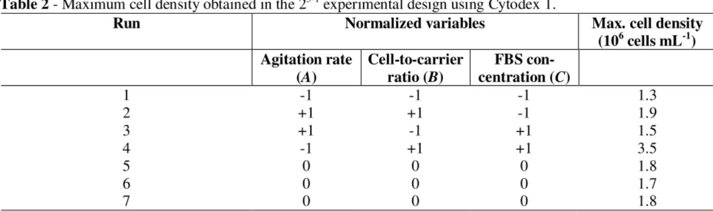
Since various factors affect mammalian cell growth on microcarriers (Sun et al., 2000; Murkhopadhyay et al., 1993; Amersham Pharmacia Biotech, 1999; Forestell et al., 1992; Ng et al., 1996), a fractional factorial experimental design was carried out to help identifying the most appropriate conditions for obtaining high cell densities on Cytodex 1 microcarriers. The experimental conditions tested (expressed as normalized values), as well as the maximum cell densities obtained in the different runs, are shown in Table 2. The lowest cell density (1.3 x 10⁶ cells mL-1) was obtained in experiment 1, when all variables were at their lowest limit (serum concentration: 1%; agitation: 40 rpm; cell-to-carrier ratio: 10 cells/µg carrier). On the other hand, cell density was highest (3.5 x 10⁶ cells mL -1
) in experiment 4, when agitation remained low (40 rpm), but serum concentration and cell-to-carrier ratio were at their highest levels (5% and 70 cells/µg carrier, respectively).
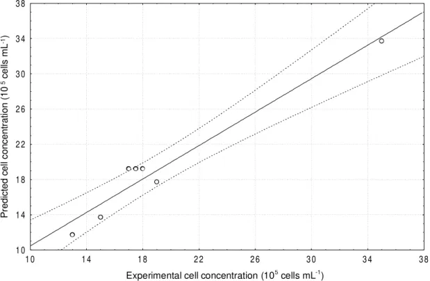
Through statistical analysis of the experimental results, it was verified that all the three variables tested were significant at a 95% confidence level. A linear model (Equation 2) was obtained to describe the experimental data (R = 0.974).
Table 2 - Maximum cell density obtained in the 23-1 experimental design using Cytodex 1.
Run Normalized variables Max. cell density (106 cells mL-1) Agitation rate
(A)
Cell-to-carrier ratio (B)
FBS con-centration (C)
1 -1 -1 -1 1.3
2 +1 +1 -1 1.9
3 +1 -1 +1 1.5
4 -1 +1 +1 3.5
5 0 0 0 1.8
6 0 0 0 1.7
7 0 0 0 1.8
C
4.5
B
6.5
A
3.5
-19.2
X
=
+
+
(2)where X is cell concentration (given in 105 cells mL-1), A is the normalized agitation rate, B is the normalized cell-to-carrier ratio, and C is the
normalized concentration of fetal bovine serum in the culture medium.
although the effect was negative (-3.5) for the agitation rate and positive for the SFB concentration (+4.5) and the cell-to-carrier ratio (+6.5). This was in agreement with the fact that the
highest cell density was obtained for a minimum agitation rate and maximum cell-to-carrier ratio and serum concentration (Table 2).
Concentração celular experimental (105 cél/mL)
Concentração celular prevista (10
5 cél/mL)
1 0 1 4 1 8 2 2 2 6 3 0 3 4 3 8
1 0 1 4 1 8 2 2 2 6 3 0 3 4 3 8
Experimental cell concentration (105 cells mL-1)
P
red
ic
ted
ce
ll
c
o
n
c
en
tr
a
ti
on
(10
5 c
e
lls
m
L
-1)
Figure 1 -Comparison of experimentally observed values and those predicted by Equation (2)
When compared to Vero cell propagation in T flasks (data not shown), the cell yield per medium volume obtained in experiment 4 was 3 times higher. Sun et al. (2000) studied the influence of the agitation rate in the range of 20 to 45 rpm on Vero cell adhesion onto Cytodex 3 microcarriers. They observed that low stirring velocities (20 rm) favoured cell adhesion, but led to a non-homogeneous cell distribution on microcarriers. At 45 rpm, cell adhesion rate was significantly decreased. They concluded that optimal agitation rate was 30 rpm, while in the present work using Cytodex 1 40 rpm was the most adequate stirring velocity.
Mukhopadhyay et al. (1993) studied the kinetics of cell adhesion on Cytodex 1, 2 and 3 microcarriers. They found the highest adhesion rate at a cell-to-carrier ratio of 15 cells per bead. Considering that (according to the microcarriers manufacturer) one gram of Cytodex 1 contained 4.37 x 10⁶ beads, the range studied in the present work was 2.3-16.3
cells per bead. Thus, the best results in the present work were obtained at 16.3 cells per bead, which were quite similar to those found by Mukhopadhyay et al. (1993).
Comparison of Vero cell growth on Cytodex 1 and Cytodex 3 microcarriers
Due to the different surface properties of different microcarriers, and due to the effects that these properties can have on growth kinetics of cell on microcarriers, in this work a comparison of Vero cell propagation on Cytodex 1 and Cytodex 3 was carried out. Cytodex 1 was formed by substituting a cross-linked dextran matrix with positively charged DEAE (N,N-diethylaminoethyl) groups. Cytodex 3 was also formed by a cross-linked dextran matrix, but was coated with a thin layer of denatured collagen, being indicated by the
manufacturer for cells with an epithelial-like morphology.
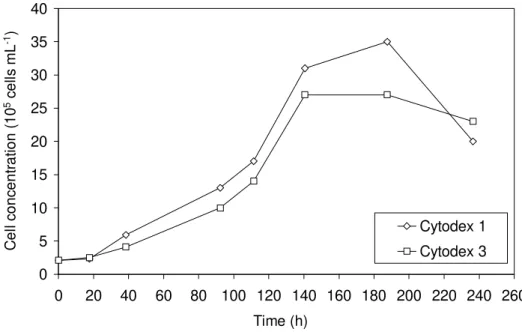
Vero cell propagation on these two microcarriers is shown in Fig. 2. For both microcarriers, under the conditions employed (agitation rate: 40 rpm; cell-to-carrier ratio: 70 cells per µg carrier; FBS concentration: 5%), a lag phase of approximately 20 h was observed. Maximum cell concentration
for Cytodex 3 (2.7 x 10⁶ cells
mL-1) was reached on day 6. For Cytodex 1, although maximum cell density (3.5 x 10⁶ cells mL-1) was reached only on day 8, it was approximately 30% higher than for Cytodex 3.
0 5 10 15 20 25 30 35 40
0 20 40 60 80 100 120 140 160 180 200 220 240 260
Tempo (horas)
Concentração celular (10
5 células/mL)
Cytodex 1
Cytodex 3
Time (h)
C
e
ll
c
on
c
en
tr
a
ti
on
(10
5 c
e
lls
m
L
-1)
Figure 2 - Total Vero cell concentration in spinner flasks with 3 mg mL-1 Cytodex 1 and Cytodex 3 microcarriers (agitation rate: 40 rpm; cell-to-carrier ratio: 70 cells per µg carrier; FBS concentration: 5%)
Although the bead mass was the same in both experiments, Cytodex 1 had a surface area of 4400 cm2 g-1, while Cytodex 3 had 2700 cm2 g-1. In order to evaluate if the earlier stationary phase and lower cell density obtained with Cytodex 3 was due to its lower surface area per gram microcarrier, the specific growth rate (µ) and duplication time (tD) of cells in both the carriers during the exponential growth phase were calculated. For Cytodex 1, the growth rate was 0.0194 h-1 and the duplication time was 35.7 h, while for Cytodex 3, µ was slightly reduced (0.0179 h-1), and tD was 39.8 h. These results indicated that cells on Cytodex 3 carriers may
have entered the stationary phase earlier due to the lower surface area available for growth.
CONCLUSIONS
It was shown that agitation rate in spinner flasks, FBS concentration in the culture medium and the ratio of inoculated cells to microcarriers exerted statistically significant effects on Vero cell growth on Cytodex 1 microcarriers. Within the ranges studied in this work, it was shown that a high cell-to-carrier ratio combined with a high FBS concentration and a low agitation rate resulted in high cell densities. Under such culture conditions, when compared to Cytodex 3, Cytodex 1 allowed a 30%-higher cell concentration to be obtained, which was probably related to the lower surface area per bead mass of Cytodex 3 microcarriers.
ACKNOWLEDGEMENTS
Thanks are due to Capes, CNPq, FAPERJ and Bio-Manguinhos/FIOCRUZ for the financial support.
RESUMO
Cultivos de células animais são amplamente utilizados para a produção de vacinas virais e para a expressão de proteínas recombinantes. A linhagem celular Vero é uma linhagem contínua, dependente de ancoragem, recomendada pela Organização Mundial de Saúde para a produção de vacinas de uso humano. Para a produção de vacinas virais em larga escala, vêm sendo cada vez mais empregados microcarregadores, que são microesferas que servem de suporte para as células. O emprego de microcarregadores em biorreatores agitados permite a obtenção de altas densidades celulares e, conseqüentemente, de altos títulos de antígenos virais. Com o objetivo de selecionar condições de cultivo adequadas, estudou-se, neste trabalho, o efeito das variáveis agitação, razão de células inoculadas por microcarregador e concentração de soro fetal bovino sobre o crescimento de células Vero em microcarregadores Cytodex 1. Nas melhores condições selecionadas, o desempenho dos microcarregadores Cytodex 1 e Cytodex 3 foi comparado.
REFERENCES
Amersham Pharmacia Biotech (1999), Microcarrier Cell
Culture: Principles and Methods. Amersham
Pharmacia Biotech, Uppsala.
Butler, M.; Burgener, A.; Patrick, M.; Berry, D.; Moffatt, D.; Huzel, N.; Barnabé, N. and Coombs, K. (2000), Application of a serum-free medium for the growth of Vero cells and production of reovirus. Biotechnol.
Prog., 16, 854-858.
Cruz, H. J.; Moreira, J. L. and Carrondo, M. J. T. (1999), Metabolic shifts by nutrient manipulation in continuous cultures of BHK cells. Biotechnol. Bioeng.,
66, 104-113.
Desmyter, J.; Melnick, J. L. and Rawls, W. E. (1968), Defectiveness of interferon production and of rubeolla virus interference in a line of African green monkey kidney cells (Vero). J. Virol., 2, 955-958.
Doyle, A. and Griffiths, B. (1998), Cell and Tissue
Culture: Laboratory Procedures in Biotechnology.
New York : John Wiley and Sons.
Forestell, S. P.; Kalogerakis, N.; Behie, L. A. and Gerson, D. F. (1992), Development of the optimal inoculation conditions for microcarrier cultures,
Biotechnol. Bioeng., 39, 305-313.
Frazzati-Gallina, N. M.; Paoli, R. L.; Mourão-Fuches, R. M.; Jorge, S. A. C. and Pereira, C. A. (2001), Higher production of rabies virus in serum-free medium cell cultures on microcarriers. J. Biotechnol., 92, 67-72. Kadouri, A. (1994), Cultivation of anchorage-dependent
mammalian cells and production of various metabolites.
Colloid Surf. B - Biointerfaces, 2, 265-272.
Mendonça, R. Z. and Pereira, C. A. (1995), High density Vero cell culture on microcarriers in a cell bioreactor.
Bioproc. Eng., 12, 279-282.
Mendonça, R. Z. and Pereira, C. A. (1998), Cell metabolism and medium perfusion in Vero cell cultures on microcarriers in a bioreactor. Bioproc. Eng., 18, 213-218.
Mukhopadhyay, A.; Mukhopadhyay, S. N. and Talwar, G. P. (1993), Influence of serum proteins on kinetics of attachment of Vero cells to Cytodex microcarriers. J.
Chem. Tech. Biotechnol., 56, 369-374.
Ng, Y. C.; Berry, J. M. and Butler, M. (1996), Optimization of physical parameters for cell attachment and growth on macroporous microcarriers.
Biotechnol. Bioeng., 50, 627-635.
Nikolai, T. J. and Hu, W. S. (1992), Cultivation of mammalian cells on macroporous microcarriers.
Enzyme Microb. Technol., 14, 203-208.
Spier, R. E. and Kadouri, A. (1997), The evolution of process for the commercial exploitation of anchorage-dependent animal cells. Enzyme Microb. Technol.,
21, 2-8.
Yasumara, Y. and Kawakita, M. (1963), The research for the SV40 by means of tissue culture technique. Nippon
Rinsho, 21, 1201-1219.