ISSN 0100-879X
CLINICAL INVESTIGATION
www.bjournal.com.br
www.bjournal.com.br
Volume 45 (9) 792-874 September 2012
Braz J Med Biol Res, September 2012, Volume 45(9) 851-855
doi:
10.1590/S0100-879X2012007500115
Mutation and genomic amplification of the
PIK3CA
proto-oncogene
in pituitary adenomas
C.B. Murat, P.B.S. Braga, M.A.H.Z. Fortes, M.D. Bronstein, M.L.C. Corrêa-Giannella and
R.R. Giorgi
Institutional Sponsors
The Brazilian Journal of Medical and Biological Research is partially financed by
Faculdade de Medicina de Ribeirão Preto Campus
Ribeirão Preto
Explore High - Performance MS Orbitrap Technology In Proteomics & Metabolomics
Mutation and genomic amplification of
the
PIK3CA
proto-oncogene in
pituitary adenomas
C.B. Murat
1, P.B.S. Braga
1, M.A.H.Z. Fortes
1, M.D. Bronstein
2,
M.L.C. Corrêa-Giannella
1and R.R. Giorgi
11Laboratório de Endocrinologia Celular e Molecular (LIM-25), Faculdade de Medicina,
Universidade de São Paulo, São Paulo, SP, Brasil
2Unidade de Neuroendocrinologia, Serviço de Endocrinologia, Hospital das Clínicas,
Faculdade de Medicina, Universidade de São Paulo, São Paulo, SP, Brasil
Abstract
The tumorigenesis of pituitary adenomas is poorly understood. Mutations of the PIK3CA proto-oncogene, which encodes the
p110-α catalytic subunit of PI3K, have been reported in various types of human cancers regarding the role of the gene in cell proliferation and survival through activation of the PI3K/Akt signaling pathway. Only one Chinese study described somatic mutations and amplification of the PIK3CA gene in a large series of pituitary adenomas. The aim of the present study was to determine genetic alterations of PIK3CA in a second series that consisted of 33 pituitary adenomas of different subtypes
diagnosed by immunohistochemistry: 6 adrenocorticotropic hormone-secreting microadenomas, 5 growth hormone-secreting macroadenomas, 7 prolactin-secreting macroadenomas, and 15 nonfunctioning macroadenomas. Direct sequencing of exons
9 and 20 assessed by qPCR was employed to investigate the presence of mutations and genomic amplification defined as a copy number ≥4. Previously identified PIK3CA mutations (exon 20) were detected in four cases (12.1%). Interestingly, the Chinese study reported mutations only in invasive tumors, while we found a PIK3CA mutation in one noninvasive corticotroph microadenoma. PIK3CA amplification was observed in 21.2% (7/33) of the cases. This study demonstrates the presence of
somatic mutations and amplifications of the PIK3CA gene in a second series of pituitary adenomas, corroborating the previously
described involvement of the PI3K/Akt signaling pathway in the tumorigenic process of this gland.
Key words: Pituitary adenomas; PIK3CA proto-oncogene; Genomic amplification; Somatic mutation
Introduction
Correspondence: R.R. Giorgi, Laboratório de Endocrinologia Celular e Molecular (LIM-25), Faculdade de Medicina, USP,
Av. Dr. Arnaldo, 455, 01246-903 São Paulo, SP, Brasil. Fax: +55-11-3061-8457. E-mail: [email protected]
Received January 14, 2012. Accepted June 28, 2012. Available online July 13, 2012. Published August 17, 2012. The tumorigenesis of pituitary adenomas is poorly
understood. The phosphatidylinositol 3-kinases (PI3Ks) are a group of heterodimeric lipid kinases composed of regulatory (85-kDa; p85) and catalytic (110-kDa; p110) subunits that play a key role in cell growth, proliferation, motility, and survival (1-3). The activation of PI3K results
in phosphorylation of phosphatidylinositol-4,5-biphosphate
(PIP2) to phosphatidylinositol-3,4,5-triphosphate (PIP3),
which acts as an anchor for Akt serine/threonine kinase and 3-phosphoinositide-dependent protein kinase-1 (PDK1),
and facilitates phosphorylation of Akt by PDK1, stimulat
-ing Akt activity and subsequent phosphorylation of several
proteins involved in a variety of intracellular processes, including antiapoptosis (4,5). PIP3 levels are tightly
regu-lated by the action of phosphatases such as phosphatase and tensin homologue (PTEN) (6).
Mutations of the PIK3CA proto-oncogene, which
en-codes the p110-α catalytic subunit of PI3K, have been
reported in various types of human cancers (7-10) and more than 80% of PIK3CA mutations are clustered in the
helical domain encoded by exon 9 and in the kinase domain
encoded by exon 20 (11), as shown with high prevalence in endometrioid, esophageal and pancreatic carcinomas (12-15).
A recent Chinese study (16) has described somatic
mutations and amplifications of the PIK3CA gene in a
852 C.B. Murat et al.
gland. Since a diversity of mutational spectra in different populations had been described for certain neoplasias (17,18), in the present study we investigated genetic altera-tions in the PIK3CA proto-oncogene in a second series of pituitary adenomas.
Material and Methods
Tissue specimens and DNA isolation
Tissue collection was carried out in compliance with the Institutional Ethics Committee (CAPPesq) and in
ac-cordance to the Declaration of Helsinki, with informed and
free written consent being required from each subject or subject’s guardian. From 1994-2009, tumor tissues were obtained from patients diagnosed with pituitary adenomas. During surgery, tumor fragments were collected into sterile containers and immediately frozen in liquid nitrogen. Thirty-three sporadic pituitary adenomas were obtained after surgery. The anatomo-pathological characteristics of these adenomas are shown in Table 1. Pituitary adenomas con-sisted of 6 adrenocorticotropic hormone (ACTH)-secreting microadenomas, 5 growth hormone (GH)-secreting mac-roadenomas, 7 prolactin (PRL)-secreting macmac-roadenomas, and 15 nonfunctioning (NF) macroadenomas. As control, four normal pituitary tissues were obtained within 8 h post mortem from subjects without endocrine diseases. The tissue fragments were fragmented in a tissue pulverizer
(Mikro-Dismembrator U, B. Braun Melsungen, Germany). Tumor DNA was extracted using the DNeasy kit (Qiagen,
USA) according to manufacturer instructions.
Mutation analysis
Exons 9 and 20 of PIK3CA were analyzed by direct
sequencing on an ABI 3130X Genetic Analyzer (Applied Biosystems, USA) after PCR amplification of genomic DNA.
PCR was carried out in 50 µL containing 100 ng genomic DNA, 0.2 mM of each primer, 200 µM deoxynucleotides, 1X buffer and 1 U DNA Taq polymerase (GE Healthcare, USA). The nucleotide sequences for the primers were designed using
the Primer3 software: exon 9 forward 5’-CAAAGCAATTTCT ACACGAGATCC-3’; exon 9 reverse 5’-GTAAAAACATGCTG AGATCAGCCACAT-3’; exon 20 forward 5’-TGGAATGCC AGAACTACAATCTTT-3’, and exon
20 reverse 5’-GGTCTTTGCCTGCTG
AGAGTT-3’ (Invitrogen, USA). Reac
-tions were carried out under the following
cycling conditions: 95°C for 5 min, 35 cycles of 95°C for 30 s, followed by 56°C (exon 9) or 62°C (exon 20) for 30 s and
72°C for 10 min. The PCR products were subjected to direct sequencing with the use of BigDye Terminator sequencing reagents (Applied Biosystems) with the following cycles: 96°C for 15 s, 50°C for
15 s and 60°C for 4 min for 35 cycles. All
mutated DNA sequences were confirmed by sequencing in
the reverse direction.
Copy number analysis
PIK3CA gene copy number amplification was assessed
by qPCR using Platinum SYBRGreen qPCR SuperMix-UDG (Invitrogen), 100 ng DNA and primers for genomic sequences (PIK3CA forward: 5’-ATCTTTTCTCAATGATGC
TTGGCT-3’ and PIK3CA reverse: 5’-CTAGGGTGTTTCG
AATGTATG-3’) and compared with the signal obtained from
COL7A1 as reference gene (COL7A1 forward: 5’-ACC
CAGTACCGCATCATTGTG-3’ and COL7A1 reverse: 5’-TC
AGGCTGGAACTTCAGTGTG-3’).
PCR was carried out on a Rotor-Gene 6000 instrument (Corbett Life Science, Australia). The reaction consisted of an initial incubation at 95°C for 10 min followed by 40 cycles of 95°C for 15 s and 55°C for 1 min. Fluorescence changes were monitored after each cycle. Melting curve analysis
was performed at the end of each reaction to confirm PCR product’s identity (72°C ramping to 99°C at 0.2°C/s with continuous fluorescence readings). Standard curves were constructed to assess the amplification efficiency of
PIK3CA and COL7A1 using duplicate serial dilutions with seven different DNA concentrations (400 to 6.25 ng) from a reference sample of DNA obtained from normal pituitary
tissue. Since equal amplification efficiencies of target and
reference genes were attained (E >0.9), the mathematical model 2-ΔΔCt was used to evaluate the relative genomic
amplification of PIK3CA. All samples were run in duplicate.
DNA obtained from a pool of four normal pituitary tissues was included in the assay as a calibrator sample and DNA extracted from the MCF-7 cell line was used as a positive control for PIK3CA amplification. DNA amplification was
defined as values ≥4 (16).
Results
PIK3CA mutations
Previously reported PIK3CA mutations were detected
in four cases (12%): 3 of 15 (20%) NF pituitary adenomas
and 1 of 6 (16.6%) ACTH-secreting tumors, both in exon 20 (Table 2). No mutations were found in exon 9.
Table 1. Demographic data and anatomo-pathological findings of 33 patients with
pituitary adenomas who participated in the present study.
Pituitary adenomas N Gender
(male/female) Age (years) (micro/macroadenoma)Tumor size
ACTH-secreting 6 4/2 32.3 (13-61) 6/0 GH-secreting 5 2/3 30.2 (23-55) 0/5 PRL-secreting 7 1/6 31 (20-64) 0/7 Nonfunctioning 15 6/9 44 (21-70) 0/15
ACTH = adrenocorticotropic hormone; GH = growth hormone; PRL = prolactin; mi
Amplification of PIK3CA
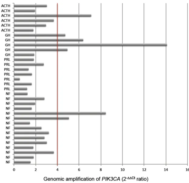
The number of copies of the PIK3CA gene in each
tumor sample is shown in Figure 1. Genomic amplification
was observed in 21.2% (7/33) of the cases; in 4 of 5
(80%) GH-secreting adenomas, 1 of 6 (16.6%)
ACTH-secreting adenomas and in 2 of 15 (13.3%) NF pitu
-Figure 1. PIK3CA gene copy number detectedby qPCR in different subtypes of pituitary adenomas. Positive amplification isdefined
as ≥4 copies. ACTH = adrenocorticotropic hormone-secreting adenoma; GH = growth hormone-secreting macroadenoma; PRL = prolactin-secreting macroadenoma; NF = nonfunctioning pituitary adenoma.
Table 2.PIK3CA mutations found in four pituitary adenomas.
Exon Functional domain PIK3CA DNA mutation Amino acid Subtype of tumor 20 Kinase domain GCC > GAC, C2994A Ala995Asp, A > D ACTH
20 Kinase domain GGC > GGT, C3031T Gly1007Gly, G > G NF
20 Kinase domain TCT > CCT, T3053C Ser1015Pro, S > P NF
20 Kinase domain CAA > CGA, A3108G Gln1033Arg, Q > R NF
854 C.B. Murat et al.
itary adenomas. No genomic amplification was detected
in PRL-secreting adenomas.
Discussion
Several studies have implicated PIK3CA somatic
muta-tions and genomic amplification in the tumorigenic process
of different human cancers (19-26). In the present study,
we investigated mutations and genomic amplification of the
PIK3CA proto-oncogene in 33 sporadic pituitary adenomas.
During our experiments, another group of investigators evaluated the association of these genetic alterations in pituitary tumors (16). Given the diversity of mutational spectra in different populations for certain neoplasias (17,18), we decided to continue to examine the incidence
of these mutations and amplifications in pituitary tumors
of Brazilian patients.
The overall frequency of PIK3CA gene mutations and
amplification in the Chinese study of 353 pituitary tumors
was 2.3% (8.8% of the invasive tumors and 0% of the non
-invasive ones) and 28% (26.3% of the non-invasive tumors and 32.9% of the invasive ones), respectively (16), while in
the current series, PIK3CA mutations and amplification were
found in 12.1 and 21.2% of the cases, respectively.
Although it is difficult to compare studies that differ so
much in the size of the evaluated population, the present study corroborates the previous report (16) that genetic alterations in the PIK3CA gene are found in pituitary tu-mors. We only detected mutations in one ACTH-secreting and three NF pituitary adenomas, while Lin et al. (16) also found mutations in two PRL-secreting tumors. Mutations in GH-secreting tumors were not found in any of the two series. Interestingly, the Chinese study reported mutations only in invasive tumors, while we found a PIK3CA mutation in one noninvasive corticotroph microadenoma. Recent reports have shown that PIK3CA knockdown reduces proliferation
and invasiveness in different cell lines (27-29).
Defining positive gene amplification as a copy number ≥4, ACTH-secreting tumors (16.6%), NF pituitary tumors (13.3%) and particularly GH-secreting tumors (80%) were
identified as harboring PIK3CA amplifications, with the
highest number being 14 copies in a GH-secreting tumor. Adding to the previous Chinese study, PIK3CA amplification
was more common than mutations. Amplification of PIK3CA
has been demonstrated to be a mechanism of resistance
for PI3K-targeted therapy in cancer (30).
The coexistence of PIK3CA mutations with amplifica
-tions in pituitary adenomas seems to be rare; only one case
(NF tumor) harbored coexisting PIK3CA copy gain and a gene mutation. These findings suggest that PIK3CA
muta-tion and amplificamuta-tion are independent oncogenic pathways
in pituitary tumors (16), as mutually exclusive mechanisms,
as already described for thyroid tumors (31); however, the
coexistence of these genetic alterations has been reported
in breast cancers (32).
The major effector of PI3K is Akt kinase, which phospho
-rylates target proteins regulating key processes such as cell
proliferation and survival. This process is counteracted by the phosphatase encoded by the PTEN tumor suppressor
gene, which opposes PI3K activity (33). A recent study has implicated phosphorylated Akt expression associated with
PIK3CA mutations in low-stage colorectal cancers (34).
In 2005, Musat et al. (6) reported a higher expression of
phosphorylated Akt in pituitary tumors in comparison to the
normal gland in the absence of PTEN mutations. Thus, it is
reasonable to suppose that increased Akt phosphorylation in pituitary adenomas could result from activation of the PI3K
pathway following PIK3CA mutation or amplification.
In summary, this study demonstrates the presence of
somatic mutations and amplifications of the PIK3CA gene
in a second series of pituitary adenomas, corroborating the
previously described involvement of the PI3K/Akt signaling pathway in the tumorigenesis of this gland (16,35,36) and its molecular potential for targeted therapies (3,27-29,37)
1. Samuels Y, Diaz LA Jr, Schmidt-Kittler O, Cummins JM, DeLong L, Cheong I, et al. Mutant PIK3CA promotes cell
growth and invasion of human cancer cells. Cancer Cell
2005; 7: 561-573.
2. Samuels Y, Ericson K. Oncogenic PI3K and its role in cancer.
Curr Opin Oncol 2006; 18: 77-82.
3. Liu P, Cheng H, Roberts TM, Zhao JJ. Targeting the phos
-phoinositide 3-kinase pathway in cancer. Nat Rev Drug Discov 2009; 8: 627-644.
4. Raimondi C, Falasca M. Targeting PDK1 in cancer. Curr Med Chem 2011; 18: 2763-2769.
5. Vivanco I, Sawyers CL. The phosphatidylinositol 3-kinase AKT pathway in human cancer. Nat Rev Cancer 2002; 2:
489-501.
6. Musat M, Korbonits M, Kola B, Borboli N, Hanson MR, Nan
-zer AM, et al. Enhanced protein kinase B/Akt signalling in
pituitary tumours. Endocr Relat Cancer 2005; 12: 423-433.
7. Gallia GL, Rand V, Siu IM, Eberhart CG, James CD, Marie
SK, et al. PIK3CA gene mutations in pediatric and adult glio -blastoma multiforme. Mol Cancer Res 2006; 4: 709-714.
8. Kozaki K, Imoto I, Pimkhaokham A, Hasegawa S, Tsuda H, Omura K, et al. PIK3CA mutation is an oncogenic aberra -tion at advanced stages of oral squamous cell carcinoma.
Cancer Sci 2006; 97: 1351-1358.
9. Woenckhaus J, Steger K, Sturm K, Munstedt K, Franke FE, Fenic I. Prognostic value of PIK3CA and phosphorylated AKT expression in ovarian cancer. Virchows Arch 2007; 450:
387-395.
10. Santarpia M, Altavilla G, Margeli M, Cirauqui B, Mesiti M,
in breast cancer: potential biomarkers for chemoresistance.
Cancer Invest 2008; 26: 1044-1051.
11. Samuels Y, Wang Z, Bardelli A, Silliman N, Ptak J, Szabo S, et al. High frequency of mutations of the PIK3CA gene in
human cancers. Science 2004; 304: 554.
12. Hayes MP, Wang H, Espinal-Witter R, Douglas W, Solomon
GJ, Baker SJ, et al. PIK3CA and PTEN mutations in uterine
endometrioid carcinoma and complex atypical hyperplasia.
Clin Cancer Res 2006; 12: 5932-5935.
13. Rudd ML, Price JC, Fogoros S, Godwin AK, Sgroi DC, Me
-rino MJ, et al. A unique spectrum of somatic PIK3CA (p110al -pha) mutations within primary endometrial carcinomas. Clin Cancer Res 2011; 17: 1331-1340.
14. Phillips WA, Russell SE, Ciavarella ML, Choong DY,
Mont-gomery KG, Smith K, et al. Mutation analysis of PIK3CA and PIK3CB in esophageal cancer and Barrett’s esophagus. Int J Cancer 2006; 118: 2644-2646.
15. Schonleben F, Allendorf JD, Qiu W, Li X, Ho DJ, Ciau NT, et
al. Mutational analyses of multiple oncogenic pathways in intraductal papillary mucinous neoplasms of the pancreas.
Pancreas 2008; 36: 168-172.
16. Lin Y, Jiang X, Shen Y, Li M, Ma H, Xing M, et al. Frequent
mutations and amplifications of the PIK3CA gene in pituitary
tumors. Endocr Relat Cancer 2009; 16: 301-310.
17. Minamoto T, Mai M, Ronai Z. Environmental factors as regulators and effectors of multistep carcinogenesis. Car-cinogenesis 1999; 20: 519-527.
18. Pernick NL, Sarkar FH, Philip PA, Arlauskas P, Shields AF, Vaitkevicius VK, et al. Clinicopathologic analysis of pancre -atic adenocarcinoma in African Americans and Caucasians.
Pancreas 2003; 26: 28-32.
19. Samuels Y, Velculescu VE. Oncogenic mutations of PIK3CA
in human cancers. Cell Cycle 2004; 3: 1221-1224.
20. Angulo B, Suarez-Gauthier A, Lopez-Rios F, Medina PP, Conde E, Tang M, et al. Expression signatures in lung cancer
reveal a profile for EGFR-mutant tumours and identify selec
-tive PIK3CA overexpression by gene amplification. J Pathol
2008; 214: 347-356.
21. Hou P, Liu D, Shan Y, Hu S, Studeman K, Condouris S, et
al. Genetic alterations and their relationship in the
phos-phatidylinositol 3-kinase/Akt pathway in thyroid cancer. Clin Cancer Res 2007; 13: 1161-1170.
22. Okudela K, Suzuki M, Kageyama S, Bunai T, Nagura K, Iga
-rashi H, et al. PIK3CA mutation and amplification in human
lung cancer. Pathol Int 2007; 57: 664-671.
23. Wu G, Xing M, Mambo E, Huang X, Liu J, Guo Z, et al. So
-matic mutation and gain of copy number of PIK3CA in human
breast cancer. Breast Cancer Res 2005; 7: R609-R616.
24. Yamamoto S, Tsuda H, Takano M, Iwaya K, Tamai S, Matsubara O. PIK3CA mutation is an early event in the
development of endometriosis-associated ovarian clear cell
adenocarcinoma. J Pathol 2011; 225: 189-194.
25. Choy E, Hornicek F, Macconaill L, Harmon D, Tariq Z, Gar -raway L, et al. High-throughput genotyping in osteosarcoma
identifies multiple mutations in phosphoinositide-3-kinase
and other oncogenes. Cancer 2012; 118: 2905-2914.
26. Ji M, Guan H, Gao C, Shi B, Hou P. Highly frequent promoter
methylation and PIK3CA amplification in non-small cell lung
cancer (NSCLC). BMC Cancer 2011; 11: 147.
27. Weber GL, Parat MO, Binder ZA, Gallia GL, Riggins GJ. Abrogation of PIK3CA or PIK3R1 reduces proliferation,
migration, and invasion in glioblastoma multiforme cells.
Oncotarget 2011; 2: 833-849.
28. Zhou XK, Tang SS, Yi G, Hou M, Chen JH, Yang B, et al. RNAi knockdown of PIK3CA preferentially inhibits invasion of mutant PIK3CA cells. World J Gastroenterol 2011; 17:
3700-3708.
29. Guerreiro AS, Fattet S, Fischer B, Shalaby T, Jackson SP, Schoenwaelder SM, et al. Targeting the PI3K p110alpha
isoform inhibits medulloblastoma proliferation, chemore-sistance, and migration. Clin Cancer Res 2008; 14:
6761-6769.
30. Liu P, Cheng H, Santiago S, Raeder M, Zhang F, Isabella A, et al. Oncogenic PIK3CA-driven mammary tumors frequent
-ly recur via PI3K dependent and PI3K
pathway-independent mechanisms. Nat Med 2011; 17: 1116-1120.
31. Wang Y, Hou P, Yu H, Wang W, Ji M, Zhao S, et al. High
prevalence and mutual exclusivity of genetic alterations in
the phosphatidylinositol-3-kinase/akt pathway in thyroid
tumors. J Clin Endocrinol Metab 2007; 92: 2387-2390.
32. Kadota M, Sato M, Duncan B, Ooshima A, Yang HH, Diaz-Meyer N, et al. Identification of novel gene amplifications in breast cancer and coexistence of gene amplification with an activating mutation of PIK3CA. Cancer Res 2009; 69:
7357-7365.
33. Tena-Suck ML, Ortiz-Plata A, de la Vega HA. Phosphatase
and tensin homologue and pituitary tumor-transforming gene in pituitary adenomas. Clinical-pathologic and immunohis-tochemical analysis. Ann Diagn Pathol 2008; 12: 275-282.
34. Baba Y, Nosho K, Shima K, Hayashi M, Meyerhardt JA, Chan AT, et al. Phosphorylated AKT expression is associated with PIK3CA mutation, low stage, and favorable outcome in 717
colorectal cancers. Cancer 2011; 117: 1399-1408.
35. Suhardja A, Kovacs K, Rutka J. Role of transcription fac -tors in the pathogenesis of pituitary adenomas: a review. J Neurooncol 2001; 55: 185-193.
36. Grossman AB, Korbonits M. Akting and cycling: a tale of the
pituitary. Horm Res 2004; 62 (Suppl 3): 117-123.
37. Jamieson S, Flanagan JU, Kolekar S, Buchanan C, Kendall JD, Lee WJ, et al. A drug targeting only p110alpha can block phosphoinositide 3-kinase signalling and tumour growth in