Sm 1 4 ge ne e xpre ssio n in diffe re nt
stage s o f the
Schisto so m a m anso ni
life
cycle and im m uno lo calizatio n o f the
Sm 1 4 pro te in within the adult wo rm
1Laboratório de Imunologia de Doenças Infecciosas, Departamento de
Bioquímica e Imunologia, Instituto de Ciências Biológicas, Universidade Federal de Minas Gerais, Belo Horizonte, MG, Brasil 2Department of Biology, University of York, York, UK
3Instituto Ludwig de Pesquisas sobre o Câncer, São Paulo, SP, Brasil 4Laboratório de Imunologia, Centro de Pesquisas René Rachou,
Belo Horizonte, MG, Brasil C.F.A. Brito1, G.C. O liveira4,
S.C. O liveira1, M. Street2, S. Riengrojpitak2, R.A. Wilson2, A.J.G. Simpson3 and R. Correa-O liveira4
Abstract
Sm14 is a 14-kDa vaccine candidate antigen from Schistosoma man-soni that seems to be involved in cytoplasmic trafficking of fatty acids.
Although schistosomes have a high requirement for lipids, they are not able to synthesize fatty acids and sterols de novo. Thus, they must
acquire host lipids. In order to determine whether Sm14 is present in different stages of the life cycle of the parasite, we performed RT-PCR. Sm14 mRNA was identified in all stages of the life cycle studied, mainly schistosomulum, adult worm and egg. Additionally, we used a rabbit anti-Sm14 polyclonal antibody in an indirect immu-nofluorescence assay to localize Sm14 in adult worm sections. The basal lamella of the tegument and the gut epithelium were strongly labeled. These tissues have a high flow of and demand for lipids, a finding that supports the putative role of Sm14 as an intracellular transporter of fatty acids from host cells.
Co rre spo nde nce
C.F.A. Brito
Laboratório de Imunologia de Doenças Infecciosas, ICB, UFMG Av. Antônio Carlos, 6627 31270-901 Belo Horizonte, MG Brasil
Fax: + 55-31-3499-2666 E-mail: cbrito@ icb.ufmg.br
Research supported by CAPES, FAPEMIG, CNPq, CPqRR and EEC. Publication supported by FAPESP.
Received September 3, 2001
Accepted January 2, 2002
Ke y wo rds
·Sm14
·Fatty acid-binding protein ·FABP
·Immunolocalization ·RT-PCR
Schistosomiasis is a chronic parasitic dis-ease which affects more than 200 million people worldwide causing extensive liver damage (1). Although drugs are available to treat schistosomiasis, the large extension of endemic areas and the constant reinfections make chemotherapy an ineffective approach to control the disease (2). Therefore, much effort has been devoted to research for vac-cine development. Recently, the World Health Organization selected six molecule candi-dates to compose a subunit vaccine against schistosomiasis (3). One of them is Sm14, a 14-kDa Schistosoma mansoni antigen that induces partial protection in mice following
vaccination and cercarial challenge (4). Sm14 is a cytoplasmic fatty acid-binding protein (FABP) and its ability to bind to palmitic and linolenic acids in vitro has been demon-strated (5).
syn-thesize fatty acids and sterols de novo, and thus must acquire lipids from host cells (7). The parasite is capable of synthesizing only phospholipids and triacylglycerols from pre-cursors obtained from the host such as the low-density lipoproteins (LDL) of the blood in which the parasites reside (8). LDL recep-tors have been identified on the surface of schistosome membranes (9). The fraction of fatty acids can subsequently be modified, mainly by chain elongation (7).
FABPs appear to be involved in the ac-quisition and utilization of fatty acids in different organisms (10). There are at least two main classes of FABPs, a cytoplasmic and a plasmalemmal FABP (11). Cytoplas-mic FABP has been identified in a variety of parasites, including Fasciola hepatica (12),
Schistosoma mansoni (5) and S. japonicum
(13). Plasmalemmal FABP seems not to be present in trematodes.
The aim of the present study was to first determine the gene expression profile of Sm14 in different stages of the life cycle of the parasite and then to localize the Sm14 protein within adult S. mansoni by indirect immunola-beling using rabbit anti-Sm14 polyclonal se-rum followed by fluorescence microscopy.
For detection of Sm14 in different stages of the parasite life cycle, we used RT-PCR. Eggs were recovered from macerated liver of infected mice and adult worms were per-fused from the portal vein of 42-day-in-fected mice. The cercariae were obtained from Biomphalaria glabrata infected with a single miracidium in vitro and recovered using artificial light. The schistosomula were mechanically transformed according to Ramalho-Pinto et al. (14) and cultured in vitro in RPMI 1640 medium (Gibco BRL, Rockville, MD, USA) until day 7 that corre-sponds to the lung stage worm.
Total RNA was extracted from newly transformed schistosomula, in vitro cultured schistosomula, adult worms, cercariae and eggs using the Rneasy total RNA system (Qiagen Inc., Valencia, CA, USA). The RNA
samples were quantified by spectrophotom-etry and 2.5 µg was used to synthesize cDNAs using oligo d(T) as a primer. cDNAs were then amplified using specific primers for Sm14 (Sm14f: 5'-CGGTGGTCGTTCAA GCGTATCAGAAGC-3' and Sm14r: 5'- CCCTCGCAAATATCAGTGTTCATTTG-3'). To standardize the quantities of cDNA used, we also amplified the cytochrome oxi-daseC1 gene (housekeeping) from S. man-soni using specific primers (CoxF: 5'-AAACTGCGAGGTTGAGCAGG-3' and CoxR: 5'-CAACACTAAACATAAAAA CAGATAG-3'). Five pmoles of each primer was used in PCR, which consisted of a dena-turation step at 90ºC for 1 min, a primer annealing step at 50ºC for 30 s, and an extension step at 72ºC for 1 min. This cycle was repeated 30 times for Sm14 and 22 times for Cox amplification. The samples were resolved on 4% acrylamide gels and stained with ethidium bromide and densi-tometry analysis of bands was carried out using the ImageMaster VDS®
videodocu-mentation system and software (1996, ver-sion 2.0; Pharmacia Biotech Inc., San Fran-cisco, CA, USA).
For immunofluorescence analysis, the specific antiserum was produced by immu-nization of two rabbits with recombinant Sm14 (4). The rabbits were injected three times at two-week intervals with 50 µg of recombinant Sm14 emulsified in Freunds complete adjuvant subcutaneously and the last immunization was performed without adjuvant. Sera were collected 10 days after the final boost. The specificity of the poly-clonal antibody was confirmed by Western blot analysis (data not shown).
S. mansoni Puerto Rican strain main-tained in BALB/c mice was used in the immunolocalization assay. Adult worms were obtained by perfusion of mice 42 days after infection (15). The recovered parasites were maintained in RPMI 1640 culture medium (Gibco BRL). About eight worms were in-cluded in resin (Tissue-Tek®
compound; Sakura Finetek, Torrance, CA, USA) and quickly frozen by immersion in liquid nitrogen. These blocks were cut with a microtome at -20ºC into sections of approxi-mately 6-7 µm. The cryosections were fixed with acetone on glass slides for 3-4 min at room temperature. Nonspecific binding sites were blocked by incubation of slides with naive goat serum diluted 1:10 in 0.15 M PBS, pH 7.2, for 30 min at room tempera-ture. A rabbit anti-Sm14 antibody diluted 1:100 in PBS plus 10% (v/v) naive goat serum was added to each slide and kept at room temperature for 1 h. As a negative control, we used normal rabbit serum as primary antibody (data not shown). Unbound antibodies were removed with three washes of 10 min each with PBS. Slides were then incubated with goat anti-rabbit IgG conju-gated with FITC (fluorescein isothiocyanate, Vector Laboratories, Burlingame, CA, USA) diluted 1:50 in 10% (v/v) naive goat serum. Slides were then washed in PBS again, dried and covered with Vectashield®
fluorescent medium (Vector Laboratories) before a cov-erslip was applied. Slides were then kept at 4ºC in the dark and immunofluorescence was observed using a fluorescence micro-scope (Nikon, Melville, NY, USA).
Using RT-PCR, we were able to identify transcripts for Sm14 in all parasite stages stud-ied (Figure 1A). Using the values of Cox amplification to normalize the Sm14 mRNA levels we plotted a graph with the mean value of two experiments performed independently to determine the relative quantities of Sm14 mRNA (Figure 1B). Higher levels of Sm14 transcripts were detected in 6- and 7-day-cultured schistosomula, adult worms or eggs compared to the other parasite life cycle stages studied. Since the lung schistosomulum of S. mansoni is the main target of protective immu-nity in vaccinated mice and there is evidence of cross-reactivity among schistosomulum, adult worm, or egg antigens, we speculate that the higher expression of Sm14 in these para-site stages may be related to the ability of this
antigen to induce protection against infection in mice. However, further studies should be performed using a more powerful technique such as real-time PCR to confirm the Sm14 expression levels found within these parasite stages.
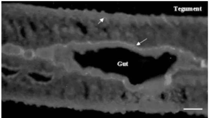
Figure 2 shows a longitudinal section of a
Figure 2. Localization of Sm14 by indirect immunofluorescence staining in adult w orm cryosections. The sections reacted w ith rabbit anti-Sm14 polyclonal antibody diluted 1:100. The arrow s indicate the basal lamella of the gut and of the tegument. Bar = 100 µm.
Sm14
Cox
R
e
la
ti
v
e
q
u
a
n
ti
ti
e
s
o
f
S
m
1
4
m
R
N
A
25
20
15
10
5
0
0 2 4 5 6 7 AW Cc Egg
Life cycle stages
A
B
Figure 1. A, Amplification of cDNA from a Schistosoma mansoni new ly transformed schistosomulum (0), and from 2, 4, 5, 6 or 7-day-cultured schistosomula; adult w orm (AW); cercariae (Cc) or egg using specific primers for Sm14 or Cox. B, The data represent relative quantities of specific Sm14 mRNA in relation to the Cox transcripts (intensities of Sm14 in pixels x 100/intensities of Cox in pixels).
male adult worm where the gut is clearly visible in the center. The major structure sharply delineated is the basal lamella of the gut which was revealed as a thin bright line in the immunofluorescence reaction. The basal lamella underlying the tegument was also strongly positive. Therefore, Sm14 was localized by anti-Sm14-specific antibodies in the basal lamella of the gut and underlying the tegument. Both tissues are related to flow of and demand for fatty acids and these results corroborate the putative role for Sm14. As a positive control, we used a rabbit anti-SWAP IgG as the primary antibody and the labeling observed was not localized to spe-cific structures, but was broadly distributed throughout all tissues (data not shown). A previous study by Moser et al. (5) demon-strated that Sm14 was present on tubercles, which are structures located on the dorsal surface of the parasite rich in lipid globules, but not in the muscle layers or the tegument. Thus, our findings bring new insights into the localization and putative function of Sm14 in S. mansoni.
The localization of Sm14 shown in this study is related to other FABPs, which are preferably localized in tissues with a high flow of or demand for fatty acids (10). In mammals, there are three major cytoplasmic
FABPs: intestinal, heart and liver FABPs. Intestinal and liver FABPs are abundant in the gut epithelium, associated with delivery and metabolism of fatty acids from the diet (16). Heart FABP is expressed at high levels in the myocardium, associated with ß-oxida-tion to generate power to the muscle (17). Although Sm14 has a higher phylogenetic relationship to heart FABP, it is known that
S. mansoni is unable to use lipids as a power source (18,19).
Furthermore, Sm14 is localized in tissues near the interfaces of parasite/host contact, such as the basal lamella of the tegument and the epithelium of the gut. This suggests a putative role for Sm14 as a second element in the uptake and transport of fatty acids, but it is not clear whether this uptake occurs through the tegument or the gut because both tissues are related to the flow of and demand for fatty acids. Hockley and McLaren (20) demonstrated that the captured fatty acids go to the basal lamella of the tegument and are stored in the gut or in the esophageal gland. Furthermore, our results are similar to Goberts findings (21), who recently local-ized S. japonicum FABP in the subtegumental region of male adult worms and vitellinic glands of female worms using electron mi-croscopy and colloidal gold staining.
Re fe re nce s
1. World Health Organization (1993). Expert Committee on the Control of Schistoso-miasis. WHO Technical Report Series No. 830, WHO, Geneva, Sw itzerland. 2. Pearce EJ (1993). Proselytizing w ith
im-munity. Nature, 363: 19-20.
3. Bergquist NR (1995). Schistosomiasis vac-cine development: approaches and pros-pects. M emórias do Instituto Osw aldo Cruz, 90: 221-227.
4. Tendler M , Brito CFA, Serra-Freire N, Diogo CM , Almeida M S, Delbem ACB, Silva JF, Savino W, Garrat RC, Katz N & Simpson AJG (1996). Evidence from mo-lecular modeling and experimental immu-nization that a Schistosoma mansoni fatty acid binding protein Sm14 is the potential
basis of a dual-purpose anti-helminth vac-cine. Proceedings of the National Acade-my of Sciences, USA, 92: 269-273. 5. M oser D, Tendler M , Griffiths G & Klinkert
M Q (1991). A 14-kDa Schistosoma man-soni polypeptide is homologous to a gene family of fatty acid binding proteins. Jour-nal of Biological Chemistry, 266: 8447-8454.
6. Furlong ST (1991). Unique roles for lipids in Schistosoma mansoni. Parasitology To-day, 7: 59-62.
7. M eyer F, M eyer H & Bueding E (1970). Lipid metabolism in the parasite and free-living flat w orms, Schistosoma mansoni
and Dugesia dorotocephala. Biochimica et Biophysica Acta, 210: 256-266.
8. Rogers M V (1991). Do parasites express receptors for host lipoproteins? Parasitol-ogy Today, 7: 117-120.
9. Rumjanek FD, Campos EG & Alonso LCC (1988). Evidence for the occurrence of LDL receptors in the extracts of schisto-somula of Schistosoma mansoni. M olec-ular and Biochemical Parasitology, 28: 145-152.
10. Kaikaus RM , Bass NM & Ockner RK (1990). Functions of the fatty acid binding proteins. Experientia, 46: 617-630. 11. Glatz JF & Van der Vusse GJ (1990).
No-menclature of fatty acid-binding proteins.
M olecular and Cellular Biochemistry, 98: 231-235.
Garcia-Blanco M A & Hillyer GV (1992).
Fasciola hepatica: molecular cloning, nu-cleotide sequence, and expression of a gene encoding a polypeptide homologous to a Schistosoma mansoni fatty acid bind-ing protein. Experimental Parasitology, 74: 400-407.
13. Becker M M , Kalinna BH, Waine GJ & M cM anus DP (1994). Gene cloning, over-production and purification of a function-ally active cytoplasmic fatty acid-binding protein (Sj-FABPc) from the human blood fluke Schistosoma japonicum. Gene, 148: 321-325.
14. Ramalho-Pinto FJ, Gazzinelli G, How ells RE, M ota-Santos TA, Figueiredo EA & Pellegrino J (1974). Schistosoma man-soni: defined system for stepw ise
trans-formation of cercaria to schistosomule in vitro. Experimental Parasitology, 36: 360-372.
15. Smithies SR & Terry RJ (1965). Naturally-acquired resistance to experimental infec-tions of Schistosoma mansoni in the rhesus monkey (M acaca mulatta). Parasi-tology, 55: 701-710.
16. Shields HM , Bates M L, Bass NM , Best CJ, Alpers DH & Ockner RK (1986). Light microscopic immunocytochemical local-ization of hepatic and intestinal types of fatty acid-binding proteins in rat small in-testine. Journal of Lipid Research, 27: 549-557.
17. M organ HE, Rannels DE & Kao RL (1974). Factors controlling protein turnover in heart muscle. Circulation Research, 35:
22-31.
18. Esteves A, Joseph L, Paulino M & Ehrlich R (1997). Remarks on the phylogeny and structure of fatty acid binding proteins from parasitic plathelminths. International Journal for Parasitology, 27: 1013-1023. 19. Beuding E (1950). Carbohydrate
metabo-lism of Schistosoma mansoni. Journal of General Physiology, 33: 475-495. 20. Hockley DJ & M cLaren DJ (1973).
Schis-tosoma mansoni: changes in the outer membrane of the tegument during devel-opment from cercaria to adult w orm. In-ternational Journal for Parasitology, 3: 13-25.