Crystallographic and Biochemical
studies on
Dissimilatory Sulfite Reductases
Thesis dissertation presented to obtain a PhD degree in
Biochemistry at Instituto de Tecnologia Química e Biológica,
Universidade Nova de Lisboa
Doctor Margarida Archer
Scientific Advisers
Doctor Inês Pereira
Doctor Amir Khan
Finantial support for this work was provided by a Fellowship
(SFRH/BD/29519/2006) from Fundação para a Ciência e Tecnologia, and partial support was also provided by Dr. Amir Khan at Trinity
I
I owe a great deal of thanks to many people. First of all, I would like
to thank my two direct supervisors, Doctor Margarida Archer and
Doctor Inês Pereira for creating the conditions which made this
work possible. For their teaching, guidance, critical discussions, and
most importantly, for being there in decisive moments and
supporting me, I convey my sincere thanks.
I want to particularly thank Dra Margarida Archer, for receiving me
in her lab in the early stages of my scientific career, and for
encouraging me through this PhD adventure. Thanks, for all the
motivation and help in overcoming the difficulties I encountered,
for your guidance and friendship and for supporting all my
decisions. It was a great privilege to work so closely with you.
I would like to thank all my colleagues in the Bacterial Energy
Metabolism laboratory, with special mention to Sofia Venceslau, for
all her teaching in the biochemistry oriented part of my work, for
her patience, motivation and friendship.
To my Membrane Protein Crystallography Lab colleagues, thanks for
all the support, friendship and work discussions, as well as for the
II
I cannot forget, Dr. Pedro Matias for the precious support in the
Linux and non-trivial crystallography software world, as well as for
the teaching and motivation during data collections at Synchrotron
sources. I really feel fortune to have been taught by you. And of
course, I cannot forget the uncountable answers to my ´help´
emails. Thanks a lot for everything.
To Diana and David, thanks for your friendship and for all the good
moments that we have shared together.
To Luisa a special thanks for her patience and for being such a nice
teacher in the crystallization/crystallography field. Moreover, I
would like to thank her for listening to me so many times and for
being my friend.
In Trinity College I really have to thank Dr. Amir Khan for receiving
me in his lab. It was a very enriching and pleasant experience to
work with you. Thanks for the opportunity to embrace new projects
and for all the advice during my work. For all the members of his
lab, most of all, thanks for making me feel home.
To Beto, thanks a lot for always being there for me, and for helping
III
for being such a good friend.
To my godparents, thanks for all the interest showed along these
years and for believing and supporting me.
A huge thanks to my parents, for always believing and supporting
me when needed, and for creating the conditions and the
environment which allowed me to pursue my education. To my
father, thanks for all the invaluable advise. Your passion for the
studies and your intense search for knowledge, created a wonderful
model for me. For my mother, more than special thanks for all her
love and supporting during my life. Thanks for understanding my
decisions and for always being there for me.
Finally, I thank Ricardo for all his patience and support, and for the
hundreds of pages that he has printed for me, but most of all, for all
IV
V
Life on earth is only possible through tightly interwoven material
transformations through various cycles. Carbon, nitrogen, fosforous
and sulfur, with a special interest in the latter, are essential
components of all living organisms and represent the most
important elements circulating within the biosphere.
During this circulation, sulfur can be found in various oxidation
states with transformations occurring both biological and
chemically. Dissimilatory sulfate reduction is one of those reactions,
where sulfate is reduced to the final product sulfide in order to
obtain energy for their metabolism. Sulfate reduction however, is
not a favourable energetic reaction, and so sulfate is initially
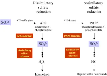
activated to adenosine-5’-phosphosulfate (APS) by ATP sulfurylase.
APS is then reduced to sulfite by APS reductase allowing the sulfite
reductase to reduce sulfite to the final product sulfide in a six
electron transfer reaction. This last step can occur in an assimilatory
or dissimilatory way.
This work was focused on the last step of sulfate reduction,
VI
dissimilatory sulfite reductases can be classified according to their
ultraviolet/visible absorption spectra and other characteristics:
desulfoviridin, desulforubidin, desulfofuscidin and P582.
Understanding the differences in terms of structures, their
assembly, cofactor content and reaction mechanisms was the main
goal of this work.
The presented PhD dissertation is divided into five chapters, in
which the first consists of a general introduction on the importance
of sulfur and sulfate reducing organisms in nature, followed by a
more detailed description of sulfite reducing organisms, their
classification and reaction mechanism.
Following this, chapters are presented based on the published
articles, with an overview of the material and methods used, the
results obtained and a discussion of the most important results
presented.
The second chapter describes the purification, crystallization and
preliminary structure characterization of dissimilatory sulfite
VII
The third chapter presents a detailed structural description of D.
vulgaris dSir. This structure revealed novel features and a
mechanism for sulfite reduction is proposed.
The fourth chapter consists of structural and biochemical studies of
a dissimilatory sulfite reductase from Desulfomicrobium norvegicum
classified as desulforubidin. A comparison between the two
structures from the different classes (desulfoviridin versus
desulforubidin) is performed with predictions on the structural
properties for the other classes - desulfofuscidin and P582. In
addition, mass spectrometry analysis identified different
stoichiometry complex arrangements of dSiRs which enhance our
understanding on the DsrC function.
Finally, in chapter five, a brief conclusion of the work is presented
with the major structural and functional features along with future
VIII
múltiplas e intensivas transformações de materiais nos diversos
ciclos biológicos. O carbono, nitrogénio, fósforo e enxofre são
componentes essenciais existentes em todas as células dos
organismos, representando os elementos mais importantes que
circulam na biosfera. No estudo seguidamente apresentado, o
enxofre e as reacções em que se encontra envolvido assumem um
interesse especial.
Durante a circulação do enxofre na biosfera, este pode ser
encontrado em diferentes estados de oxidação, sendo que a
transformação entre estes mesmos estados pode ocorrer tanto
biologica como quimicamente. A redução dissimilativa do sulfato é
uma das reacções mais importantes, em que o sulfato é reduzido a
sulfureto de modo a que os organismos consigam obter energia
para manter o seu metabolismo activo.
No entanto, a redução do sulfato é uma reacção energeticamente
desfavorável, pelo que o sulfato tem de ser primeiramente activado
a adenosina-5’-fosfosulfato (APS) pela enzima APS sulforilase. O APS
IX
sulfureto. Esta última reacção pode ocorrer assimilativa ou
dissimilativamente. Embora estas enzimas (sulfite reductase)
tenham sido alvo de intensivos estudos nas últimas décadas, muitas
questões continuam ainda por esclarecer relativamente às
proteínas envolvidas na reacção.
As sulfito reductases dissimilativas podem ser classificadas com
base nos máximos de absorção no espectro ultravioleta/vísivel e
algumas características moleculares nas seguintes classes:
Desulfoviridina, Desulforubidina, Desulfofuscidina e P582.
No trabalho de doutoramento aqui apresentado o principal
objectivo consistiu na determinação das estruturas tri-dimensionais
de duas sulfito reductases de diferentes classes, que permitiu
elucidar várias questões como a sua arquitectura, caracterização
dos cofactores (natureza e número), tendo sido proposto
mecanismo para a redução do sulfito.
O trabalho apresentado na presente dissertação encontra-se
dividido em cinco capítulos, sendo que o primeiro consiste numa
X
sobre os organismos redutores de sulfito, sua classificação e
mecanismo de reacção.
Nos capítulos seguintes é feita uma apresentação do trabalho
realizado, baseado nos artigos publicados com uma descrição dos
materiais e métodos, seguida dos resultados obtidos e discussão de
questões mais relevantes.
No 2ª capítulo é descrito o trabalho de purificação, cristalização e
resolução preliminar da estrutura da sulfito reductase (dSiR) de
Desulfovibrio vulgaris Hildenborough, que pertencente à classe das
desulfoviridinas. Esta estrutura encontra-se ligada à DsrC, uma
proteína que assume um papel importante no mecanismo de
redução do sulfito.
No 3º capítulo é apresentada uma descrição detalhada da estrutura
com destaque para as suas características mais relevantes. Ao longo
deste capítulo é efectuada uma comparação entre as diferentes
classes desulfoviridina e desulforubidina, com uma previsão das
propriedades estruturais das classes desulfofuscidina e P582.
No 4º capítulo é feita a caracterização da dSiR isolada de
XI
estequiométricas das subunidades do complexo que ajudam a
compreender a importância e envolvimento da proteína DsrC na
redução de sulfito.
Para finalizar, é efectuado um breve resumo do trabalho realizado
com ênfase nas características estruturais e funcionais mais
relevantes, com sugestões de trabalho importante a realizar nesta
XII
SRB – sulfate reducing bacteria SRP – sulfate reducing prokaryotes alSiR - assimilatory sulfate reductase type SiR – sulfite reductase
rSiR – reverse sulfite reductase asrC – anaerobic sulfite reductases aSiR – assimilatory sulfite reductase dSiR – dissimilatory sulfite reductase
dsr – gene coding for the dissimilatory sulfite reductase Dsr – dissimilatory sulfite reductase
DsrA – alpha subunit of dissimilatory sulfite reductase DsrB - beta subunit of dissimilatory sulfite reductase
DVir – dissimilatory sulfite reductase from Desulfovibrio vulgaris Hildenborough
Drub - dissimilatory sulfite reductase from Desulfomicrobium (Dm)
Norvegicum
SRH – sirohydrochlorin SRM – siroheme FDX – ferredoxin
APS – adenosine-5’-phosphosulfate ATPS - ATP sulfurylase
XIII
SDS – sodium dodecyl sulfate
XIV
Chapter 1
–
Introduction
11.1 Sulfur in the Environment – A General Introduction 2
1.2 The Sulfur Cycle 4
1.3 Sulfate Reducing Bacteria 8
1.4 Sulfate Reduction and Evolution 11
1.5 Sulfite Reductase and Cofactors 20
1.6 Sulfite Reduction 24
1.6.1 Assimilatory Sulfite Reductases 23
1.6.2 Dissimilatory Sulfite Reductases 25
1.7 dSiR Classification 27
1.8 The Mechanism of Sulfate Reduction 29
1.8.1 Sulfate Reduction 31
1.8.2 Sulfite Reduction Pathway 32
1.9 Environmental and Biotechnological Importance of SRB 35
1.10 References 38
Chapter 2
–
Purification, Crystallization and Preliminary
Crystallographic analysis of a Dissimilatory DsrAB Sulfite
Reductase in complex with DsrC
2.1 Abstract 522.2 Introduction 53
2.3 Protein Purification 55
2.4 Crystallization 57
XV
2.8 References 65
Chapter 3 –
The Crystal Structure of
Desulfovibrio vulgaris
Dissimilatory Sulfite Reductase Bound to DsrC Provides Novel
Insights into the Mechanism of Sulfite Respiration
3.1 Abstract 733.2 Introduction 74
3.3 Experimental Procedures 3.3.1 Protein Crystallization and X-Ray Data 79
3.3.2 Structure Determination and Refinement 80
3.4 Results 3.4.1 The Crystal Structure of DVir 82
3.4.2 DsrAB Structure and Cofactor Binding 86
3.4.3 The Catalytic Site 92
3.4.4 Structure of DsrC Bound to DsrAB 96
3.5 Discussion 99
3.6 Acknowledgements 108
3.7 References 109
Chapter 4 –
Structural Insights into Dissimilatory Sulfite
Reductases. Structure of desulforubidin from
Desulfomicrobium norvegicum
4.1 Abstract 1214.2 Introduction 122
XVI
4.3.4 Mass Spectrometry Studies 128
4.3.5 Electron Transfer Assays 129
4.4 Results and Discussion 4.4.1 Drub Crystal Structure 131
4.4.2 Overall Drub Architecture and Cofactors 136
4.4.3 The Catalytic Site 139
4.4.4 DsrC fold and complex Interaction 140
4.4.5 Structural Comparison of Drub with other dSiRs 141
4.4.6 Analysis of D. vulgaris and Dm. norvegicum dSir oligomeric states 144
4.4.7 Search for the dSiR electron donor 151
4.5 Concluding Remarks 153
4.6 Acknowledgements 154
4.7 References 155
1
Chapter 1
2
1.1
Sulfur in the Environment – A General
Introduction
Everything has a beginning, and when the earth was formed around 4.5 billion years ago [1], the scene was set for the fascinating story of the evolution of the species. All life requires energy, and through the eons organisms have had to adapt to diverse and changing environments, to eke out energy from sometimes very limited sources and with varying degrees of success. Initially, the environmental conditions were very anoxic with atmospheric gases such as ammonia, methane, hydrogen, nitrogen, sulfur and carbon dioxide predominating, and it is hard to comprehend how our ‘aerobic’ life emerged from such a different world [2].
3
metabolic opportunities, which in turn promoted further evolutionary progression [5].There is still uncertainty amongst biologists regarding the origin and position of eukaryotes in the overall scheme of evolution, with three different hypotheses being put forward; a) eukaryotes evolved from prokaryotes; b) were of contemporaneous origin; c) prokaryotes evolved from eukaryotic ancestors through a process of simplification [6]. Despite all the controversy, since the 1990’s, organisms have been classified into three principal domains: Archaea (archaebacteria), Bacteria, and the Eucarya [7].
4
1.2
The Sulfur Cycle
On Earth, tectonics and atmospheric photochemical processes are continuously supplying substrates and removing products based on redox reactions, successive transfers of electrons and protons from a relatively limited set of chemical elements in a cyclical manner. The six major elements – H, C, N, O, S and P constitute the major building blocks for all biological macromolecules and their biological fluxes are driven largely by microbially catalyzed, thermodynamically constrained redox reactions which are important components of the Earths elemental cycles [2].
Sulfur is among the most abundant elements on the earth and an essential element for maintaining life. The ocean is a major reservoir for sulfur, having large quantities in the form of dissolved sulfate and sedimentary minerals such as gypsum (CaSO4) and
pyrite (FeS2) in rocks and sediments (7.8x10¹⁸ g) and seawater
(1.28x10¹⁸
In the Earth´s crust, sulfur is cycled by biological processes on such a profound scale that the effects are evident globally. It has been estimated that at least 75% of crustal sulfur has been
5
biologically cycled and that 4 to 5x10¹²During sulfur circulation, it can be found in various oxidation states ranging from S
kg of sulfate is cycled through living cells every year [11].
2- (completely reduced) in sulfide and reduced
organic sulfur, to S6+
The biological sulfur cycle consists of oxidative and reductive sides, where sulfate on the reductive side functions as an electron acceptor and is converted to sulfide, and on the oxidative side, reduced sulfur compounds like sodium sulfide serve as an electron donor for phototrophic or chemolithothrophic bacteria which convert these compounds to elemental sulfur or sulfate [13]. Apart from the above reactions, an energy generating process – sulfur disproportionation, can occur with elemental sulfur or thiosulfate serving both as electron donor and electron acceptor and resulting in the formation of sulfate and sulfide [12]. A simplified scheme of the microbial sulfur cycle demonstrating the fundamental reactions is presented in Figure 1.
6
Figure 1. Schematic representation of sulfur transformations (Adaptation of the sulfur cycle image from [9]).
7
land, where plants and microorganisms take up sulfate via anaerobic assimilatory sulfate reduction, and animals are only able to take up reduced sulfur compounds through their diet. In the atmosphere, DMS is oxidized to acidic aerosol particles which affects cloud properties and the amount of solar radiation reflected back into space, thereby influencing the atmospheric chemistry and the climate system [10].8
reacts with water which forms sulfuric acid and results in acid rain. Image taken from [9].
1.3
Sulfate Reducing Bacteria
Anaerobic sulfate reduction represents an ancient, but evolutionary successful metabolic system in some prokaryotes. Molecular evidence has suggested that dissimilatory sulfate reduction is ancient [15] and geochemical data indicate the occurrence of microbial sulfate reduction 3.47 billion years ago [16].
Despite its long evolutionary history, the anaerobic sulfate respiration pathway seems to be restricted to a rather small group of very specialized microbes, termed the sulfate-reducing prokaryotes (SRP).
SRP obtain their energ
molecular2 whil
mainly found in the Bacteria and Archaea domains, with a greater incidence falling into the phylogentic lineages of mesophilic delta-proteobacteria and thermophilic gram-positive bacteria, this being the reason why SRP are commonly refered to as sulfate reducing bacteria (SRB) [9].
10
the inorganic or organic character of the energy source there are two types of anaerobic respiration of sulfates [13]:
1 – Autotrophic reduction of sulfates – the energy source is gaseous hydrogen, and the carbon source is CO2, and the reaction proceeds
according to the equation 4H2 + SO42- -> S2- + 4H2
2 –
O
Heterotrophic reduction of sulfates – the energy sources are simple organic substances, such as lactate, fumarate, pyruvate and some alcohols. Depending on the final product (oxidation state of organic substrate), the reduction process can be classified as incomplete or complete with the final products being acetate (CH3COO-)or carbon dioxide (CO2) plus H2O respectively [13].
2CH3CHOHCOO- + SO42- -> 2CH3COO- + 2HCO3- + H2S
4CH3COCOONa + 5MgSO4 -> 5MgCO3 + 2Na2CO3 + 5H2S + 5CO2 +
H2O
These organisms are probably responsible for most of the H2S
11
indicates the importance of sulfate reducers in both sulfur and carbon cycles, and consequently, why SRB are under extensive scrutiny [8-9].1.4
Sulfate Reduction and Evolution
It appears to be clear from geochemical (sulfur isotopic) evidence that the process of biological sulfate reduction is evolutionary ancient, being one of the oldest microbial pathways on earth [22],[11]
A variety of techniques have been used to understand the evolutionary relationships among organisms, their diversity and activity. One of the oldest techniques used for this purpose is cultivation. This technique, although useful has limitations, as only a small fraction (less than 1%) of naturally occurring SR organisms can be cultured [9]. Another technique used, but having taxonomic resolution limitations is the phospholipid fatty acids analysis [9]. To overcome these problems, the application of culture independent molecular methods for SRP detection in environmental samples was applied [23].
12
13
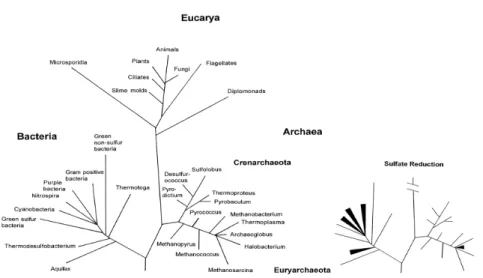
Figure 3. Principal lineages within the ‘Tree of Life’ determined from the comparison of 16S rRNA sequences, with particular relevance on the lineages within Bacteria and Archaea domains involved in the sulfate reduction. Figure was taken from [4].14
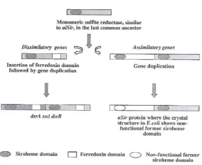
Consequently is necessary to identify and exploit additional phylogenetic genetic markers which allow one to specifically detect and identify SRP. The dsrA and dsrB genes code for two subunits (DsrAB) of the dissimilatory sulfite reductase and are well-suited phylogenetic marker molecules for dissimilatory sulfate reducing organisms, since the DsrAB is present in all dissimilatory sulfate-reducing organisms investigated so far [24],[23]. The genes encoding the two subunits are found adjacent to each other in the respective genomes and probably arose from the duplication of an ancestral gene [25]. Comparative amino acid sequences of the dissimilatory sulfite reductase genes (dsrAB) have then been used to investigate the evolutionary history of anaerobic sulfate (sulfite) respiration, suggesting a single ancestral progenitor present before the split between the Bacteria, Archaea and Eucarya domains [25].
The comparison of results from phylogenetic analysis of 16sRNA sequences and dsrAB databases yielded similar tree topologies, suggesting that comparative DsrAB sequence analysis allows specific yet independent identification of SRB. Based on the phylogenetic analysis, SRB can be organized into five major branches as presented in Figure 4. There are 3 branches within the Bacteria and 2 branches within the Archaea domains, where in the Bacteria most of the sulfate reducers belong to the delta-proteobacteria class (more than 35 genera), followed by the gram-positive SRB genus (Desulfotomaculum, Desulfitobacterium and
15
Thermodesulfobacterium phylum (Thermodesulfobacterium genus) and Thermodesulfobiaceae (Thermodesulfobium genus). Separately from the Bacteria domain, SR can also be found within the Archaea domain, with organisms belonging to the genus Archaeoglobus in the Euryarchaeota, and to the genera Thermocladium andCaldirvirga in the Crenarchaeota [26],[9], [12].
Figure 4. 16S rRNA gene based tree containing all recognized phyla of sulfate-reducing organism. Different phyla are color coded. Figure accordingly with [27] and [28].
16
--1
---1~::~:~
- :;;==~
..:::::..
- -
- -
-- --
_ .-
-,,"=,... - -
..
..::::.. -=-=
-- --
--~
- -
i::§~= i::§~=
-
--- ---
---
--
---_.
---
--
--
----
--
----
--
-.~----_.
--
--
---
--
,
---
----
--
----
-
----
--
-
---"'.,
---
-
--t
;::=:-"'::---'
--
---
--"r'--
""'=----=_
----
--
--
---
----
---
----
~-==--
-
--'--
--,--
- -
--- ---
--
----
-
- -
...
--
--
---"\'I,
-- --
- -
-- --
---
- -
-,-_J-
-- ----_.
- -
-
----
---===_ ..
- -
---
--- ---
--
--
-
--- ---
- -
--~ ~~~~~~~
~
---""'"
- -
---
---
---
---
--==--=
--==
=
--
= ~ _.J17
Figure 5: Phylogenetic tree based on comparative 16S rRNA gene sequence anddsrAB sequence analysis. Phylogenetic groups are color coded: Thermodesulfovibrio – red, Archaeoglobus – magenta, Thermodesulfobacterium – yellow, Gram-positives (Desulfotomaculum, Desulfitobacterium, Desulfosporosinus) – green and
Deltaproteobacteria – blue. Figure adapted from [29].
Some discrepant 16S rRNA and dsrAB gene phylogenies are found for the thermophilic bacterial genus Thermodesulfobacterium
and several, mostly thermophilic, gram-positive sulfate-reducing
18
amino acids and acetyl-coA [26]. After the separation of dissimilatory and assimilatory pathways, a deep archaeal/bacterial divergence probably occurred. According to a study performed by Dhillon et al. on phylogenetic topologies of dsrAB genes, the symmetry shown between domains indicate that ancestral gene duplication occurred within or prior to the last common ancestor of Bacteria and Archaea [26].
20
1.5
Sulfite Reductase and Cofactors
Cofactors are used for a variety of functions in a diverse set of biological scenarios. One of the most familiar examples of these molecules is the protoporphyrin IX-derived macrocycles like the iron-containing hemes [11].
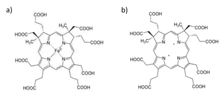
Siroheme (Figure 7a) is an isobacteriochlorin, meaning that its central ring is more reduced than the protoporphyrin IX-derived macrocycles like heme, and more closely related to cobinamide, the corrin ring of cobalamin (vitamin B12) [31].This molecule, derived from early intermediates in heme synthesis and most likely evolved before the cytochromes, is commonly characterized as
21
Figure 7: Schematic representation of (a) siroheme and (b) sirohydrochlorin molecules. Images were adopted from [31].A derivative of the first intermediate in heme biosynthesis, is the iron tetrahydroporphyrin of the isobacteriochlorin type (where adjacent pyrrole rings are reduced) which contain eight carboxylic acid groups, and is named sirohydrochlorin (Figure 7b) [33]. This prosthetic group has been detected in Desulfovibrio vulgaris and has been associated with a UV-visible maximum absorbance at 628 nm [41]. Remarkably, in dissimilatory and assimilatory reductases and nitrate reductases the siroheme/sirohydrochlorin molecules are bridged to an iron sulfur-cluster [4Fe4S] by a sulfur atom forming a characteristic complex cofactor as shown in Figure 8 [42],[43],[44]. The active redox center comprising the two metallo-cofactor complexes is involved in the transfer of electrons to the substrate [14].
+
a)
b)
22
23
1.6
Sulfite Reduction
Sulfite reduction to sulfide is a widespread reaction in nature, and is performed by different types of sulfite reductases, like, the assimilatory sulfite reductase (aSiR), dissimilatory sulfite reductase (dSiR), nitrite reductases, anaerobic sulfite reductase (asrC) (for example Salmonella typhimurium and Clostridium species) and assimilatory sulfate reductase type alSiR. There is also a reverse dissimilatory sulfite reductase which can be found in some sulfide and sulfur oxidizing bacteria[26].
Despite having different biological roles, dSiR, aSiR and NiRs (nitrite reductases) belong to a super family of enzymes, where a strictly conserved functional unit called the SNiRR (sulfite or nitrite reductase repeat), plays a crucial role in the six-electron reduction [14],[31],[45-46].
1.6.1 Assimilatory Sulfite Reductases
Assimilatory sulfite reductases (aSiRs) can be isolated from both Prokaryotic (bacteria) and Eucaryotic (plants and fungi) organisms, and is primarily concerned with the assimilation of sulfur into cellular material, for the biosynthesis of organosulfur compounds, such as sulfur-containing amino acids and enzyme cofactors [26].
24
containing the characteristic chromophore with an absorption peak at 580-590 nm, due to the siroheme- [4Fe4S] cluster coupled cofactor per polypeptide chain. In addition to the UV/visible maximum peak observed due to the presence of the SRM molecule, the aSiRs are characterized by UV/visible spectra with maximums also at 545 and 405 nm [47]. Electro Paramagnetic Resonance (EPR) and Mossbauer studies on aSiRs have shown that the siroheme has a low-spin ferric status, S=1/2, exhibiting EPR resonances at g=2.44, 2.36 and 1.77 [47]. Regarding the sulfite reduction reaction, these enzymes produce sulfide in a single six-electron step, with no intermediate sulfur compounds being released and form a complex with carbon monoxide (CO) or cyanide which inhibits activity [48],[31],[49].
25
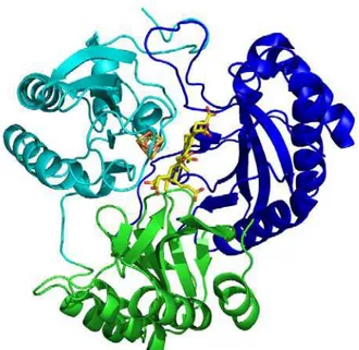
Figure 11. Cartoon representation of aSiR with the tri-lobed domains colored differently. Domain 1 is green, domain 2 blue, and domain 3 cyan. A stick representation of the cofactor content is also shown, SRM (C- yellow, O- red, Fe-brown) and [4Fe4S] (Fe- orange, S- gold). The figure was generated using Pymol [50].1.6.2 Dissimilatory Sulfite Reductases
26
reductase activity occurs in the terminal step of the respiratory electron transfer chain [51]. They are distinguished from aSiRs by their molecular composition (larger molecular mass and subunit composition) and their propensity to primarily produce incomplete reduced sulfur species in the form of trithionate and to a lesser extent thiosulfate [51]. Additionally, dSiRs usually do not form complexes with CO and cyanide and so are not inhibited by these small molecules [49]. There are some exceptions however, such as the Desulfotomaculum nigrificans and Desulfovibrio desulfuricans
Norway 4 strains, which readily form complexes resulting in activity inhibition by CO and cyanide [52].
The physical properties of the various dissimilatory sulfite reductases are overall quite similar, although they differ in finer details. They form large oligomers assembled in α2β2
Analysis of the cofactor content by heme extraction experiments result in a characteristic siroheme type spectrum with a ratio of two sirohemes per 240 kDa molecular mass. The iron content, determined calorimetrically, was shown to be much higher than that observed in aSiRs, with each enzyme containing multiple [Fe-S] clusters, resulting in 14 to 21 nonheme irons, plus equivalent sulfide content per molecule [54],[57],[58].
arrangements with molecular masses ranging from 145 to 225 kDa [14]. Their optical spectra show typical siroheme bands in the region of 540-580 nm and around 400 nm with a high-spin ferric state of S=5/2 [49],[53],[54],[55] and S = 9/2 in Desulfovibrio vulgaris
27
1.7
dSir Classification
On the basis of UV/visible absorption spectroscopy, and other molecular properties, four major types of dissimilatory sulfite reductases are distinguished in sulfate reducing-bacteria [14]. In 1965, desulfoviridins were the first class to be described with the identification of a green pigment [59]. It was isolated from several species such as Desulfovibrio (D.) gigas, D.salexigens and D.vulgaris
and is easily distinguishable from the other dSiR classes by the characteristic sirohydrochlorin maximum spectra at 628 nm [49],[53],[60]. The second class was named Desulforubidin [52] and when isolated displayed a characteristic reddish brown color, with characteristic maximum absorption at 545 nm. Proteins from this class have been isolated from Desulfovibrio desulfuricans strain Norway 4, which has been renamed to Desulfomicrobium baculatus
Norway 4 [61], and Desulfovibrio DSM 1743 [62]. The two remaining classes, classified as Desulfofuscidin and P582, have a characteristic brown color, and are distinguished by the maximum absorption spectra at 576 and 582 nm, respectively. In 1965 the Desulfofuscidin protein was first identified in the Desulfomaculum
genus [63], and has now been isolated from a host of different
species, such as Desulfovibrio thermophilus and
Thermodesulfobacterium commune, both thermophilic sulfate reducers [58]. Finally from the P582 class, proteins from
28
archaeal dSiR has been purified [25]. The various dissimilatory sulfite reductases share many similarities. Besides their high molecular weight and α2β2
Despite years of intensive analysis on cofactor content of dSiRs there is still some disparity for the different classes (see Table 1).
assembly [51], they all contain a reduced porphyrin of the isobacteriochlorin class – SRM which is covalently coupled to an iron-sulfur cluster [4Fe4S] to form the electronically integrated metallocofactor for delivering electrons to substrate at the active site [65].
Table 1. Physico-chemical and composition of dissimilatory sulfite reductases [58].
A third subunit (γ) has been observed in a desulfoviridin-type of dSiR from D.vulgaris [66],[67] and D. desulfuricans strain Essex [68], corresponding to the dsrC gene. This protein was suggested to be a subunit the dSiR protein and to be arranged as α2β2γ2
Property
. The γ -Desulfofuscidin Desulfoviridin (D. gigas) Desulforubidin (Dsm. Baculatum DSM 1741) P582 (Dm. Nigrificans)
T. commune T. mobile
Molecular Mass
(kDa) 167 190 200 225 194
Subunit structure α2β2 α2β2 α2β2 α2β2 α2β2
Absorption maxima
(nm) 389, 576, 693 392, 578, 700 390, 408, 580, 628 392, 545, 580 392, 582, 700
Iron content 20-21 32 16.5 16.6 16
Labile sulfide 16 ND 14 14.7 14
Siroheme 4 4 2 1.3
Sirohydroporphyrin 2
[4Fe4S] 4 8 4 4 4
29
subunit however, is not encoded on the same operon as the α andβ subunits and there is no coordinated expression with the αand β
subunits [67], which is an interesting feature.
1.8
The Mechanism of Sulfate Reduction
1.8.1
Sulfate Reduction
The reduction of sulfate can be divided in two phases; the reduction of sulfate to sulfite and the reduction of sulfite to sulfide [69],[14]. However, from a chemical point of view, sulfate is an unfavorable electron acceptor for microorganisms. The sulfate-sulfite couple redox potential (E0´) is -516mV, too negative to allow reduction by the intracellular electron mediators present in sulfate reducers such as ferredoxin or NADH (E0´ of -398 mV and -314 mV, respectively). As a result, prior to reduction, inorganic sulfate (SO42-)
30
reductase presents some differences. In the dissimilative reduction, the sulfate moiety of APS is reduced directly to sulfite with concomitant release of AMP by the enzyme APS reductase [71], [72]
In both cases the product of sulfate reduction is sulfite, which is further reduced to sulfide by the assimilatory or dissimilatory sulfite reductases, with an E
. On the other hand, in the assimilative reduction, another phosphorous atom is added to APS by APS kinase phosphorylase to form phosphoadenosine-5´-phosphosulfate (PAPS), before reduction by PAPS reductase to sulfite [9, 12] (see Figure 9).
0´of the redox couple sulfitesulfide of
32
1.8.2
Sulfite Reduction Pathway
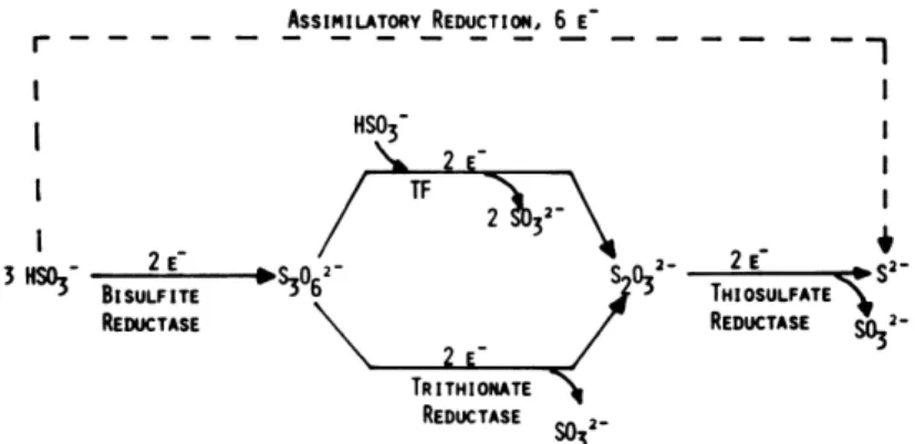
The assimilatory reduction occurring in bacteria involves the direct reduction of sulfite to sulfide without the formation of any detectable intermediates by aSiR. In contrast, a series of complex reactions have been proposed for the dissimilatory pathway leading to the formation of a mixture of products, such as sulfide, trithionate and thiossulfate.
In 1969, the finding of a thiosulfate producing system during reduction experiments in D. vulgaris [74],[75],[76], led to a period of intense work on sulfite reduction and its products, with an initial suggestion of bisulfite (HSO3-) as the actual substrate instead of
sulfite (SO32-) [74]. Around the same period, it was reported that
33
reduced by bisulfate reductase to trithionate, which remains in the active site while being reduced by thritionate reductase to thiosulfate, which is then further reduced to sulfide by thiosulfate reductase (Figure 10).Figure 10: The proposed pathway for the reduction of bisulfate to sulfide occurring in three consecutive two electron steps, with the formation of trithionate and thiossulfate as reaction intermediates. Diagram was adopted from [75].
34
However, if the concentration of the electron donor is high enough, the two-sulfur intermediate may undergo a reduction to thiosulfate. As the reaction proceeds and bisulfite is depleted, site A becomes empty and the reduction of the two-sulfur intermediates predominates with the formation of thiossulfate. When the bisulfite concentration is almost entirely depleted, the sulfoxilate intermediate is reduced to sulfide [83] (Figure 11).
Figure 11: The proposed pathway of bisulfate reduction at the active site of bisulfate reductase [86].
35
1.9
Environmental and Biotechnological importance of
SRB
In addition to their relevance in the biogeochemical sulfur cycle, the sulfate reducing bacteria (SRB) have considerable economic and environmental impact. As a result of their metabolic actions, SRB can have positive and negative impacts on the environment, with significant economic losses, environmental dangers, and health and safety risks [12],[13],[88]. The next few paragraphs are dedicated to the presentation of some of these impacts.
In the process of anaerobic respiration SRB produce a considerable amount of the toxic, odorous and corrosive gas hydrogen sulfide (H2S) which reacts in an aqueous medium with
36
Other negative effects of SRB activity can be found in paper industries, where as a result of SRB activity, iron sulfides contaminate water processing and causes paper blackening [92].
Moreover, SRB can also be a major problem in health. Sulfate reducing bacteria are normal inhabitants of the intestine in humans and animals. SRB have been implicated in a number of gastrointestinal diseases, such as cholecystitis and abdominal abscesses. In some cases, the increase of SRB in the human tract is related with other illnesses, like spondylitis and colorectal cancer [20]. Regarding the latter, it is suggested that the formation of sulfide activates a number of biochemical pathways believed to be involved in the initiation of the disease [20]. Moreover, at physiological concentrations, sulfide has been shown to protect colon cancer cells from drugs such as β-phenyl ethyl isocyanate for the promotion of tumor genesis. Intense inflammation of the large bowel mucosa (underlying epithelial cell surfaces) may also develop. Symptoms vary between individuals, but in general, the disease is associated with bloody diarrhea, abdominal pain, weight loss, urgency to defecate, and arthritic [20]. Ulcerative colitis is one of the two major forms of idiopathic inflammatory bowel disease and represents a highly disabling incurable condition. Current maintenance therapies rely on anti-inflammatory drugs and steroids, but in severe cases, partial or complete surgical removal of the bowel is necessary [20].
38
1.10
References:
1. Newman, W., Age of the Earth. Publications Services, USGS, 2007.
2. Falkowski, P.G., T. Fenchel, and E.F. Delong, The microbial engines that drive Earth's biogeochemical cycles. Science, 2008. 320(5879): p. 1034-9.
3. Rabus, R., T. Hansen, and F. Widdel, Dissimilatory Sulfate- and Sulfur-Reducing Prokaryotes, in The Prokaryotes, M.e.a. Dworkin, Editor. 2007, Springer-Verlag, New York. p. 659-768.
4. Canfield, D.E. and R. Raiswell, The Evolution of the Sulfur Cycle. American Journal of Science, 1999. 299: p. 627-723.
5. Canfield, D.E., M.T. Rosing, and C. Bjerrum, Early anaerobic metabolisms. Philos Trans R Soc Lond B Biol Sci, 2006.
361(1474): p. 1819-34; discussion 1835-6.
6. Martin W., Who is the Tree of Life. In Microbial Phylogeny and Evolution. Concepts and Controversies. Oxford University Press, 2005. 139.
7. Woese C., Kandler O., and W. M., Towards a natural system of organisms: proposal for the domains Archaea, Bacteria and Eucarya. Proc Natl Acad Sci USA 1990. 87(12): p. 4576-9.
39
9. Muyzer, G. and A.J. Stams, The ecology and biotechnology of sulphate-reducing bacteria. Nat Rev Microbiol, 2008.6(6): p. 441-54.
10. Sievert, S.M., R.P. Kiene, and H. Schiulz-Vogt, Microbes and major elemental cycles. Oceanography, 2007. 20(2): p. 117-123.
11. Skyring, G.W. and T.H. Donnelly, Precambrian sulfur Isotopes and a possible role for sulfite in the evolution of biological sulfate reduction. Precambrian Research, 1982.
17: p. 41-61.
12. Tang, K., V. Baskaran, and M. Nemati, Bacteria of the Sulphur Cycle: An overview of microbiology, biokinetics and their role in petroleum and mining industries. Biochemical Engineering Journal, 2009. 44: p. 73-94.
13. Luptakova, A., Importance of Sulphate-Reducing Bacteria in Environment. Nova Biotechnologica, 2007. VII-I: p. 17-22. 14. Rabus, R., T. Hansen, and F. Widdel, The Prokaryotes.
Dissimilatory Sulfate- and Sulfur- Reducing Prokaryotes
Springer New York, 2006.
15. Wagner, M., et al., Phylogeny of dissimilatory sulfite reductases supports an early origin of sulfate respiration. J Bacteriol, 1998. 180(11): p. 2975-82.
16. Shen, Y.N. and R. Buicks, The antiquity of microbial sulphate reduction. Earth-Science Reviews, 2004. 64: p. 243-272.
17. Lobo, S.A., et al., The anaerobe Desulfovibrio desulfuricans ATCC 27774 grows at nearly atmospheric oxygen levels.
FEBS Lett, 2007. 581(3): p. 433-6.
40
19. Peck, H.D. and J. LeGall, Biochemistry of dissimilatory sulphate reduction. Philos Trans R Soc Lond B Biol Sci, 1982.
298(1093): p. 443-66.
20. Barton, L.L. and W.A. Hamilton, Sulphate-reducing Bacteria. Cambridge University Press, 2007.
21. Trudinger, P.A. and D.J. Swaine, eds. The biological sulfur cycle. Biogeochemical Cycling of Mineral-Forming Elements. Vol. 293-313. 1979 Elsevier: Amsterdam. 22. Stahl, D.A., et al., Origins and diversification of
sulfate-respiring microorganisms. Antonie Van Leeuwenhoek, 2002. 81(1-4): p. 189-95.
23. Wagner, M., et al., Functional marker genes for identification of sulfate-reducing prokaryotes. Methods Enzymol, 2005. 397: p. 469-89.
24. Loy, A., S. Duller, and M. Wagner, Evolution and Ecology of Microbes Dissimilating Sulfur Compounds: Insights from Siroheme Sulfite Reductases, in Microbial Sulfur Metabolism, C. Dahl and C. Friedrich, Editors. 2007, Springer Berlin Heidelberg. p. 46-59.
25. Dahl, C., et al., Dissimilatory sulphite reductase from Archaeoglobus fulgidus: physico-chemical properties of the enzyme and cloning, sequencing and analysis of the reductase genes. J Gen Microbiol, 1993. 139(8): p. 1817-28. 26. Dhillon, A., et al., Domain evolution and functional
diversification of sulfite reductases. Astrobiology, 2005.
5(1): p. 18-29.
27. Woese, C.R., Bacterial evolution. Microbiol Rev, 1987.
51(2): p. 221-71.
41
29. Klein, M., et al., Multiple lateral transfers of dissimilatory sulfite reductase genes between major lineages of sulfate-reducing prokaryotes. J Bacteriol, 2001. 183(20): p. 6028-35.30. Zverlov, V., et al., Lateral gene transfer of dissimilatory (bi)sulfite reductase revisited. J Bacteriol, 2005. 187(6): p.
2203-8.
31. Stroupe, M.E., Making and Using Siroheme: the X-ray Crystallographic Structures of Siroheme Synthase and Sulfite Reductase. 2002, The Scripps Research Institute: California.
32. Murphy, M.J. and L.M. Siegel, Siroheme and
sirohydrochlorin. The basis for a new type of porphyrin-related prosthetic group common to both assimilatory and dissimilatory sulfite reductases. J Biol Chem, 1973. 248(19):
p. 6911-9.
33. Murphy, M.J., et al., Reduced nicotinamide adenine dinucleotide phosphate-sulfite reductase of enterobacteria. II. Identification of a new class of heme prosthetic group: an iron-tetrahydroporphyrin (isobacteriochlorin type) with eight carboxylic acid groups. J Biol Chem, 1973. 248(8): p. 2801-14.
34. Schedel, M., J. Legall, and J. Baldensperger, Sulfur metabolism in Thiobacillus denitrificans evidence for the presence of a sulfite reducatase activity. Arch Microbiol, 1975. 105(3): p. 339-41.
42
36. Hucklesby D. P., et al., Properties of nitrite reductase from Cucurbita Pepo Phytochemistry, 1976. 15: p. 599-603.
37. Vega, J.M. and R.H. Garrett, Siroheme: a prosthetic group of the Neurospora crassa assimilatory nitrite reductase. J Biol Chem, 1975. 250(20): p. 7980-9.
38. Tan, J. and J.A. Cowan, Enzymatic redox chemistry: a proposed reaction pathway for the six-electron reduction of SO3(2-) to S2- by the assimilatory-type sulfite reductase from Desulfovibrio vulgaris (Hildenborough). Biochemistry, 1991. 30(36): p. 8910-7.
39. Schedel, M. and H.G. Truper, Purification of Thiobacillus denitrificans siroheme sulfite reductase and investigation of some molecular and catalytic properties. Biochim Biophys Acta, 1979. 568(2): p. 454-66.
40. Schedel, M., M. Vanselow, and H.G. Trüper, Siroheme-sulfite reductase isolated from Chromatium vinosum. Purification and investigation of some molecular and catalytic properties. Archives of Microbiology, 1979.
121(1): p. 29-36.
41. Kobayashi, K., E. Takahashi, and M. Ishimoto, Biochemical studies on sulfate-reducing bacteria. XI. Purification and some properties of sulfite reductase, desulfoviridin. J Biochem, 1972. 72(4): p. 879-87.
42. Christner, J.A., et al., Mossbauer evidence for exchange-coupled siroheme and [4Fe-4S] prosthetic groups in Escherichia coli sulfite reductase. Studies of the reduced states and of a nitrite turnover complex. J Biol Chem, 1983.
258(18): p. 11147-56.
43
Enzyme State and Complexes with Sulfide. Journal of the American Chemical Society, 1984. 106: p. 6786-6794.
44. Crane, B.R., L.M. Siegel, and E.D. Getzoff, Structures of the siroheme- and Fe4S4-containing active center of sulfite reductase in different states of oxidation: heme activation via reduction-gated exogenous ligand exchange.
Biochemistry, 1997. 36(40): p. 12101-19.
45. Postgate, J.R., Sulphate Reduction by Bacteria. Annual Review of Microbiology, 1959. 13: p. 505-520.
46. Crane, B.R., L.M. Siegel, and E.D. Getzoff, Sulfite reductase structure at 1.6 A: evolution and catalysis for reduction of inorganic anions. Science, 1995. 270(5233): p. 59-67.
47. Moura, I., et al., Low-spin sulfite reductases: a new homologous group of non-heme iron-siroheme proteins in anaerobic bacteria. Biochem Biophys Res Commun, 1986.
141(3): p. 1032-41.
48. Murphy, M.J., L.M. Siegel, and K. H., An Iron
Tetrahydroporphyrin Prosthetic group common to both Assimilatory and Dissimilatory Sulfite Reductases.
Biochemical and Biophysical Research Communications, 1973. 54(1): p. 82-88.
49. Lee, J.P., J. LeGall, and H.D. Peck, Jr., Isolation of assimilatroy- and dissimilatory-type sulfite reductases from Desulfovibrio vulgaris. J Bacteriol, 1973. 115(2): p. 529-42. 50. DeLano, W.L., The PyMOL Molecular Graphics System,
Version 1.2r3pre, Schrödinger, LLC. 2002.
51. Crane, B.R. and E.D. Getzoff, The relationship between structure and function for the sulfite reductases. Curr Opin Struct Biol, 1996. 6(6): p. 744-56.
44
Role in Sulfite Reduction Journal of Bacteriology, 1973.
115(1): p. 453-455.
53. Lui, S.M., A. Soriano, and J.A. Cowan, Electronic properties of the dissimilatory sulphite reductase from Desulfovibrio vulgaris (Hildenborough): comparative studies of optical spectra and relative reduction potentials for the [Fe4S4]-sirohaem prosthetic centres. Biochem J, 1994. 304 ( Pt 2): p. 441-7.
54. Wolfe, B.M., S.M. Lui, and J.A. Cowan, Desulfoviridin, a multimeric-dissimilatory sulfite reductase from Desulfovibrio vulgaris (Hildenborough). Purification, characterization, kinetics and EPR studies. Eur J Biochem, 1994. 223(1): p. 79-89.
55. Huynh, B.H., et al., Characterization of a sulfite reductase from Desulfovibrio vulgaris. Evidence for the presence of a low-spin siroheme and an exchange-coupled siroheme-[4Fe-4S] unit. J Biol Chem, 1984. 259(24): p. 15373-6. 56. Pierik, A.J. and W.R. Hagen, S = 9/2 EPR signals are
evidence against coupling between the siroheme and the Fe/S cluster prosthetic groups in Desulfovibrio vulgaris (Hildenborough) dissimilatory sulfite reductase. Eur J Biochem, 1991. 195(2): p. 505-16.
57. Seki, Y., K. Kobayashi, and M. Ishimoto, Biochemical studies on sulfate-reducing bacteria. XV. Separation and comparison of two forms of desulfoviridin. J Biochem, 1979.
85(3): p. 705-11.
58. Hatchikian, E.C., Desulfofuscidin: dissimilatory, high-spin sulfite reductase of thermophilic, sulfate-reducing bacteria.
Methods Enzymol, 1994. 243: p. 276-95.
45
desulphuricans. 1956. 14(Journal of General Microbiology): p. 545-572.
60. Kobayashi, K., Y. Seki, and M. Ishimoto, Biochemical studies on sulfate-ruducing bacteria. 8. Sulfite reductase from Desulfovibrio vulgaris--mechanism of trithionate, thiosulfate, and sulfide formation and enzymatic properties. J Biochem, 1974. 75(3): p. 519-29.
61. Lee, J.P. and H.D. Peck, Purification of the Enzyme Reducing Bisulfite to Trithionate from Desulfovibrio gigas and its Identification as Desulfoviridin. Biochemical and Biophysical Research Communications, 1971. 45(3): p. 583-589.
62. Moura, I., et al., Characterization of two Dissimilatory Sulfite Reductases (Desulforubidin and Desulfoviridin) from the Sulfate-Reducing Bacteria. Mössbauer and EPR Studies.
Journal of the American Chemical Society, 1988. 110: p. 1075-1082.
63. Campbell, L.L. and J.R. Postgate, Classification of the spore-forming sulfate-reducing bacteria. Bacteriol Rev, 1965.
29(3): p. 359-63.
64. Akagi, J.M. and V. Adams, Isolation of a bisulfite reductase activity from Desulfotomaculum nigrificans and its identification as the carbon monoxide-binding pigment P582. J Bacteriol, 1973. 116(1): p. 392-6.
65. Miller, J.D. and A.M. Saleh, A Sulphate-Reducing Bacterium Containing Cytochrome C3 but Lacking Desulfoviridin. J Gen Microbiol, 1964. 37: p. 419-23.
66. Pierik, A.J., et al., The third subunit of desulfoviridin-type dissimilatory sulfite reductases. Eur J Biochem, 1992.
46
67. Karkhoff-Schweizer, R., R.M. Bruschi, and G. Voordouw,
Expression of the gama-subunit gene of Desulfoviridin-type dissimilatory sulfite reductase and of the alfa and beta subunit gene is not coordinatellt regulated. European Journal of Biochemistry, 1993. 211: p. 501-507.
68. Steuber, J., et al., Molecular properties of the dissimilatory sulfite reductase from Desulfovibrio desulfuricans (Essex) and comparison with the enzyme from Desulfovibrio vulgaris (Hildenborough). Eur J Biochem, 1995. 233(3): p.
873-9.
69. Postgate, J.R., Recent advances in the study of the sulfate-reducing bacteria. Bacteriol Rev, 1965. 29(4): p. 425-41.
70. Akagi, J.M. and L.L. Campbell, STUDIES ON THERMOPHILIC SULFATE-REDUCING BACTERIA III. : Adenosine Triphosphate-sulfurylase of Clostridium nigrificans and Desulfovibrio desulfuricans. J Bacteriol, 1962. 84(6): p. 1194-201.
71. Ishimoto, M. and T. Yagi, Biochemical studies on sulphate-reducing bacteria. IX. Sulfite reductase. The Journal of Biochemistry, 1961. 49: p. 103-109.
72. Fritz, G., et al., Structure of adenylylsulfate reductase from the hyperthermophilic Archaeoglobus fulgidus at 1.6-A resolution. Proc Natl Acad Sci U S A, 2002. 99(4): p. 1836-41.
73. Lengeler, J.W., Drews G., and S.H. G., Biology of the prokaryotes. 1999, Stuttgart, Germany: Thieme Verlag. 74. Suh, B. and J.M. Akagi, Formation of thiosulfate from sulfite
by Desulfovibrio vulgaris. J Bacteriol, 1969. 99(1): p. 210-5.
47
76. Findley, J.E. and J.M. Akagi, Evidence for thiosulfateformation during sulfite reduction by Desulfovibrio vulgaris.
Biochem Biophys Res Commun, 1969. 36(2): p. 266-71. 77. Chambers, L.A. and P.A. Trudinger, Are thiosulfate and
trithionate intermediates in dissimilatory sulfate reduction?
J Bacteriol, 1975. 123(1): p. 36-40.
78. Findley, J.E. and J.M. Akagi, Role of Thiosulfate in Bisulfite Reduction as Catalyzed by Desulfovibrio vulgaris. Journal of Bacteriology, 1970. 103(3): p. 741-744.
79. Akagi, J.M., Reduction of bisulfite by the trithionate pathway by cell extracts from Desulfotomaculum nigrificans. Biochem Biophys Res Commun, 1983. 117(2): p.
530-5.
80. Akagi, J.M., M. Chan, and V. Adams, Observations on the bisulfite reductase (P582) isolated from Desulfotomaculum nigrificans. J Bacteriol, 1974. 120(1): p. 240-4.
81. Haschke, R.H. and L.L. Campbell, Thiosulfate reductase of Desulfovibrio vulgaris. J Bacteriol, 1971. 106(2): p. 603-7. 82. Drake, H.L. and J.M. Akagi, Product analysis of bisulfite
reductase activity isolated from Desulfovibrio vulgaris. J Bacteriol, 1976. 126(2): p. 733-8.
83. Drake, H.L. and J.M. Akagi, Characterization of a novel thiosulfate-forming enzyme isolated from Desulfovibrio vulgaris. J Bacteriol, 1977. 132(1): p. 132-8.
84. Hatchikian, E.C., Purification and properties of thiosulfate reductase from Desulfovibrio gigas. Arch Microbiol, 1975.
105(3): p. 249-56.
48
86. Drake, H.L. and J.M. Akagi, Bisulfite reductase of Desulfovibrio vulgaris: explanation for product formation. J Bacteriol, 1977. 132(1): p. 139-43.
87. Kim, J.H. and J.M. Akagi, Characterization of a trithionate reductase system from Desulfovibrio vulgaris. J Bacteriol, 1985. 163(2): p. 472-5.
88. Hamilton, W.A., Sulphate-reducing bacteria and anaerobic corrosion. Annu Rev Microbiol, 1985. 39: p. 195-217. 89. Seth, A.D. and R.G.J. Edyvean, The function of
sulfate-reducing bacteria in corrosion of potable water mains. 2006, Department of Chemical and Process Engineering, University of Sheffield, Sheffield, UK.
90. Pereira, A.S., et al., Characterization of representative enzymes from a sulfate reducing bacterium implicated in the corrosion of steel. Biochem Biophys Res Commun, 1996. 221(2): p. 414-21.
91. Beech, I.B., Sulfate-reducing bacteria in biofilms on metallic materials and corrosion. Microbiology, 2003. 30: p. 115-117.
92. Soimajärvi J., Pursiainen M., and K. J., Sulphate-reducing bacteria in paper machine waters and in suction roll perforations. Applied Microbiology and Biotechnology, 1978. 5(2): p. 87-93.
49
Chapter 2
Purification, crystallization and
preliminary crystallographic
analysis of a dissimilatory
DsrAB sulfite reductase in
50
This Chapter was published in:
Oliveira, T. F.; Vonrhein, C.; Matias, P. M.; Venceslau, S. S.;
Pereira, I. A. C.; (2008), Purification, crystallization and
preliminary crystallographic analysis of a dissimilatory DsrAB
sulfite reductase in complex with DsrC. Journal of Structure
Biology, 164, 236-239
Note:
SSV and
TFO purified the protein samples, TFO
51
2.1 Abstract 52
2.2 Introduction
53
2.3 Protein Purification 55
2.4 Crystallization
57
2.5 Data Collection 59
2.6 Preliminary Structure Determination 62
52
2.1 Abstract
Dissimilatory sulfite reductase (dSiR, DsrAB) is a key protein in several types of dissimilatory sulfur metabolism, one of the earliest types of energy metabolism to be traced on earth. dSirs are large
oligomeric proteins around 200 kDa forming an α2β2 arrangement
and including a unique siroheme-[4Fe-4S] coupled cofactor. Here, we report the purification, crystallization and preliminary X-ray diffraction analysis of dSir isolated from Desulfovibrio vulgaris
Hildenborough, also known as desulfoviridin. In this enzyme the DsrAB protein is associated with DsrC, a protein of unknown function that is believed to play an important role in the sulfite reduction. Crystals belong to the monoclinic space group P21 with
53
2.2. Introduction
Reduction of sulfite is a crucial biological reaction both in assimilatory pathways, which lead to incorporation of sulfur into amino-acids and other cellular metabolites, or in dissimilatory pathways used by anaerobic microorganisms that respire sulfur compounds [1]. Both assimilatory and dissimilatory sulfite reductases (SiR) contain in the active site a unique combination of cofactors that includes a reduced porphyrin of the isobacterichlorin class, termed siroheme, that is coupled through its cysteine axial ligand to a [4Fe4S] iron-sulfur cluster [2-4]. Furthermore, these two classes of enzymes belong to a superfamily of coupled siroheme-[4Fe4S] containing enzymes that include also assimilatory and dissimilatory nitrite reductases that reduce nitrite to ammonia [1]. The assimilatory sulfite reductases (aSiRs) have been extensively investigated and serve as models for this family of proteins since they are structurally well characterized [2, 5], In contrast, many questions remain regarding dissimilatory sulfite reductases dSiRs, which are apparently more diverse based on distinct spectroscopic properties [6]. One of the most distinguishing factors of dSiRs include the fact that in vitro they reduce sulfite incompletely to mainly trithionate and some thiosulfate and sulfide, whereas aSiRs reduce sulfite completely to sulfide [7]. The aSiRs are monomeric and share low sequence similarity to dSiRs, which are large oligomeric proteins with a molecular mass in the order of 200 kDa and minimally composed of two different types of subunits in a
54
also not clear with studies reporting from two to four sirohemes and 10 to 32 non-heme irons per α2β2
The dSiRs are very important enzymes for our understanding of biological evolution, since they catalyze a key energy-conserving module [4, 9-11]. Although some authors propose that the cubane-siroheme cofactor arrangement of dSiRs is similar to that found in aSiRs [4, 10], other authors have proposed the presence of higher nuclearity high-spin iron-sulfur clusters [10-12].
One of the most studied dSiRs is the protein desulfoviridin (Dvir) present in Desulfovibrio spp. [7, 13]. This protein is particularly intriguing since it is reported that up to 80% of its siroheme is not metallated but is in the form of sirohydrochlorin [4, 11, 14], although this has been disputed [10]. Another interesting aspect of Dvir is that it forms a stable complex with a smaller protein of 11 kDa, initially classified as a third subunit of dSiR [15], but later identified as the DsrC protein. DsrC is encoded separately from DsrAB, the two subunits of dSiR and their expression is not coordinately regulated [16]. It is actually one of the most highly expressed proteins in Desulfovibrio vulgaris Hildenborough, with twice the expression level of DsrAB [17]. This indicates that it plays a crucial role in energy metabolism, but that it is more than a subunit of the dSiR complex. DsrC is also found in sulfite or thiosulfate reducing organisms and is one of the few genes of the
55
step in organisms that metabolise sulfur compounds. Phylogenetic analysis supports an ancient origin of dSiR [20-22], and evidence for the existence of sulfur-metabolizing organisms dates back to 3.49 Gyr ago in the Early Archaean period [23-24]. It is thus be very important to obtain structural information for dSiR to provide insights into these open questions. We have concentrated our efforts in trying to crystallize the Dvir from D. vulgaris as this protein forms a stable complex with DsrC, and its structure will thus not only resolve the questions about its cofactor composition, but will also yield evidence for the mode of interaction between DsrAB and DsrC, and the possible role of this protein in the reduction of sulfite.2.3 Protein Purification
56
57
Figure 1 - A) 9% Native-PAGE of Dvir (20 μg) showing two bands: “slow” (S) and“fast” form (F). B) 12% SDS-PAGE of the desulfoviridin “slow” form (20 µg) after the electroelution.
2.4. Crystallization
Some initial crystallization screenings from Qiagen and Emerald BioSystems were performed in parallel with both forms (slow and fast) of Dvir at 4 and 20 ºC, using 0.1 μl nanodrops in a
58
quality crystals. After several crystal optimization experiments,
small green crystals were grown by mixing 2 μl of protein with 1 μl
of reservoir solution containing 12.5% PEG 4000, 0.1 M Tris-HCl pH 8.5, 0.2 M MgCl2. The crystals appeared within two days and grew
to maximum dimensions of 0.1x0.1x0.1 mm in one week (Fig. 2). An SDS-PAGE confirmed the presence of DsrA, DsrB and DsrC proteins in the crystals (data not shown). Crystals were cryoprotected with the reservoir solution supplemented with 20% glycerol and were directly flash cooled in liquid nitrogen. However, it was extremely difficult to get suitable crystals for X-ray diffraction experiments, due to the low reproducibility of the crystallization conditions and very poor diffraction quality shown by most screened crystals. Many similar crystallization conditions were set up yielding quite different results, and most of the obtained crystals were pre-screened in our in-house generator prior to shipment to synchrotron sources.
![Figure 1. Schematic representation of sulfur transformations (Adaptation of the sulfur cycle image from [9])](https://thumb-eu.123doks.com/thumbv2/123dok_br/15769924.641174/25.629.104.460.118.378/figure-schematic-representation-sulfur-transformations-adaptation-sulfur-cycle.webp)

![Figure 8: Schematic representation of siroheme-[4Fe4S] cluster covalently coupled via a sulfur bridge](https://thumb-eu.123doks.com/thumbv2/123dok_br/15769924.641174/41.629.196.359.88.354/figure-schematic-representation-siroheme-cluster-covalently-coupled-sulfur.webp)
![Figure 11: The proposed pathway of bisulfate reduction at the active site of bisulfate reductase [86]](https://thumb-eu.123doks.com/thumbv2/123dok_br/15769924.641174/53.629.97.486.286.394/figure-proposed-pathway-bisulfate-reduction-active-bisulfate-reductase.webp)
