Ana Raquel Martinho Ramos
Dissertation presented to obtain the Ph.D degree in Biochemistry
Instituto de Tecnologia Química e Biológica António Xavier | Universidade Nova de LisboaOeiras,
July, 2014
Ana Raquel Martinho Ramos
Dissertation presented to obtain the Ph.D degree in Biochemistry
Instituto de Tecnologia Química e Biológica António Xavier | Universidade Nova de LisboaOeiras, July 2014
pathways in anaerobic bacteria
Supervisor
: Dr. Inês A. C. Pereira
Opponents: Prof. Uwe Deppenmeier
Dr. Wolfgang Nitschke
Prof. Carlos Salgueiro
Manuela Pereira.
July 15, 2014
Bacterial Energy Metabolism Laboratory
Instituto de Tecnologia Química e Biológica António Xavier
Universidade Nova de Lisboa
Av. República, Estação Agronómica Nacional
2780-157 Oeiras, Portugal
http://www.itqb.unl.pt
ACKNOWLEDGEMENTS ... XI
THESIS OUTLINE ... XV
LIST OF PUBLICATIONS... XVII
DISSERTATION SUMMARY ... XIX
SUMÁRIO DA DISSERTAÇÃO ... XXV
LIST OF ABBREVIATIONS ...XXXI
CHAPTER 1-INTRODUCTION ... 1
1.1-ENERGY CONSERVATION IN ANAEROBIC BACTERIA... 3
1.1.1-ELECTRON BIFURCATION, FLAVOPROTEINS AND FERREDOXIN ... 10
1.1.2-THE FLAVIN BASED ELECTRON BIFURCATION MECHANISM ... 14
1.1.2.1-FBEB AND METHANOGENS ... 19
1.1.2.2-THE BIFURCATING [FEFE]-HYDROGENASES ... 23
1.1.2.3-OTHER EXAMPLES OF FBEB ENZYMES ... 26
1.1.2-IMPORTANT CONSIDERATIONS ON FBEB MECHANISMS ... 28
1.2-THE SULFUR CYCLE AND SULFATE REDUCING BACTERIA ... 31
1.2.1-PHYLOGENETIC AND PHYSIOLOGICAL DIVERSITY OF SRB ... 32
1.2.2-THE GENUS DESULFOVIBRIO ... 37
1.2.3-THE IMPACT OF SRB METABOLISM ... 39
1.2.4-SULFATE REDUCTION:THE CENTRAL METABOLIC PATHWAY OF SRB ... 44
1.2.5-SULFATE REDUCTION, MEMBRANE PROTEINS AND HDR-LIKE PROTEINS ... 51
SULFATE REDUCTION ... 69
SECTION 2.1-A COMPARATIVE GENOMIC ANALYSIS OF ENERGY METABOLISM IN SULFATE REDUCING BACTERIA AND ARCHAEA ... 71
2.1.1–SUMMARY ... 72
2.1.2-INTRODUCTION ... 73
2.1.3-PROTEINS ESSENTIAL FOR SULFATE REDUCTION ... 75
2.1.3.1-THE QMOABC COMPLEX ... 76
2.1.3.2-THE DSRMKJOP COMPLEX ... 78
2.1.3.3–DSRC ... 80
2.1.4-CYTOPLASMIC ELECTRON TRANSFER ... 84
2.1.4.1-CYTOPLASMIC HASES ... 85
2.1.4.2-ELECTRON BIFURCATING TRANSHYDROGENASE ... 88
2.1.4.3-HETERODISULFIDE REDUCTASE-LIKE PROTEINS ... 90
2.1.5-CONCLUDING REMARKS ... 94
2.1.6-ACKNOWLEDGMENTS ... 96
2.1.7–SUPPLEMENTARY MATERIAL ... 96
SECTION 2.2-UNIFYING CONCEPTS IN ANAEROBIC RESPIRATION: INSIGHTS FROM DISSIMILATORY SULFUR METABOLISM ... 97
2.2.1-SUMMARY ... 98
2.2.2–INTRODUCTION ... 98
2.2.3-THE APRBA TERMINAL REDUCTASE AND ITS EVOLUTION ... 101
2.2.4-MODULARITY OF SIMPLE RESPIRATORY MEMBRANE COMPLEXES ... 106
2.2.5-CYTOCHROME C-ASSOCIATED MEMBRANE COMPLEXES OF DELTAPROTEOBACTERIAL SRO ... 109
2.2.5.1-THE QRCABCD COMPLEX ... 109
2.2.6-HDR-RELATED PROTEINS AS WIDESPREAD REDOX MODULES IN ANAEROBIC RESPIRATION ... 112
SECTION 2.3-REFERENCES ... 119
CHAPTER 3-STUDIES OF THE PHYSIOLOGICAL ROLE OF A CONSERVED MEMBRANE-BOUND COMPLEX IN SRP:THE QMOABC COMPLEX FROM DESULFOVIBRIOSP. ... 133
SECTION 3.1-THE MEMBRANE QMOABC COMPLEX INTERACTS DIRECTLY WITH THE DISSIMILATORY ADENOSINE 5´-PHOSPHOSULFATE REDUCTASE IN SULFATE REDUCING BACTERIA ... 135
3.1.1-SUMMARY ... 136
3.1.2-INTRODUCTION ... 137
3.1.3-MATERIALS AND METHODS ... 139
3.1.3.1-PROTEIN PURIFICATION ... 139
3.1.3.2-APS REDUCTASE ACTIVITY ... 140
3.1.3.3-CO-IMMUNOPRECIPITATION ... 141
3.1.3.4-SURFACE PLASMON RESONANCE ... 142
3.1.3.5-CROSS-LINKING FAR-WESTERN BLOT ... 143
3.1.3.6-D. VULGARISHILDENBOROUGH STRAINS AND GROWTH CONDITIONS . 144 3.1.3.7-PULL DOWN ASSAY ... 147
3.1.3.8-ELECTRON TRANSFER EXPERIMENTS ... 148
3.1.4-RESULTS ... 149
3.1.4.1-CO-IMMUNOPRECIPITATION EXPERIMENTS ... 150
3.1.4.2-SURFACE PLASMON RESONANCE EXPERIMENTS ... 151
3.1.4.3-CROSS-LINKING FAR-WESTERN BLOTTING ... 153
3.1.4.4-PULL DOWN ASSAY ... 155
3.1.4.5-ELECTRON TRANSFER EXPERIMENTS ... 157
3.1.5-DISCUSSION ... 158
3.1.6-ACKNOWLEDGEMENTS ... 165
3.2.3-MATERIAL AND METHODS ... 170
3.2.3.1-BIOCHEMICALS... 170
3.2.3.2-PREPARATION OF CELL EXTRACTS AND PROTEINS PURIFICATION ... 170
3.2.3.3-BIOCHEMICAL ANALYSIS ... 171
3.2.3.4-ENZYME ACTIVITY ASSAYS ... 172
3.2.3.5-SPECTROSCOPIC TECHNIQUES ... 172
3.2.3.6-ELECTRON TRANSFER ASSAYS ... 173
3.2.3.7-COLORIMETRIC DETERMINATION OF SULFITE ... 174
3.2.3.8-HPLC DETERMINATION OF SULFITE ... 174
3.2.4-RESULTS ... 175
3.2.4.1-DIRECT ELECTRON TRANSFER ... 176
3.2.4.2-REVERSE DIRECT ELECTRON TRANSFER ... 180
3.2.4.3–BIFURCATION/CONFURCATION ELECTRON TRANSFER ... 181
3.2.5-DISCUSSION ... 186
3.2.6-AKNOWLEDGEMENTS ... 195
SECTION 3.3-REFERENCES ... 196
CHAPTER 4-THE HDRABC-FLOXABCD GENE CLUSTER ENCODES A NOVEL NADH DEHYDROGENASE/HETERODISULFIDE REDUCTASE WIDESPREAD IN ANAEROBIC BACTERIA AND INVOLVED IN ETHANOL METABOLISM IN DESULFOVIBRIO VULGARISHILDENBOROUGH ... 203
4.2-INTRODUCTION ... 206
4.3-MATERIALS AND METHODS ... 209
4.3.1-GENOME AND SEQUENCE ANALYSIS ... 209
4.3.2-STRAINS AND MEDIA... 210
4.3.3-GROWTH CURVES ... 215
4.3.6-QUANTITATIVE REAL-TIME PCR ... 217
4.3.7-PROTEIN PURIFICATION ... 218
4.3.8-ENZYMATIC ASSAYS ... 220
4.4 -RESULTS ... 221
4.4.1-THE FLOXABCD PROTEINS ... 221
4.4.2-THE HDRCBA-FLOXDCBA GENE CLUSTER IS WIDESPREAD IN BACTERIA ... 224
4.4.3-CO-EXPRESSION OF FLOX AND HDR GENES ... 227
4.4.4-EXPRESSION STUDIES IN DIFFERENT GROWTH CONDITION ... 228
4.4.5–GROWTH STUDIES OF MUTANT STRAINS ... 231
4.4.6-PROTEIN PURIFICATION ... 234
4.5-DISCUSSION ... 236
4.6-ACKNOWLEDGEMENTS ... 246
4.7–SUPPLEMENTARY MATERIAL ... 247
4.8-REFERENCES ... 248
CHAPTER 5-CONCLUDING REMARKS ... 255
APPENDICES ... 261
APPENDIX I-SUPPLEMENTARY MATERIAL OF CHAPTER 2 ... 262
collaboration of several people to whom I wish to thank.
First of all, I would like to thank the mentor of my work and PhD thesis, Dr. Inês
C. Pereira, without whom the work would not be possible. I have to thank Inês
for the opportunity of working in her lab, first as a research student and later as
a PhD student. In these last years I had the opportunity of get to know the
world of sulfate reducing bacteria and to make my contribution through the
work developed during my thesis. I am grateful to Inês for all the opportunities
that occurred during my PhD, especially the possibility of learning new skills in
Prof. Judy D. Wall and Prof. Wolfgang Buckel. I also express my gratitude for all
her friendship, support, guidance and revision of my work.
I thank Prof. Judy D. Wall that welcomed me in her lab in the University of
Missouri and for the opportunity to learn the genetic tools to manipulate
Desulfovibrio sp. that allowed me to accomplish the Chapter 3 and 4 of my thesis. A special thank to Kimberly L. Keller, who helped me before, during and
after my stay in Missouri, for all the guidance and support that allowed me to
learn everything in a short period of time. To Grant and Geoffrey for driving me
every day from the hotel to the University. To Tom Juba and Barbara Rapp-Giles
for all the help and support during my visit, and to all the people from the Wall
laboratory that directly or indirectly contributed to my work.
I thank Prof. Wolfgang Buckel from the Max Planck Institute for Terrestrial
Microbiology, for all the help and support in the last years of my PhD work,
especially for welcoming me in his lab where I had the opportunity of learning
how to do bifurcating assays. A special thank to Huan Li (“Happy”) and Nilanjan
I thank all the current and former members from the Bacterial Energy
Metabolism lab. A special thank to Sofia Venceslau, my dearest friend and
colleague who guided me since I started in the lab. Sofia introduced me to the
membrane protein world and taught me the daily routine of the lab. She was
always open for discussion of my experiments, failures and successes. I thank
her for all the friendship and support during these last years. I thank also to
Fabian Grein who worked with us for one year in the lab, and made possible
part of the work presented in this thesis. It was really great to share the
knowledge and assemble our work together. I thank to Marta, Mónica and
Cláudia for all the support during my thesis; Gonçalo for his contribution on the
work of Flox-Hdr and André for all the help in the HPLC measurements. I thank
also Sofia Silva for all the help and assistance that she gave me with the
antibodies and anaerobic growths. Will Leavitt, who stayed in our lab for a
short period of time, I am grateful for all the helpful discussions on sulfite
quantification. More recently, I have to thank to Américo Duarte for all the
support in the last year of my PhD.
I thank to Isabel Pacheco for all her friendship and also for all the support in the
lab, especially with the anaerobic chamber.
I thank to João Carita from the Fermenting Unit, for the large scale growths and
for helping us breaking cells whenever we needed.
I thank Prof. Claudina R. Pousada from the Genomic and Stress lab, for allowing
us to use the electroporator, without which it would not be possible to
generate the mutants in D. vulgaris. I thank to Fábio Silva and Cátia Santos for all the help and support and also for helpful discussions during my work. I also
To all the people from the third floor, especially to the Molecular Genetics of
Microbial Resistance (in particular to Susana Lobo, who so many times shared
with me the anaerobic chamber) and to the Biological Energy Transduction lab
(Metalloproteins and Bioenergetics Unit), in particular to Ana Paula Batista and
Manuela Pereira for sharing with me some ideas.
To all my PhD Course colleagues for all the funny moments we lived that made
possible the accomplishment of the course units. A special thank to João
Cardoso that help me with NMR spectra acquisition.
To ITQB for providing such good facilities to perform my work and to all the
people from ITQB in general that made possible the achievement of my work.
To my closest friends Sofia Fragoso, Ana Almeida and Marta Simões for all the
friendship and support during all this years. I thank to Cristiana, Rita, Joana, and
Rute whose friendship accompanied me in these last years, a special thank to
Rute who got me several articles for my thesis writing.
I thank my Yoga Master H.H. Jagat Guru Amrta Súryánanda Mahá Rája and my
teacher Master Chandra Dévi for all the love, support and friendship in
particular in the last months. To Luís Vicente, Bárbara Fernandes and Alexandra
Pinto, for all the friendship, support and comprehension especially in the last
months.
I thank my parents and sister. My parents, because without them it would not
had been possible to obtain my graduations, for all the love, support and
encouragement they always provide me, and which made me the person I am
To my beloved husband Jaime, whom I met during my PhD and which life we
now share. I am grateful for all the moments we shared; I thank you all the
love, support, friendship, comprehension during these years. I especially thank
all the help and support during my thesis writing and for always discussing with
me my ideas. I love you.
Fundação para a Ciência e Tecnologia is acknowledge for financial support of
my PhD fellowship (SFRH/BD/60500/2009).
metabolism pathways in anaerobic bacteria focusing on the investigation
of flavin-based electron bifurcation in the energy metabolism of sulfate
reducing bacteria.
The thesis starts with an introductory chapter which is divided in two
parts. In the first part a description of the mechanism of flavin-based
electron bifurcation is presented, with examples of protein complexes
that perform this mechanism. The second part describes the principal
characteristics of sulfate reducing organisms, with a special attention to
Desulfovibrio sp. and to proteins that are involved in sulfate reduction.
Chapter two describes a genomic analysis to 25 genomes of sulfate
reducers, focusing on proteins essential for sulfate reduction, proteins
involved in cytoplasmic electron transfer and heterodisulfide
reductase-like proteins, as well as a structural and evolutionary insight on proteins
involved in dissimilatory sulfate reduction. Chapters three and four
describe experimental results obtained during this work. Chapter three
consists on the investigation of the physiological role of a conserved
membrane-bound complex in sulfate reducing prokaryotes, QmoABC.
Chapter four describes a new protein complex from Desulfovibrio
vulgaris, the Flavin oxidoreductase, which together with Heterodisulfide
reductase is involved in ethanol metabolism, possibly through
flavin-based electron bifurcation. The last chapter consists of general
reducing bacteria and archaea.
Pereira IAC, Ramos AR, Grein F, Marques MC, Marques da Silva S and Venceslau SS
Front. Microbio. (2011) 2:69. doi: 10.3389/fmicb.2011.00069
The membrane QmoABC complex interacts directly with the dissimilatory adenosine 5´-phosphosulfate reductase in sulfate reducing bacteria.
Ramos AR, Keller KL, Wall JD and Pereira IC
Front. Microbio. (2012) 3:137. doi: 10.3389/fmicb.2012.00137
Unifying concepts in anaerobic respiration: Insights from dissimilatory sulfur metabolism.
Grein F, Ramos AR, Venceslau SS, Pereira IA.
Biochim. Biophys. Acta. (2013) Feb;1827(2):145-60. doi: 10.1016/j.bbabio.2012.09.001.
The hdrABC-floxABCD gene cluster encodes a novel NADH dehydrogenase/heterodisulfide reductase widespread in anaerobic bacteria and involved in ethanol metabolism in Desulfovibrio vulgaris
Hildenborough
Ramos AR, Grein F, Oliveira G, Venceslau SS, Keller KL, Wall JD, Pereira IAC
two possible processes, substrate level phosphorylation (SLP) and
electron transfer phosphorylation (ETP). This second mechanism, also
known as respiration, involves chemiosmotic coupling. However, a third
mechanism for energy coupling was recently proposed: the flavin-based
electron bifurcation (FBEB). The FBEB mechanism is characterized by
coupling unfavorable reactions to favorable ones, and it has been
demonstrated experimentally in acetogens, methanogens and
fermentative organisms. It is also believed that this mechanism was
present in the early stages of life as an ancestral mechanism to obtain
energy. The protein complexes involved in FBEB are cytoplasmic and
contain a flavin cofactor (FMN or FAD), and the reaction can be
bifurcating if there are two different electron acceptors or confurcating if
there are two different electron donors. A common feature is that one of
the electron acceptor/donor is usually ferredoxin (Fd).
Sulfate reducing prokaryotes (SRP) are found ubiquitously in anaerobic
environments and are metabolically versatile, capable of metabolizing a
wide range of substrates. Despite their environmental importance, the
mechanism of energy conservation in sulfate respiration remains to be
fully elucidated. Moreover, the occurrence of Heterodissuldide
reductase-like proteins (Hdr) in sulfate reducers, especially homologous
to HdrA, the flavin containing subunit proposed to carry FBEB in
methanogens, suggests that FBEB may also occur in sulfate reducers.
membrane bound, QmoABC, and another cytoplasmic,
HdrABC-FloxABCD.
This work starts with a genomic analysis of 25 available genomes of
sulfate reducers and a structural and evolutionary overview of proteins
involved in dissimilatory sulfate reduction. The genes coding for all the
proteins already identified as directly involved in sulfate reduction are
present in all SRO analysed: sulfate transporters, ATP sulfurylase,
pyrophosphatase, APS reductase, DsrAB, DsrC, DsrMK and Fd1. The Qmo
complex is also present in the majority of the organisms, except in
Caldivirga maquiligensis and in Gram-positive bacteria where the QmoC
subunit is missing. We found several proteins related to Hdr of
methanogens, in particular HdrA, which points to the occurrence of
flavin-based electron bifurcation mechanisms. Additionally, we identified
a large number of cytoplasmic hydrogenases, formate dehydrogenases
and other proteins as possible candidates for electron bifurcation
involving diverse electron donors such as H2, formate, pyruvate and
NAD(P)H. We also identified a new redox protein, the Flavin
oxidoreductase (FloxABCD) that together with HdrABC is probably
involved in FBEB with NAD(P)H, Fd and DsrC. Thus, it seems that SRO
conserve energy with membrane-based chemiosmotic energy coupling,
1 Rabus, R., T. Hansen and F. Widdel (2006). Dissimilatory Sulfate- and
In the first part of the experimental work, we investigated the
physiological role of the membrane complex QmoABC (Quinone
interacting membrane-bound oxidoreductase). Qmo was proposed to be
electron donor to APS reductase, since the qmo genes are usually found
next to aprBA genes. A direct connection between QmoABC and sulfate
reduction was established when a Desulfovibrio vulgaris Hildenborough
mutant lacking the qmoABC genes was not able to grow with sulfate, but
grew well with sulfite or thiosulfate as electron donor2. This
demonstrated that the Qmo complex is involved in electron flow
between the menaquinone pool and adenosine 5’-phosphosulfate (APS)
reduction. However, direct electron transfer between the Qmo complex
and Apr could not be detected, which could suggest the involvement of
third partners in the process.
The protein-protein interaction studies reported herein provided the
first direct evidence that QmoABC interacts with AprBA in Desulfovibrio
spp. in vitro and also in vivo. The interaction was characterized as strong
but with a transient character, as is typical of electron transfer proteins,
and the QmoA subunit was identified as the subunit most involved in the
interaction. Since no direct electron transfer between menaquinol
reduced Qmo and APS through AprBA was observed, an alternative
2 Zane, G. M., H. C. Yen and J. D. Wall (2010). "Effect of the deletion of qmoABC
and the promoter-distal gene encoding a hypothetical protein on sulfate reduction in
that menaquinol (E0’ − 75 mV) can probably not serve as sole electron
donor to APS reduction (E0’ APS/SO32- = − 60 mV). Additionally the
membrane potential (~150 mV) has to be overcome when transferring
electrons from the quinone binding site in QmoC to AprBA in the
cytoplasm. The proposal involves a reverse electron bifurcation, i.e.
electron confurcation. The electron confurcation mechanism considers
that menaquinol and a cytoplasmic reductant with low redox potential
(probably Fd) could both donate electrons to the Qmo complex, which
would confurcate electrons to the APS reductase. Thus, coupling APS
reduction with menaquinone pool oxidation through electron
confurcation, could contribute to chemiosmotic energy conservation
during sulfate reduction. We investigated possible mechanisms of
electron confurcation by in vitro assays followed by spectrophotometry
or by sulfite quantification, but unfortunately could not obtain evidence
for confurcation. We propose future experiments involving the
reconstitution of the system in lipossomes.
In the second part of the work we performed a detailed characterization
of a new NADH oxidoreductase, the Flavin oxidoreductase (FloxABCD)
from Desulfovibrio vulgaris that is also widespread among anaerobic
bacteria. FloxA is composed of a FAD binding domain, a NAD(P)-binding
domain and a [2Fe-2S] cluster binding site, and is similar to γ subunit of
Pyrococcus furiosus soluble Hases (SH) I and II. FloxB is an iron-sulfur
protein constituted by a binding site for two canonical [4Fe-4S] centers
subunit is similar to MvhD subunit of methanogens, the subunit that
binds a [2Fe-2S] cluster and is responsible for electron transfer to
HdrABC in Methanothermobacter marburgensis. In some organism, like
in D. vulgaris Hildenborough, the FloxC and FloxD are fused in a single
protein (FloxCD).
We investigated the physiological function of flox-hdr genes in
D. vulgaris through the generation of two mutants strains, one with a
Ω kanamycin cassette in hdrC (IPFG01) that induces the premature
termination of the transcription of hdrC and the downstream genes of
the same transcriptional unit, and another strain lacking the floxA gene
(IPFG02). In the first strain, we could not detect FloxA confirming that
flox genes are in the same transcriptional unit of hdr genes. Gene and
protein expression of wild type cells grown with different electron
donors for sulfate or sulfite reduction revealed that floxA and hdrA are
more expressed with ethanol as electron donor. Additionally, the
neighbouring gene for an alcohol dehydrogenase (adh1, DVU2405) is
also highly expressed in the same conditions, but is much more
expressed than hdrA and floxA genes demonstrating that adh1 is not in
the same operon region as the flox-hdr genes. Phenotypic
characterization of the mutant strains revealed that both mutant strains
were unable to grow with ethanol as electron donor for sulfate
reduction, while the complemented strain (IPFG03) grew similarly to wild
type. In pyruvate fermentation, the two mutant strains produced much
ethanol metabolism in Desulfovibrio vulgaris. We propose that the
FloxABCD-HdrABC complex can perform FBEB coupling Fd reduction with
NADH to DsrCox reduction also with NADH.
Overall, this work contributed to a better understanding of how energy
can be conserved in sulfate reducing bacteria, with special attention to a
novel mechanism of energy conservation, FBEB, which seems to be
que a BEBF pode também ocorrer em redutores de sulfato. Assim, o objetivo deste trabalho foi investigar mecanismos de conservação de energia em organismos redutores de sulfato (ORS) que possam envolver BEBF, e ao mesmo tempo estudar a função fisiológica de dois complexos proteicos, um membranar, o QmoABC, e outro citoplasmático, o HdrABC-FloxABCD.
O trabalho começa com uma análise genómica de 25 genomas de organismos redutores de sulfato e com uma caracterização estrutural e evolutiva de proteínas envolvidas na redução dissimilatória do sulfato. Os genes que codificam para as proteínas já identificadas como envolvidas diretamente na redução de sulfato estão presentes em todos os ORS analisados: transportadores de sulfato, ATP sulfurilase, pirofosfatase, APS redutase, DsrAB, DsrC, DsrMK e Fd. O complexo Qmo está presente na maioria dos organismos, excepto em Caldivirga maquiligensis e em bactérias Gram-positivas a subunidade QmoC está ausente. Foram encontradas várias proteínas relacionadas com Hdr’s de metanogénicos, em particular HdrA, que sugere a ocorrência de mecanismos de bifurcação de electrões com flavinas. Além disso, foram também identificados várias hidrogenases citoplasmáticas, formato desidrogenases e outras proteínas como possíveis candidatos para a bifurcação de electrões envolvendo diversos dadores de electrões, tais como H2, formato, piruvato e NAD(P)H. Identificámos também uma nova
mecanismos envolvendo BEBF.
Na primeira parte do trabalho experimental, investigou-se a função fisiológica do complexo membranar QmoABC (oxidoreductase ligada à membrana que interage com quinonas). O Qmo foi proposto ser o dador de electrões da APS redutase, uma vez que os genes do qmo são normalmente encontrados próximos dos genes da aprBA. Uma ligação direta entre o QmoABC e a redução de sulfato foi estabelecida quando um mutante sem os genes qmoABC de Desulfovibrio vulgaris
Hildenborough foi incapaz de crescer em sulfato, mas cresceu em sulfito ou tiossulfato como dadores de electrões. Isto demonstrou que o Qmo está envolvido na transferência de electrões entre a menaquinona e a adenosina 5'-fosfosulfato (APS). No entanto, a transferência directa de electrões entre o Qmo e Apr não foi detectada, o que pode sugerir o envolvimento de terceiros elementos no processo.
redução da APS (E0’ APS/SO32- = – 60 mV). Além disso, o potencial de
membrana (~150 mV) tem de ser superado durante a transferência de electrões do QmoC para AprBA no citoplasma. A proposta envolve uma bifurcação reversa de electrões, ou seja, confurcação de electrões. O mecanismo de confurcação de electrões considera que tanto o menaquinol como um redutor citoplasmático com baixo potencial redox (provavelmente Fd) podem ambos transferir electrões para o Qmo, que por sua vez os transfere para a APS reductase. Assim, o acoplamento da redução de APS com a oxidação do menaquinol através da confurcação de electrões, pode contribuir para a conservação de energia quimiosmótica durante a redução do sulfato. Foram investigados possíveis mecanismos de confurcação de electrões por ensaios in vitro, seguidos por espectrofotometria ou por quantificação do sulfito formado, mas infelizmente não foi possível obter evidências para a confurcação. Propomos assim em futuras experiências a reconstituição do sistema em lipossomas.
Na segunda parte do trabalho foi realizado uma caracterização detalhada de uma nova oxidoreductase de NADH, a oxidoreductase de flavina (FloxABCD) de Desulfovibrio vulgaris, que está também presente em outras Bactérias anaeróbias. A FloxA é composta de um domínio de ligação FAD, um domínio NAD(P) e um domínio de ligação de um centro [2Fe-2S], e é semelhante à subunidade γ da Hase solúvel (SH) I e II de
com um domínio de ligação para dois centros [4Fe-4S]. Finalmente, a subunidade FloxD é semelhante à subunidade MvhD de metanogénicos, a subunidade que possui um centro [2Fe-2S] e é responsável pela transferência de electrões para a HdrABC em Methanothermobacter marburgensis. Em alguns organismos, como em D. vulgaris
Hildenborough, a FloxC e FloxD estão fundidos numa só proteína (FloxCD).
mutantes produziram níveis muito mais baixos de etanol do que o WT, indicando que nestas condições de crescimento, a FloxABCD está envolvida na redução de NAD+ para a produção de etanol. Os nossos resultados mostram que a proteína FloxABCD está envolvida no metabolismo de etanol em Desulfovibrio vulgaris. Assim, propomos que o complexo FloxABCD-HdrABC pode realizar BEBF acoplando a redução de Fd com NADH à redução de DsrCox também com NADH.
ADP − Adenosine diphosphate
AMP − Adenosine monophosphate
Apr − adenosine-5’-phosphosulfate reductase
APS – Adenosine 5´-phosphosulfate
ATP – Adenosine triphosphate
Bcd/Etf − butyryl-CoA dehydrogenase/electron transfer complex
BCIP − 5-bromo-4-chloro-3-indolyl phosphate
Car – caffeyl-CoA reductase
CISM − Complex iron–sulfur molybdoenzymes
CoA – Coenzyme A
CoB-SH – coenzyme B, N-7-mercaptoheptanoyl-L-threonine phosphate
Co-IP – Co-imunoprecipitation
CoM-SH – Coenzyme M, 2-mercaptoethanesulfonate
CoM-S-S-CoB – Heterodisulfide
D. – Desulfovibrio
D. desulfuricans – Desulfovibrio desulfuricans ATCC 27774
D. vulgaris – Desulfovibrio vulgaris Hildenborough
DDM − n-Dodecyl-β-D-maltoside
DMN − 2,3-dimethyl-1,4-naphthoquinone
DMNH2− 2,3-dimethyl-1,4-naphthoquinol
Dsr – Dissimilatory sulfite reductase
E0’ − Standard electrode potential
EDC − N-ethyl-N’-(3-dimethylaminopropyl) carbodiimide hydrochloride
ETP – Electron Transport Phosphorylation
FAD – Flavin Adenine Dinucleotide
FBEB – Flavin Based Electron Bifurcation
Flox – Flavin oxidoreductase
FMN – Flavin mononucleotide
G20 – Desulfovibrio alaskensis G20
Hase − Hydrogenase
Hdr – Heterodisulfide reductase
Hmc – High molecular weight complex
HPLC – High Performance Liquid Chromatography
Hyd – [Fe]-only-hydrogenase
Hyt − NADP-specific bifurcating hydrogenase
INT − iodonitrosotetrazolium chloride
Km – Kanamycin
Ldh – Lactate dehydrogenase
LGT – Lateral Gene Transfer
LUCA – Last Universal Common Ancestor
mBBr − monobromobimane
MFR – methanofuran
MOY – MO basal medium with yeast extract
MQ − menaquinone
MQH2− menaquinol
Mtr − methyl-coenzyme M reductase
Mvh – F420 non reducing hydrogenase
NAD+− nicotinamide adenine dinucleotide
NADH − nicotinamide adenine dinucleotide reduced form
NBT − nitro-blue tetrazolium chloride
Nfn −NADH-dependent reduced ferredoxin:NADP+ oxidoreductase
Nhc – Nine heme cytochrome complex
Nox − NADH oxidoreductase
pmf – proton motive force
POR – pyruvate:ferredoxin oxidoreductase
PPi – inorganic pyrophosphate
PVDF − polyvinylidene difluoride
Qrc – Quinone reductase complex
Rnf – Rhodobacter nitrogen fixation
RPG – Robert P. Gunsalus
SLIC – Sequence Ligation Independent Cloning
SLP – Substrate Level Phosphorylation
SOB – Sulfur Oxidizing Bacteria
sp. – specie
spp. - species
SPR – Surface Plasmon Resonance
SRB – Sulfate Reducing Bacteria
SRO – Sulfate Reducing Organism
SRP – Sulfate Reducing Prokaryotes
TBS – Tris buffered saline
TBST – Tris buffered saline Tween 20
Tmc – Transmembrane complex
TMH – Transmembrane Helix
TpIc3 – Type I cytochrome c3
TPP – thiamine pyrophosphate cofactor
Tris − tris(hydroxymethyl)aminomethane
UV − Ultraviolet
Vho − methanophenazine-reducing [NiFe] hydrogenase
et al. – et alia, and other people
etc. – et cetera, and other things
i.e. – id est, that is to say
Amino acids
Ala A Alanine Leu L Leucine
Arg R Arginine Lys K Lysine
Asn N Asparagine Met M Methionine
Asp D Aspartate Phe F Phenylalanine
Cys C Cysteine Pro P Proline
Gln Q Glutamine Ser S Serine
Glu E Glutamate Thr T Threonine
Gly G Glycine Trp W Tryptophan
His H Histidine Tyr Y Tyrosine
C
HAPTER
1
C
h
a
p
te
r
1
1.1-ENERGY CONSERVATION IN ANAEROBIC BACTERIA
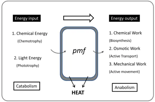
Energy is the engine of life and without it living organisms would not
exist. The energy conservation mechanisms are the chemical processes
by which cells produce ATP, the universal molecular currency of energy,
which is then used for the generation of chemical processes, movement,
heat generation and transport across membranes (Figure 1.1). In
chemotrophic organisms energy conservation is coupled to redox
reactions in catabolic pathways. The catabolic pathways can be linear
with a constant ATP output, like in aerobic respiration, or can be
branched, with several possible alternative electron acceptors, as in
anaerobic respiration, where each branch can lead to different ATP gains
and thermodynamic efficiency of ATP synthesis. Organisms that live
under anaerobic conditions are extremely diverse and versatile,
exhibiting a great metabolic diversity as a reflex of their adaptation to
different environmental conditions (temperature, pH, salinity, oxygen,
electron acceptor, etc.) (Thauer et al. 1977; Schmitz et al. 2006).
In chemotrophic bacteria, ATP can be produced by two energy
conservation mechanisms: substrate-level phosphorylation (SLP), in
which ATP is generated from energy-rich intermediates; and oxidative
phosphorylation or electron-transfer phosphorylation (ETP), where
electron carriers are reoxidized by a terminal electron-acceptor with
formation of an electrochemical gradient (ΔpH for protons or ΔpNa for
sodium ions) across the cytoplasmic membrane that is used by ATP
energy conservation, oxidative phosphorylation, is also known as
respiration and involves chemiosmotic coupling (Mitchell 1961).
Figure 1.1 - Energetical conversions inside the cell. Adapted from (Thauer et al. 1977).
Despite the large availability of carbon substrates there are only a few
reactions in anaerobes that conserve energy through SLP (Table 1.1) by
comparison to the amount of electron donors/acceptors that can be
used to generate energy by ETP (Table 1.2).
Respiratory organisms conserve energy through both pathways, SLP and
ETP, using a diverse range of organic and inorganic substrates as electron
donors/acceptors in aerobic or anaerobic respiration (Table 1.2) (Thauer
et al. 1977; Müller 2003; Herrmann et al. 2008). Fermentative organisms
C
h
a
p
te
r
1
process where organic compounds (sugars and amino acids) function as
both electron donors and acceptors and the excess reductants are
removed as reduced compounds (ethanol or H2, for example).
Table 1.1 - Reactions that yield ATP by substrate-level phosphorylation in anaerobes, adapted from (Schmitz 2006).
Reaction Enzyme ΔGabs
0
(kJ/mol)
1,3-Biphosphoglycerate + ADP ↔
3-phosphoglycerate + ATP
Phosphoglycerate
kinase
–24.1
Phosphoenol pyruvate + ADP ↔ pyruvate + ATP Pyruvate kinase –23.7
Acetyl phosphate + ADP ↔ acetate + ATP Acetate kinase –12.9
Butyryl phosphate + ADP ↔ butyrate + ATP Butyrate kinase –12.9
Carbamoyl phosphate + ADP ↔ carbamate + ATP Carbamate kinase –7.5
N-Formyl FH4 + ADP + Pi↔ formate + FH + ATP
Formyl-FH4
synthetase +8.32
Glycine + 2H+ + ADP + Pi ↔ acetate + NH3 + ATP Glycine reductase ~ –46.0
FH4, tetrahydrofolic acid
However, the recent identification of complex chemiosmotic
mechanisms in fermentative organisms, such as electrogenic transport in
lactic acid bacteria (Lolkema et al. 1995), electron transfer through
energy conserving hydrogenases in fermentative hyperthermophiles
(Sapra et al. 2003) or sodium translocating NADH dehydrogenases in
glutamate fermenting bacteria (Boiangiu et al. 2005), indicate that
fermentative organisms are more versatile in the way they conserve
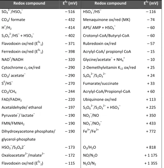
Table 1.2 - Redox potential of electron donors/electron acceptors involved in electron
transport phosphorylation, adapted from (Thauer et al. 1977; Sato et al. 1999).
Redox compound E0’ (mV) Redox compound E0’ (mV)
SO42–/HSO3– – 516 HSO3–/HS– – 116
CO2/ formate – 432 Menaquinone ox/red (MK) – 74
H+/H2 – 414 APS/ AMP + HSO3– – 60
S2O32-/HS– + HSO3– – 402 Crotonyl-CoA/Butyryl-CoA – 60
Flavodoxin ox/red (E0’1) – 371 Rubredoxin ox/red – 57
Ferredoxin ox/red (E0’1) – 398 Acrylyl-CoA/ propionyl CoA – 15
NAD+/NADH – 320 Glycine/acetate– + NH4+ – 10
Cytochrome c3 ox/red – 290 2-Demethylvitamin K12 ox/red + 25
CO2/ acetate– – 290 S4O62–/S2O32– + 24
S0/HS– – 270 Fumarate/succinate + 33
CO2/CH4 – 244 Acrylyl-CoA/Propionyl-CoA + 60
FAD/FADH2 – 220 Ubiquinone ox/red + 113
Acetaldehyde/ ethanol – 197 S3O62–/S2O32– + HSO3– + 225
Pyruvate–/ lactate– – 190 NO2–/NO + 350
FMN/FMNH2 – 190 NO3–/NO2– + 433
Dihydroxyacetone phosphate/
glycerol-phosphate
– 190 Fe3+/Fe2+ + 772
HSO3–/S3O62– – 173 O2/H2O + 818
Oxaloacetate2–/malate2– – 172 NO/N2O + 1 175
Flavodoxin ox/red (E0’2) – 115 N2O/N2 + 1 355
The advent of genomic information in the last years has also provided
invaluable information about the evolution of respiratory systems and
also of the origin of life itself. In fact, the study of the biology and the
processes involved in chemotrophic anaerobic bacteria have contributed
C
h
a
p
te
r
1
(Nitschke and Russell 2009; Martin 2012; Schoepp-Cothenet et al. 2013;
Sousa et al. 2013). In the past, fermentations were believed to be the
ancestral mechanisms for energy conservation. But in terms of evolution,
enzymes involved in SLP have no signs of antiquity, whereas
chemiosmotic coupling systems are found ubiquitously in all living
organisms, such as ATP synthase, which was present in the Last Universal
Common Ancestor (LUCA) of Bacteria and Archaea (Lane et al. 2010;
Schoepp-Cothenet et al. 2013).
Today, Earth’s atmosphere is completely different from the early days of
our planet when oxygen was absent, and high temperatures were
present together with a highly reducing environment with abundant H2
and CO2 (Liu et al. 2012; Martin 2012; Poehlein et al. 2012;
Schoepp-Cothenet et al. 2013). Methanogens and acetogens have been proposed
to be the most ancestral life forms, as their energy metabolism relies on
the electron transfer from H2 to CO2, with formation of methane and
acetate, respectively (Martin 2012). For this reason methanogens and
acetogens are good candidates to study ancestral pathways of energy
and carbon metabolism and understand how life forms have evolved
within Archaea and Bacteria, respectively (Müller 2003; Liu et al. 2012;
Martin 2012; Sousa et al. 2013). Both archaeal methanogens and
acetogenic bacteria reduce CO2 by the Acetyl-CoA or Wood-Ljungdahl
pathway (Scheme 1.1), and both cases present groups that lack
cytochromes and quinones (acetogens) or quinone analogs
(methanophenazine in methanogens) (Martin 2012).
enzymes (Simon et al. 2008; Schoepp-Cothenet et al. 2013). Different
kinds of chemiosmotic systems are coupled to different types of
quinones, which is also associated with their midpoint redox potential.
Generally, quinones can be divided in low-potential carriers such as
menaquinone (E0’ (MK/MKH2) = – 70 mV) for anaerobic conditions, and
high redox potential quinones like ubiquinone (E0´ (UQ/UQH2) = ~+ 100 mV) for aerobic conditions (Simon et al. 2008; Schoepp-Cothenet
et al. 2013). Their redox potential also reflects evolutionary aspects of
chemiosmotic mechanisms as the Earth evolved from an ancient anoxic
atmosphere to one containing oxygen (Liu et al. 2012; Schoepp-Cothenet
et al. 2013). The absence of quinones or quinone analogs in acetogens
and methanogens is unique among autotrophs, and indicates they still
conserve energy with chemiosmosis without liposoluble hydrogen
carriers (Martin 2012). The first step in methanogenesis and
acetogenesis, reduction of CO2 from H2 is an endergonic reaction, so how
is this reaction possible and at the same time allowing energy
conservation? This is a key question in the bioenergetic metabolism of
anaerobes, that until recently remained unanswered. The answer to this
question was provided by a recent energy metabolism process,
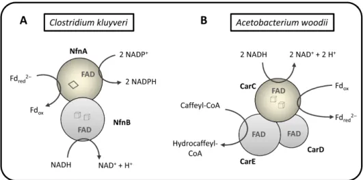
described by Buckel, Thauer and co-workers (Herrmann et al. 2008; Li et
al. 2008), named Flavin-based electron bifurcation (FBEB). This process,
first described to occur in the homoacetogenic clostridial organism
Clostridium kluyvery (Li et al. 2008), couples an endergonic to an
exergonic reaction, where the low redox potential reduced ferredoxin
C
h
a
p
te
r
1
Scheme 1.1 - Acetyl-CoA or Wood-Ljungdahl pathway for carbon dioxide fixation and respective enzymes involved in acetogenesis (A) and in methanogenesis (B). Fhd,
formate dehydrogenase; Fts, Formyl-THF synthetase; Mch, methenyl-THF
cyclohydrolase; Mhd, methylena-THF dehydrogenase; Mhr, methylene-THF reductase; Mtt, methyl-transferase; CODH, CO dehydrogenase; ACS, Acetyl-CoA synthase; Ppt, phosphotransacetylase; Ack, acetate kinase. Fwd, formyl-MFR dehydrogenase; Hmd,
H2-dependent methylene-H4MPT dehydrogenase; Mtd, F420-dependent
methylene-H4MPT dehydrogenase; Mcr, methyl-CoM reductase; Mtr, methyl-H4MPT-CoM
methyltransferase; Hdr, heterodisulfide reductase. Adapted from (Müller 2003; Costa
1.1.1-ELECTRON BIFURCATION, FLAVOPROTEINS AND FERREDOXIN
The concept of electron bifurcation was first proposed in 1976 by Peter
Mitchell to explain the energy conservation in the protonmotive Q-cycle
in cytochrome bc1 (Mitchell 1976). The cytochrome bc1 complex, also
known as Complex III, is present in the mitochondrial inner membrane of
eukaryotic cells and in several bacterial electron transfer chains that use
oxygen, nitrogen or sulfur compounds as terminal electron acceptors. It
is also part of the photosynthetic purple bacteria electron transfer chain
and belongs to the larger family of bc-type complexes that includes the
cytochrome bf complex present in chloroplasts, algae and some
gram-positive bacteria (Trumpower 1990; Brandt 1996b; Hunte et al. 2003).
The general complex is composed of three electron transfer proteins: a
cytochrome b subunit with two heme b groups (one with a low redox
potential - bL, and the other with a high redox potential - bH); a
cythocrome c1 and a [2Fe-2S]2+/1+ Rieske protein (Figure 1.2). In the
protonmotive Q cycle, ubiquinol oxidation is linked to proton release in
the positive side (center P) of the membrane and ubiquinone is reduced
in the negative side (center N) of the membrane with proton uptake.
Electron bifurcation takes place at center P during ubiquinol oxidation
with electron flow into the high redox potential Rieske [2Fe-2S]2+/1+ cluster (E0’ = + 290 mV; exergonic reaction) coupled to the low redox cytochrome bL reduction (E0’ ≈ – 20 mV; endergonic reaction) by the
ubisemiquinone radical. This electron bifurcation allows vectorial proton
C
h
a
p
te
r
1
conservation in mitochondrial and many bacterial respiratory chains –
Figure 1.2 (Brandt 1996a; Brandt 1996b).
Figure 1.2 - Homodimeric organization of the cytochrome bc1 complex with
representation of the proton-motive Q-cycle mechanism. Ubiquinol is oxidized at
center P (QP site) and one electron goes to the Rieske Fe-S protein (ISP) generating a
low potential ubisemiquinone anion, which immediately reduces heme bL and
subsequently heme bH with two protons being released at the P site of the membrane.
At the same time the electron transferred to the iron-sulfur protein is transferred to
cytochrome c1 and then to cytochrome c. The Q-cycle is complete when a second
ubiquinol is oxidized in center P and the electrons transferred to bH end up reducing the
ubisemiquinone radical at QN with proton uptake from the cytoplasm and ubiquinol
release. One complete Q-cycle requires one ubiquinol molecule oxidation at QP site,
two cytochrome c molecules reduced, four H+ release at P site and two H+ uptake at N
The FBEB mechanism was proposed by similarity to the electron
bifurcation that takes place in the center P of the mitochondrial
cytochrome bc1 complex. But in FBEB, flavin cofactors are responsible for
the bifurcation reaction in which the formation of a low redox potential
flavin semiquinone (“hot flavosemiquinone”) radical can be responsible
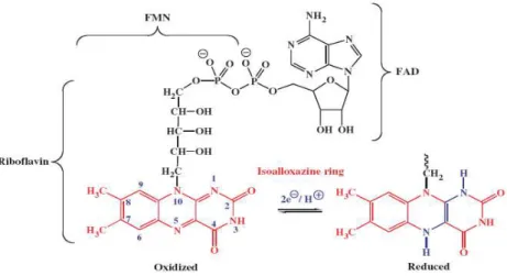
for ferredoxins reduction. Flavins are biological cofactors derived from
riboflavin (vitamin B2) and they are constituted by a redox-active
isoalloxazine ring system capable of one electron transfer in two steps or
two electron transfers at once. The flavin cofactors found in enzymes,
which are called flavoproteins or flavoenzymes, can be classified as flavin
mononucleotide (FMN) or flavin-adenine dinucleotide (FAD), according
to the group attached to the reactive ring (Figure 1.3). These kind of
proteins are very important in biological systems because they are
involved in diverse flavin dependent reactions, such as,
dehydrogenations, oxidations, monooxygenations, halogenations,
reductions and biological sensing (Macheroux et al. 2011). Additionally, if
combined with other redox-active centers, like iron-sulfur clusters
([2Fe-2S]2+/1+, [3Fe-4S]1+/0 and/or [4Fe-4S]2+/1+), they can be involved in more complex electron transfer reactions, as we can confirm from FBEB
C
h
a
p
te
r
1
Figure 1. 3 - Representation of riboflavin, FMN and FAD structures, in which the isoalloxazine ring is responsible for the redox activity of the cofactor. The isoalloxazine ring is represented in the oxidized and in the two electron reduced state. From
(Macheroux et al. 2011).
Ferredoxins (Fd) are cytoplasmic iron-sulfur proteins found in all living
organisms from archaea and bacteria to higher plants and animals. These
acidic, electron transfer proteins contain one [2Fe-2S]2+/1+ cluster or one, two or more [4Fe-4S]2+/1+ clusters (Sticht and Rosch 1998; Buckel and Thauer 2013). The redox potential of Fd is very low (E0’ = – 450 mV), and inside cells they are more than 90% reduced, which makes them good
electron donors in reactions with redox potentials as low or even
below – 500 mV. They are involved in several processes such as
hydrogen metabolism, nitrogen and CO fixation, and nitrite and sulfite
reduction among others (Sticht and Rosch 1998). But the most
interesting aspect of Fds is their antiquity reflected in their widespread
present since the primordial forms of life (Figure 1.4) (Eck and Dayhoff
1966; Kim et al. 2012; Sousa et al. 2013).
Figure 1.4 - Hypothesized iron-sulfur cluster evolution from hydrothermal vents to life.
a) Iron-sulfur minerals such as pyrite (FeS2) could spontaneously catalyze carbon
fixation to generate essential organic molecules for life; b) the organic molecules or amino acids formed allows new chemistry or enhances the existing reactions; c) synthesis of small polypeptides occurs at the water-mineral interface; d) small polypeptides constitute ferredoxin-like proteins; e) ferredoxin is preserved in all forms
of life constituting a large domain of redox proteins necessary for life. From (Kim et al.
2012).
1.1.2-THE FLAVIN BASED ELECTRON BIFURCATION MECHANISM
The Gram-positive bacterium Clostridium kluyvery is unique among
clostridia as it can grow anaerobically on ethanol and acetate as sole
energy sources (Seedorf et al. 2008). The energy metabolism of
C
h
a
p
te
r
1
how the organism ferments ethanol and acetate to butyrate, caproate
and H2, according to the equation:
(1.1) 6 ethanol + 3 acetate–→ 3 butyrate – + caproate – + H+ + 4 H2O +
2 H2
(ΔG0’ = – 183 kJ/mol)
An important question regarding the C. kluyvery metabolism is how does
it produce H2?
In 2008, the work of Herrmann et al. and Li et al. (Herrmann et al. 2008;
Li et al. 2008) could explain the mechanism of H2 formation from
reduced Fd and NADH, in this anaerobic bacterium. Their work
demonstrated that H2 formation was Fd-dependent for reduction of
crotonyl-CoA to butyryl-CoA; additionally H2 was generated during
fermentation from NADH in an endergonic reaction:
(1.2) NADH + H+→ H2 + NAD+ (ΔG0’ = 20 kJ/mol)
Herrmann et al. suggested that the highly exergonic reaction of
crotonyl-CoA reduction (E0’ = – 10 mV) with NADH (E0’ = – 320 mV) could be involved in energy conservation in this organism. During butyrate
synthesis, crotonyl-CoA is reduced to butyryl-CoA and this reaction is
NADH dependent (Herrmann et al. 2008). The work of Li et al. (Li et al.
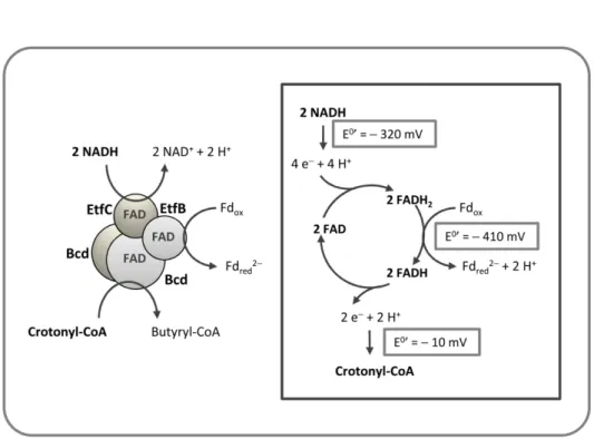
2008) demonstrated that Fd reduction (E0’ = – 410 mV) with NADH was coupled to reduction of crotonyl-CoA to butyryl-CoA with NADH. The
dehydrogenase/electron transfer complex (BcdA/EtfBC complex) –
Figure 1.5, in the following reaction:
(1.3) Fdox + 2 NADH + crotonyl-CoA → Fdred2- + 2 NAD+ + butyryl-CoA
(ΔG0’ = – 44 kJ/mol)
Figure 1.5 - Flavin based electron bifurcation mechanism by Bcd/EtfCB from Clostridium
kluyveri. The exergonic reduction of crotonyl-CoA by NADH is coupled to the
endergonic reduction of ferredoxin with NADH. Adapted from (Li et al. 2008).
The BcdA/EtfBC complex contains four FAD cofactors and no other
prosthetic group, which is why the investigators named the bifurcation
as flavin-based, as FAD is probably involved. Flavins are two electron
carriers that in some cases can be reduced by one electron to a stable
C
h
a
p
te
r
1
reduced to the fully reduced flavin nucleotide (FADH2 or FMNH2).
Generally the first one electron reduction has a much more positive
redox potential than the second electron reduction (Li et al. 2008). The
Bcd/Etf protein complex is currently one of the best well studied
examples of FBEB, and the mechanism of electron bifurcation has been
characterized through structural, biochemical, spectroscopic and kinetic
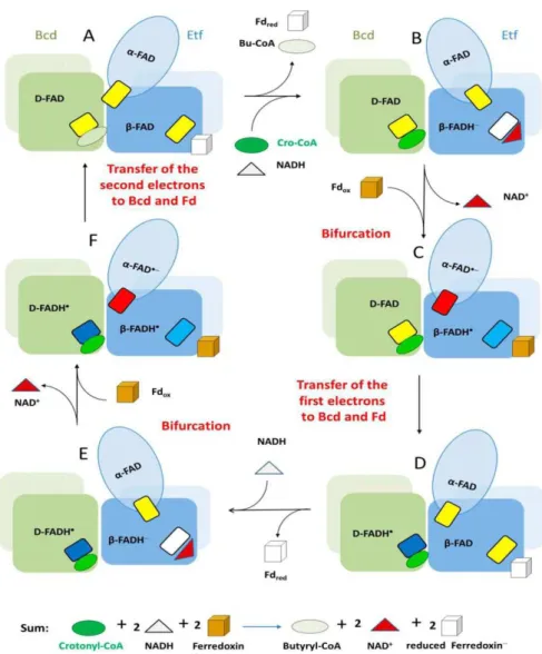
studies (Chowdhury et al. 2014). In a more recent study with Bcd/Etf
from Acidaminococcus fermentans, Chowdhury and coworkers proposed
the mechanism in Figure 1.6. The reaction starts with a two electron
transfer, in the form of hydride, from NADH to FAD in Etf generating
β-FADH– [E0’ (β-FAD/β-FADH–) = – 280 mV] (Figure 1.6A and 1.6B). The electron bifurcation in β-FADH– could generate a one electron transfer with a redox potential downhill to α-FAD generating α-FAD•– (– 60 mV), accompanied of domain reorientation, and the remaining β-FADH• with a low redox potential (– 500 mV) reduces Fd (Figure 1.6C and 1.6D). The
domain position change in Etf prevents the more favorable reduction of
α-FAD•–, and instead the electron is transferred to the D-FAD in Bcd. In the second cycle, Figure 1.6E-F-A, Fd is reduced and D-FADH– generated in Bcd is used for crotonyl-CoA reduction to butyryl-CoA (Chowdhury et
al. 2014). This concerted mechanism is similar to what happens in the
[2Fe-2S]2+/1+ Rieske protein in Complex III of the mitochondrial respiratory chain. Structural studies have demonstrated that movement
of the iron-sulfur center is necessary for the bifurcation reaction to be
possible, coupling the unfavorable reaction to the favorable one in the Q
Figure 1. 6 - Mechanism of flavin-based electron bifurcation proposed to operate at the Bcd/Etf complex based on structural considerations. Bcd dimers are represented interacting with Etf domains. The small rectangules represent FAD: yellow at the
quinone state (FAD), red at the anionic semiquinone state (FAD•−), light blue and dark
blue at the neutral semiquinone state (FADH•) and in white at the hydroquinone state
C
h
a
p
te
r
1
Since the reaction components are not directly involved in proton or ion
gradients, how can FBEB be considered a mechanism of energy
conservation? The Fdred can be considered as energy currency and
function like ATP plus NAD(P)H, contributing to energy conservation in
acetogenic organisms by two possible routes: by reducing protons to H2
increasing SLP in the oxidative branch of fermentation or by generating
an electrochemical gradient via the Rnf membrane complex (Herrmann
et al. 2008). The Rnf complex was first discovered in Rhodobacter
capsulatus and since the genes were involved in nitrogen fixation the
complex was named Rnf for Rhodobacter nitrogen fixation (Rnf). Rnf is an H+/Na+-pumping ferredoxin:NAD+ oxidoreductase that can be found in many anaerobes (see (Biegel et al. 2011) for a review).
Soon after the first publication regarding the FBEB mechanism in
Clostridium, a review article about energy conservation in methanogens
proposed that FBEB would also operate in methanogens that also lack
cytochromes (Thauer et al. 2008).
1.1.2.1-FBEB AND METHANOGENS
In methanogens, methane formation from CO2 reduction with H2 is
coupled to the formation of heterodisulfide (CoM-S-S-CoB) in a reaction
catalyzed by methyl-coenzyme M reductase (Mtr). The heterodisulfide
functions as the terminal electron acceptor of an energy-conserving
electron transport chain, and its reduction mechanism is distinct in
methanogens with and without cytochromes. Heterodisulfide reductase
membrane bound subunit with hemes
containing subunit (HdrD) (Ide et al.
2008). HdrDE receives electrons fro
through heme b, which are then
heterodisulfide reduction takes pla
membrane methanophenazine-reducin
contributes to energy conserva
(Deppenmeier 2004; Hedderich et al.
1.7A).
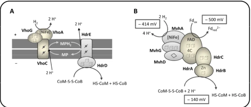
Figure 1.7 - Schematic representation of h reactions in methanogens. (A) HdrDE/VhoACG methanogens, in which heterodisulfide reductio
from (Thauer et al. 2010). (B) HdrABC/MvhAD
cytochromes. The complex is responsible for coupling it to an endergonic reaction, Fd re
Adapted from (Thauer et al. 2008). Cubes
cluster; - heme b.
es b (HdrE) and a soluble iron-sulfur
al. 1999; Deppenmeier and Müller
from the methanophenazine pool
then conducted to HdrD where
place. HdrDE together with the
ucing [NiFe] hydrogenase (VhoACG)
ervation during methanogenesis
al. 2005; Thauer et al. 2008) (Figure
of heterodisulfide reduction and coupled oACG complex from cytochrome-containing uction is coupled to chemiosmosis. Adapted hADG complex from methanogens that lack
for heterodisulfide reduction with H2 while
d reduction by H2, in a FBEB mechanism.
C
h
a
p
te
r
1
In methanogens that lack cytochromes heterodisulfide reductase is a
soluble protein complex composed of three subunits: HdrB with one
[4Fe-4S]2+/1+ cluster, HdrC containing two [4Fe-4S]2+/1+ clusters, with both subunits sharing homology with HdrD (which is like a hypothetical fusion
protein of HdrB and HdrC); and HdrA that contains four [4Fe-4S]2+/1+ clusters and one FAD (Hedderich et al. 2005; Thauer et al. 2008). HdrABC
forms a tight complex with F420-non-reducing hydrogenase (MvhADG)
and together they are responsible for heterodisulfide reduction by H2
(Figure 1.7B).
The catalytic subunits in heterodisulfide reductases are HdrB in
hydrogenotrophic methanogens and HdrD in methylotrophic
methanogens. The subunits HdrA and HdrE are most likely the contact
points with the physiological electron donors. In the case of
membrane-associated HdrDE, electron transfer from methanophenazine is linked to
energy conservation in heterodisulfide reduction, but HdrA is a
cytoplasmic protein with no membrane association of any kind, so how is
heterodisulfide reduction coupled to energy conservation in this
situation?
The answer to this question relies in the FBEB mechanism proposed by
Thauer and co-workers (Thauer et al. 2008) to operate in
hydrogenotrophic methanogens, by similarity to the Bcd/Etf complex of
clostridia. Since HdrA is a flavin containing protein, the two electron
carrier FAD could bifurcate electrons from H2 (E0’ = – 414 mV) for
S-CoB reduction by H2 was shown to be dependent on Fd reduction by
H2:
(1.4) Fdox + 2H2 + CoM-S-S-CoB → Fdred2–+ CoM-SH + CoB-SH + 2H+
(ΔG0’ = – 55 kJ/mol)
The mechanism in Figure 1.7B can explain a long observed effect (RPG
effect) (Gunsalus and Wolfe 1977). Costa and coworkers (Costa et al.
2010; Costa et al. 2013b), also proved by protein-protein interaction
studies that in hydrogenotrophic methanogens heterodisulfide
reductase (Hdr), formylmethanofuran dehydrogenase (Fwd), F420
-nonreducing hydrogenase (Vhu) and formate dehydrogenase (Fdh) can
interact in vivo and thus in methanogenesis, H2 or formate can be used
as electron donors and conserve energy by FBEB, and that the first and
last step of methanogenesis are physically connected (RPG effect). The
energy is conserved because the exergonic heterodisulfide reduction is
coupled to reduction of Fd, which is then used for the endergonic
reduction of CO2 to formylmethanofuran (formyl-MFR) (Scheme 1.1).
The fundamental part of this mechanism is HdrA that is responsible for
electron bifurcation and generation of low-potential electrons. Costa et
al. demonstrated that either with H2 or formate, electrons flow from
hydrogenases or formate dehydrogenases through VhuD (homologous to
MvhD) to HdrA, where bifurcation takes place. HdrA and MvhD are fused
in some species stressing that MvhD subunit is involved in electron
transfer from an electron donor to HdrA (Hedderich et al. 2005). Another
C
h
a
p
te
r
1
proteins in methanogens, but it is also found in non-methanogens like
sulfate reducers, suggesting that this bifurcation mechanism can also be
found in other contexts beyond methanogenesis (Stojanowic et al. 2003;
Buckel and Thauer 2013; Costa et al. 2013b).
1.1.2.2-THE BIFURCATING [FEFE]-HYDROGENASES
Right after the publication of the first FBEB mechanism performed by
BcdA-EtfBC other FBEB protein complexes were characterized. In
common they have Fd-dependent reactions and the presence of FAD or
FMN cofactors. One of these proteins was isolated from the
hyperthermophilic Thermotoga maritima, the heterotrimeric [FeFe]
hydrogenase (HydABC), which coupled the oxidation of Fd (E0’ = – 453 mV) and oxidation of NADH (E0’= – 320 mV) to generate H2 (E0’ = – 420
mV) – Figure 1.8A (Schut and Adams 2009):
(1.5) NADH + 2Fdred2- + 3H+↔ 2H2 + NAD+ + 2Fdox
Figure 1.8 - Schematic representation of the structure and function of two electron
bifurcating [FeFe] hydrogenases. (A) HydABC from Thermotoga maritima is responsible
for coupling Fdred and NADH oxidation in a reverse flavin-based electron bifurcation
(confurcation) reaction for H2 generation. Adapted from (Schut and Adams 2009). (B)
HydABCD from Acetobacterium woodii couples Fd reduction by H2 with NAD+ reduction
by H2 in a FBEB reaction. Adapted from (Schuchmann and Müller 2012). Cubes –
[4Fe-4S] cluster; diamonds – [2Fe-2S] cluster.
HydABC is a trimeric protein with subunits in a 1:1:1 ratio; HydA is
predicted to contain three [4Fe-4S]2+/1+ clusters, two [2Fe-2S]2+/1+ clusters and the H cluster where H2 is formed; HydB is predicted to bind
three [4Fe-4S]2+/1+ and one [2Fe-2S]2+/1+ clusters and the FMN cofactor; HydC is supposed to bind one [2Fe-2S]2+/1+ cluster (Buckel and Thauer 2013).
In the energy metabolism of T. maritima glucose is fermented with
generation of NADH and Fdred, whose oxidation in turn must be coupled
to H2 formation. The mechanism of H2 production was solved with the
characterization of the bifurcating FMN-containing hydrogenase,
C
h
a
p
te
r
1
generation through a reverse flavin based electron bifurcation, also
named as confurcation (Schut and Adams 2009) (Figure 1.8A).
Another electron-bifurcating hydrogenase was isolated and
characterized from the acetogenic bacterium Acetobacterium woodii
(Schuchmann and Müller 2012). A. woodii is a model organism to study
acetogenesis without cytochromes and by extension to understand
ancient metabolisms, as it contains only one site for electrochemical ion
gradient generation (sodium-motive ferredoxin: NAD+-oxidoreductase, the Rnf complex). The HydABCD complex was purified and characterized
and it was demonstrated that the [FeFe]-hydrogenase uses electron
bifurcation. The HydABC from T. maritima shares similarity to the
bifurcating hydrogenase subunits from A. woodii. The endergonic
reduction of Fd by H2 could be explained due to the presence of FBEB
performed by the HydABCD complex that couples this reaction to the
exergonic reduction of NAD+ by H2 (Figure 1.8B). A similar bifurcating
[FeFe]-hydrogenase was also isolated and characterized from Moorella
thermoacetica (Wang et al. 2013a), that performs the coupled Fd and
NAD+ reduction with H2.
A novel type of NADP-specific bifurcating hydrogenase (HytA-E) was
isolated from Clostridium autoethanogenum grown on CO. This
hydrogenase forms a functional complex with a formate dehydrogenase
(FdhA) (Wang et al. 2013a). Similarly to methanogens (Costa et al. 2010;
Costa et al. 2013a; Costa et al. 2013b), this bifurcating hydrogenase can
perform electron bifurcation with H2 or formate as electron donors. The
complex was shown to perform the reversible reaction, coupling both

![Figure 1.8 - Schematic representation of the structure and function of two electron bifurcating [FeFe] hydrogenases](https://thumb-eu.123doks.com/thumbv2/123dok_br/15770055.641192/60.748.116.614.115.380/figure-schematic-representation-structure-function-electron-bifurcating-hydrogenases.webp)