BioMedCentral
Page 1 of 12
(page number not for citation purposes)
BMC Cardiovascular Disorders
Open Access
Research article
RAS gene polymorphisms, classical risk factors and the advent of
coronary artery disease in the Portuguese population
Ana I Freitas
1, Isabel Mendonça
2, Maria Brión
3, Miguel M Sequeira
4,
Roberto P Reis
5, Angel Carracedo
3and António Brehm*
1Address: 1Human Genetics Laboratory, University of Madeira, Portugal, 2Research Unit, Central Hospital of Funchal, Portugal, 3Centro Nacional de Genotipado (CEGEN), Instituto de Medicina Legal, Universidad de Santiago de Compostela, Spain, 4Department of Biology, University of Madeira, Portugal and 5Medical Sciences Faculty, Universidade Nova de Lisboa and Pulido Valente Hospital, Lisbon, Portugal
Email: Ana I Freitas - [email protected]; Isabel Mendonça - [email protected]; Maria Brión - [email protected];
Miguel M Sequeira - [email protected]; Roberto P Reis - [email protected]; Angel Carracedo - [email protected]; António Brehm* - [email protected]
* Corresponding author
Abstract
Background: Several polymorphisms within the renin-angiotensin system cluster of genes have
been associated with the advent of coronary artery disease (CAD) or related pathologies. We investigated the distribution of 5 of these polymorphisms in order to find any association with CAD development and distinguish if any of the biochemical and behavioural factors interact with genetic polymorphisms in the advent of the disease.
Methods: ACE I/D (rs4340), ACE A11860G (rs4343), AT1R A1166C (rs5186), AGT T174M (rs4762)
and AGT M235T (rs699) gene polymorphisms were PCR-RFLP analysed in 298 CAD patients and 510 controls from Portugal. Several biochemical and behavioural markers were obtained.
Results: ACE I/D DD and ACE11860 GG genotypes are risk factors for CAD in this population.
The simultaneous presence of ACE I/D I and ACE11860 A alleles corresponds to a significant trend towards a decrease in CAD incidence. We found several synergistic effects between the studied polymorphisms and classical risk factors such as hypertension, obesity, diabetes and dyslipidaemia: the presence of the DD genotype of ACE I/D (and also ACE11860 GG) increases the odds of developing CAD when associated to each one of these classical risk factors, particularly when considering the male and early onset CAD subgroup analysis; AGT235 TT also increases the CAD risk in the presence of hypertension and dyslipidaemia, and AT1R1166 interacts positively with hypertension, smoking and obesity.
Conclusion: ACE polymorphisms were shown to play a major role in individual susceptibility to
develop CAD. There is also a clear interaction between RAS predisposing genes and some biochemical/environmental risk factors in CAD onset, demonstrating a significant enhancement of classical markers particularly by ACE I/D and ACE11860.
Published: 17 July 2008
BMC Cardiovascular Disorders 2008, 8:15 doi:10.1186/1471-2261-8-15
Received: 8 February 2008 Accepted: 17 July 2008
This article is available from: http://www.biomedcentral.com/1471-2261/8/15 © 2008 Freitas et al; licensee BioMed Central Ltd.
BMC Cardiovascular Disorders 2008, 8:15 http://www.biomedcentral.com/1471-2261/8/15
Page 2 of 12
(page number not for citation purposes)
Background
Cardiovascular diseases represent today the main cause of death in human adults in western populations. A reason-able number of studies have focused in testing specific genetic markers among groups of ischemic coronary artery disease (CAD) patients and control groups, aiming to find a correlation between these gene polymorphisms and dis-ease. The renin-angiotensin system (RAS) has been shown to play a key role in the regulation of blood pressure and influence the cardiovascular system [1], and several genes belonging to this system have been associated with CAD. Two of the most intensively investigated genetic polymor-phisms are the insertion/deletion (I/D) alleles of the
angi-otensin I-converting enzyme (ACE) gene and mutations at
the angiotensin II AT1 receptor. Several mutations at the
angiotensinogen (AGT) gene have also been studied as
candidates in essential hypertension [2,3] or myocardial infarction (MI) [4].
Albeit all these and other markers that have been identi-fied in latter years as possible risk factors for several pathologies associated to CAD, a considerable number of
opposing results also exist. The DD genotype of ACE for
instance has been found to be linked to MI [5,6] but con-trary reports also exist [7,8]. There are even conflicting reports on the assessment of risk profiles depending on
the populations studied [6,9]. The AT1R A1166C
poly-morphism has also been subjected to opposite reports as to its role in CAD, particularly when it is related to the patient's geographic origin: it was found to be signifi-cantly associated with CAD in Caucasians [9,10] but not
in Asians [11]. The AGT M235T polymorphism has been
shown to be positively related to essential hypertension [12] and MI [9] but other studies found no relation at all between the marker and disease [13,14] and few reports exist to confirm an association with plasmatic AGT con-centration level [14].
In this report we have studied the distribution of geno-typic and allelic frequencies at 5 loci, in two groups of individuals from Madeira Island (Portugal), an island set-tled mainly by the Portuguese. A group of patients with a known CAD history and a control group with no CAD related pathologies were selected. The main focus was to analyse these genetic polymorphisms, alone or combined in haplotypes, and clarify their potential association with CAD related conditions. Knowing that in most cases CAD has a multifactorial basis, involving a number of genes and environmental factors interacting to determine whether or not the disease will develop, we also tried to determine a possible interaction between the five gene polymorphisms and several well known CAD linked fac-tors.
Methods
The total population of this study consisted of 808 Cauca-sian individuals (aged between 18 and 70 years old) divided in two groups: 510 subjects without a history of CAD, MI, or stroke – excluded after a medical examina-tion and interview – randomly selected from the electoral rows, who participated as controls, plus 298 individuals recruited from patients admitted to the Cardiology Care Unit of the Main Hospital of Funchal (Hospital Central do Funchal, Madeira Island, Portugal). Patients' recruit-ment satisfied the following criteria: stable coronary dis-ease suggested by clinical analysis and proved by angiographic exam (a significant lesion was considered
when ≥ 75% of luminal narrowing was observed in at
least one of the major arteries) or occurrence of MI as defined by the WHO criteria. This investigation is in con-formity with the principles outlined in the Declaration of Helsinki and was approved by the Hospital's Ethics Com-mittee; all subjects gave their informed consent. Cases and controls filled in a questionnaire about their personal his-tories – age, sex, essential hypertension, diabetes mellitus, smoking habits, overweight, sedentary habits, alcohol ingestion and family medical history – and provided blood samples for genotype analysis and biochemical measurements. The presence of traditional risk factors in both groups was determined using the criteria standard-ized by the European Society of Cardiology: an hyperten-sive condition was attributed when systolic blood
pressure values were ≥ 139 mm Hg and/or diastolic blood
pressure values ≥ 89 mm Hg in at least two separate meas-urements or when being medicated against hypertension; subjects were considered smokers when consuming more than five cigarettes per day or non-smokers when never smoked or had stopped smoking at least one year before sample collection; obesity was defined for BMI values ≥ 30 kg/m2; dyslipidaemia was considered for plasmatic values
of total cholesterol ≥ 200 mg/dl, triglycerides ≥ 150 mg/dl,
LDL ≥ 130 mg/dl and HDL ≤ 40 mg/dl. Table 1 gives the
basic characteristics of the studied population.
Genetic and biochemical analyses
Genomic DNA was extracted from an 80 µl aliquot of
whole blood using standard phenol/chloroform method-ologies with ethanol precipitation. The ACE I/D gene alle-les (D and I) were identified by PCR amplification as previously described [15]. In order to reduce mistyping of ID heterozygotes as DD homozygotes, a re-amplification was carried out in all identified DD homozygotes using an internal primer specific for the I allele [15]. The other SNPs were identified following previously established
PCR-RFLP conditions: AT1R A1166C [16], AGT M235T
[17], AGT T174M [18] and ACE A11860G [19].
BMC Cardiovascular Disorders 2008, 8:15 http://www.biomedcentral.com/1471-2261/8/15
Page 3 of 12
(page number not for citation purposes) Statistical analysis
In order to analyze the relative influence of genotypes and biochemical parameters among gender, the two groups (controls and patients) were each subdivided into male and female subgroups. Further grouping according to age (45 or less years old) was performed, aiming to analyse the association between genetics and early onset CAD. Individuals aged ≥ 45 years were not included in this sub-analysis because no data about the age of CAD onset was obtained. Basic genetic parameters such as allele and gen-otype frequencies at each locus, proportion of individual heterozygous samples (direct count heterozygosity as well as the unbiased estimate) and population differentiation were calculated using Genepop v3.1d [20] and Arlequin [21]. Haplotype frequencies were calculated using the PHASE 2.0 software [22]. The overall genetic diversity, within each group diversity and the amount among groups were also calculated. Deviation from Hardy-Wein-berg equilibrium per population and locus was calculated according to Weir and Cockerham FIS estimator using FSTAT v.2.9.3., with a Bonferroni correction for all signif-icance levels [23]. Within all groups of subjects, distribu-tion of allele and genotype frequencies and their differences were calculated using χ2 tests. Associated
prob-abilities (P) were calculated applying Fisher's exact test adjusted for multiple comparisons of associated geno-types. To test the significance of association between gen-otypes at pairs of loci in each sample we used a log-likelihood ratio G-statistic as implemented in Genepop. The relative odds ratio (OR) and 95% confidence interval of relative CAD risk for any of the genetic polymorphisms and biochemical and behaviour markers, was assayed by
logistic regressions using the SPSS package. To analyse possible positive or negative interactions between classi-cal risk factors of CAD and genetic polymorphisms we used a 4 × 2 table approach and epiInfo 3.4.3 to calculate ORs, respective 95% confidence intervals and two-tailed p values, as well as synergy measures in additive (SI) and multiplicative models (SIM) [24-26]. It was assumed that unexposed individuals without the susceptibility geno-type have a certain background risk for disease (OR00 is assumed to be 1); OR10 refers to the relative risk for
dis-ease among people without the susceptibility genotype for disease but exposed to the environmental risk factor relative to those with neither the susceptibility genotype nor exposure; OR01 refers to the relative risk among peo-ple with the susceptibility genotype who are not exposed to the risk factor relative to those with neither the suscep-tibility genotype nor exposure; OR11 is the ratio of disease risk among exposed people with susceptibility genotype to diseased risk among unexposed people without the sus-ceptibility genotype. These ORs were then used in the cal-culation of synergy indexes: SI = (OR11-1)/(OR10+OR01 -2), SIM = OR11/(OR10 × OR01) [25,26]; the relative excess risk due to interaction, RERI = OR11-OR10-OR01+1 [26]; and the attributable proportion of the disease due to inter-action, AP = RERI/OR11 [26].
Results
Classical risk factors
As expected, both the control and patient group showed differences in the biochemical markers and other conven-tional risk factors analysed (Table 1). Systolic and diasto-lic blood pressure, dyslipidaemia, arterial hypertension,
Table 1: Baseline characteristics and clinical data for conventional risk factors
Control CAD P
n 510 298
Gender (Male/Female, %) 57.84/42.16 78.86/31.82 ***
Age (years, mean) 47.47 ± 12.55 54.96 ± 10.40 ***
Familiar CAD history (%) 12,16 56,04 ***
Sedentarism (%) 61.96 58.39 NS
Smoking habit (%) 26.86 38.93 ***
Systolic blood pressure (mm Hg) 127.54 ± 17.43 134.63 ± 20.43 ***
Diastolic blood pressure (mm Hg) 75.83 ± 10.69 79.10 ± 10.46 ***
Arterial hypertension (%) 22.35 58.72 ***
PWV (m/s) 8.80 ± 1.89 10.26 ± 2.13 ***
Body mass index (kg/m2) 26.41 27.80 ***
Glycaemia (mg/dl) 97.96 ± 24.47 119.94 ± 50.60 ***
Diabetes mellitus (%) 3.14 23.15 ***
Total cholesterol (mg/dl) 217.72 ± 44.73 205.39 ± 48.14 **
HDL (mg/dl) 57.15 ± 17.04 39.74 ± 9.75 ***
LDL (mg/dl) 114.70 ± 37.27 119.02 ± 44.08 NS
Triglycerides (mg/dl) 130.73 ± 85.71 192.97 ± 142.96 ***
Dyslipidaemia (%) 11.76 70.81 ***
BMC Cardiovascular Disorders 2008, 8:15 http://www.biomedcentral.com/1471-2261/8/15
Page 4 of 12
(page number not for citation purposes)
diabetes mellitus, triglycerides and a previous record of CAD in the family are much higher in CAD patients then in controls. In average, LDL was also higher in the CAD group and HDL values were lower in the patients group, as well as total cholesterol. We performed a multivariate logistic regression analysis using the variables included in table 1 and also the putative risk genotypes of RAS poly-morphisms. We found HDL (OR = 0.91 95%CI: 0.89– 0.93, p < 0.0001), glycaemia (OR = 1.01 95%CI: 1.01– 1.02, p < 0.0001), CAD history (OR = 2.14 95%CI: 1.36– 3.35, p = 0.001), smoking habit (OR = 1.81 95%CI: 1.11– 2.93, p = 0.017), dyslipidaemia (OR = 13.18, 95%CI:
8.32–22.14, p < 0.0001) and ACE I/D DD polymorphism
(OR = 1.72, 95%CI: 1.08–2.75, p = 0.022) to be inde-pendently related to CAD.
Allele and genotype distribution
Table 2 presents the distribution of genotypes for the 5 loci in controls and patients. Genotypes at both ACE loci show statistically different distributions for both the over-all analysis and male subgroup. Only the group of patients is in Hardy-Weinberg equilibrium at each locus and overall as the control group is not at HWE at locus ACE I/D (χ2df
1 = 7.55, P < 0.01). As expected, mutations
at the AGT and ACE loci show strong significant linkage disequilibrium within each gene (P < 0.0001) for both groups, even after Bonferroni correction. The group of patients also shows significant genotypic disequilibrium at AGT235/AT1R (P < 0.03). An exact G-test for popula-tion differentiapopula-tion gave an overall value of 0.00238 in which the loci contributing significantly to the
differenti-ation between the two samples are both ACE
polymor-phisms (χ2 test, P < 0.0001 and P < 0.01 for ACE I/D and
ACE11860, respectively). Thus taking in consideration all
loci combined, both populations are significantly differ-ent concerning the genic differdiffer-entiation (χ2 df
10 = 24.082,
P = 0.007).
Male patient and control groups show significant
differ-ences in allele content at both ACE I/D and ACE11860
loci (P < 0.0001 and P < 0.05, respectively). This is also reflected on the distribution of genotypes across ACE I/D which is highly significant in a G-like test (P < 0.003).
Analysing possible genotype associations of ACE
poly-morphisms and CAD development we obtained
signifi-cant results when testing ACE I/D DD and ACE11860 GG,
while ACE I/D ID genotype showed to decrease CAD risk
(Table 3). The male population analysis resulted even more significant for these polymorphisms and no signifi-cant results where found in the female subgroup. ACE I/D DD genotype was shown to increase the risk of early onset CAD.
Haplotype analysis
Within the ACE gene, the haplotype ACE11860 A/ACE I/ D I was found to provide a decreased risk of developing
CAD, except for the ≤ 45 subgroup analysis. An
associa-tion between ACE11860 G/ACE I/D D and CAD was
found in the whole population and male subgroup anal-ysis, while ACE11860 G/ACE I/D I was associated with the disease in females (Table 3). No association was found
when analysing the AGT gene haplotypes (results not
shown). Considering all genes involved in the RAS sys-tem, only five out of nineteen obtained combinations yielded significant associations with CAD (Table 3),
espe-Table 2: Distribution of genotypes between patients and controls
Genotype Whole population (n = 808) Males (n = 530) Females (n = 278) ≤ 45 subgroup (n = 275) CAD
(n = 298)
Control (n = 510)
P CAD
(n = 235)
Control (n = 295)
P CAD
(n = 63)
Control (n = 215)
P CAD
(n = 64)
Control (n = 111)
P
AGT235 MM 86(28.86) 148(29.02) NS 63(26.81) 87(29.49) NS 23(36.51) 61(28.37) NS 19(29.69) 63(29.85) NS MT 155(52.01) 275(53.92) 124(52.77) 155(52.54) 31(49.21) 120(55.81) 31(48.44) 111(52.61) TT 57(19.13) 87(17.06) 48(20.42) 53(17.97) 9(14.28) 34(15.81) 14(21.87) 37(17.54)
AGT174 TT 235(78.86) 400(78.43) NS 185(78.72) 229(77.63) NS 50(79.36) 171(79.53) NS 47(73.44) 160(75.83) NS TM 59(19.80) 107(20.98) 46(19.57) 65(22.03) 13(20.63) 42(19.53) 16(25.00) 48(22.75)
MM 4(1.34) 3(0.59) 4(1.70) 1(0.34) 0(0) 2(0.93) 1(1.56) 3(1.42)
AT1R1166 AA 175(58.72) 291(57.06) NS 137(58.30) 179(60.68) NS 38(60.32) 112(52.09) NS 35(54.69) 110(52.13) NS AC 106(35.57) 193(37.84) 84(35.74) 104(35.25) 22(34.92) 89(41.40) 24(37.50) 86(40.76)
CC 17(5.70) 26(5.10) 14(5.96) 12(4.07) 3(4.76) 14(6.51) 5(7.81) 15(7.11)
ACE I/D II 38(12.75) 84(16.47) ** 29(12.34) 47(15.93) *** 9(14.29) 37(17.21) NS 8(12.50) 33(15.64) NS ID 137(45.97) 282(55.29) 108(45.96) 178(60.34) 29(46.03) 104(48.37) 28(43.75) 119(56.40) DD 123(41.28) 144(28.24) 98(41.70) 70(23.73) 25(39.68) 74(34.42) 28(43.75) 59(27.96)
ACE11860 AA 54(18.12) 113(22.16) ** 41(17.45) 65(22.03) * 13(20.63) 48(22.33) NS 11(17.19) 43(20.38) NS AG 135(45.30) 255(50.00) 109(46.38) 155(52.54) 26(41.27) 100(46.51) 28(43.75) 111(52.61) GG 109(36.58) 142(27.84) 85(36.17) 75(25.42) 24(30.10) 67(31.16) 25(39.06) 57(27.01)
BMC Cardiovascular Disorders 2008, 8:15 http://www.biomedcentral.com/1471-2261/8/15
Page 5 of 12
(page number not for citation purposes)
cially a male driven decreased risk provided by the
ACE11860 A/ACE I/D I/AGT174T/AGT235 M/AT1R A
combination and an increased probability of early onset
CAD provided by ACE11860 G/ACE I/D D/AGT174 T/
AGT235T/AT1R A.
Interaction between RAS polymorphisms and classical risk factors
The analysis of the possible positive/negative association between genotypes and classical risk factors is expressed in
Tables 4 and 5, except for polymorphism ACE11860 GG,
which rendered highly similar results to the ACE I/D anal-ysis, so the results are not shown, and AGT174, with insuf-ficient polymorphic data to perform this analysis.
Regarding AGT235 TT or AT1R CC, we analysed only the
whole population data, because the subdivision in sex or age classes rendered insufficient number of individuals to
perform statistical analysis. Regarding the conventional risk of subjects unexposed to both classical risk factor and genetic risk (reference category) as being 1.0, the OR esti-mating the effect of joint exposure to hypertension and
ACE I/D DD, AGT235 TT or AT1R CC was significantly
higher than the ORs estimating the effect of each factor in the absence of the other. The synergy index in early onset
CAD analysis (ACE I/D DD) was above 9, indicating a
departure from an additive relation. In this group the pro-portion of CAD attributable to the interaction of
hyper-tension and ACE I/D DD was as high as 85%. The
genotypes AT1R CC in the overall analysis, or ACE I/D DD in all subgroups, interact with smoking habit to develop CAD, showing more than a multiplicative effect in females (SI = 0.86, SIM = 1.77, AP = 0.13). The risk pro-vided by obesity was found to be positively reinforced by AT1R CC in the whole population analysis and ACE I/D
Table 3: Allele, genotype and haplotype association between ACE polymorphism and CAD and RAS combined set of alleles association with CAD
Whole population Males Females ≤ 45 subgroup
Allele
ACE11860 G 1.312(1.07–1.60)** 1.387(1.09–1.77)** NS NS
ACE I/D D 1.414(1.15–1.74)*** 1.562(1.22–2.00)*** NS NS
Genotype
ACE11860 GG 1.560(1.16–2.11)** 1.713(1.18–2.49)** NS NS
ACE I/D DD 1.795(1.34–2.41)*** 2.283(1.57–3.31)*** NS 1.880(1.05–3.36)*
ACE haplotype (ACE11860/ACEI/D)
A/I 0.670(0.54–0.83)*** 0.660(0.51–0.85)** 0.653(0.43–1.00)* NS
CAD = 191, C = 425 CAD = 155, C = 251 CAD = 39, C = 175 CAD = 42, C = 179
A/D 1.526(1.03–2.27)* NS 2.434(1.20–4.94)* NS
CAD = 47, C = 55 CAD = 35, C = 34 CAD = 14, C = 21 CAD = 8, C = 18
G/I NS NS 8.372 (2.13–32.87)** NS
CAD = 20, C = 23 CAD = 12, C = 21 CAD = 7, C = 3 CAD = 3, C = 6
G/D 1.256(1.03–1.53)* 1.440(1.13–1.84)** NS NS
CAD = 335, C = 520 CAD = 269, C = 283 CAD = 66, C = 231 CAD = 75, C = 219
RAS combined set of alleles1(ACE11860/ACEI/D/AGT174/AGT235/AT1R)
A/I/T/M/A 0.698(0.53–0.93)* 0.663(0.50–0.88)** NS NS
CAD = 76, C = 179 CAD = 96, C = 164
A/I/T/T/A 0.727(0.55–0.97)* NS NS NS
CAD = 76, C = 173
A/D/M/T/A NS 3.804(1.02–14.13)* NS NS
CAD = 9, C = 3
A/I/M/T/C 0.324(0.11–0.95)* NS NS NS
CAD = 4, C = 20
G/D/T/T/A 1.459(1.06–2.02)* 1.400(1.01–1.95)* NS 2.468(1.21–5.04)*
CAD = 71, C = 88 CAD = 87, C = 82 CAD = 14, C = 20
Values presentation: OR(95% CI); *P < 0.05, **P < 0.005, ***P < 0.0001, NS not significant P > 0.05; CAD, number of CAD patients; C, number of control subjects.
BMC
Ca
rdiova
scu
lar
Disorde
rs
20
08,
8
:15
h
ttp
://www.bio
m
e
d
cent
ral.com/147
1-226
1/8/1
5
Pa
ge 6 of
1
2
(page nu
mber not
for
cit
a
ti
on pur
poses)
Table 4: Synergistic effect of ACE I/D DD genotype and classical risk factors in CAD patients and controls
Classical risk factor
ACE I/D DD Whole population Males Females ≤ 45 subgroup
CAD Control OR
(95%CI)
CAD Control OR
(95%CI)
CAD Control OR
(95%CI)
CAD Control OR
(95%CI)
Hypertension
0 0 63 256 1 54 143 1 9 113 1 24 132 1
0 1 43 99 1.76(1.10–2.84)* 39 43 2.40(1.36–4.25)** 4 56 0.90(0.22–3.38)NS 11 55 1.10(0.47–2.55)NS
1 0 112 110 4.14(2.78–6.17)*** 83 82 2.68(1.69–4.25)*** 29 28 13.00(5.16–33.64)*** 12 20 3.30(1.32–8.25)*
1 1 80 45 7.22(4.46–11,73)*** 59 27 5.79(3.22–10.46)*** 21 18 14.65(5.33–41.40)*** 17 4 23.38(6.59–90.92)***
SI = 1.59; SIM = 0.99; RERI = 2.32; AP = 0.32
SI = 1.56; SIM = 0.90; RERI = 1.71; AP = 0.30
SI = 1.15; SIM = 1.25; RERI = 1.75; AP = 0.12
SI = 9.32; SIM = 6.44; RERI = 19.98; AP = 0.85
Smoking
0 0 112 269 1 76 153 1 36 116 1 11 106 1
0 1 70 104 1.62(1.09–2.39)* 48 47 2.06(1.23–3.45)* 22 57 1.24(0.64–2.41)NS 9 37 2.34(0.81–6.72)NS
1 0 63 97 1.56(1.04–2.34)* 61 72 1.71(1.07–2.71)* 2 25 0.26(0.04–1.21)NS 25 46 5.24(2.24–12.46)***
1 1 53 40 3.18(1.95–5.21)*** 50 23 4.38(2.40–8.02)*** 3 17 0.57(0.12–2.23)NS 19 22 8.32(3.21–21.96)***
SI = 1.85; SIM = 1.26; RERI = 1.00; AP = 0.31
SI = 1.91; SIM = 1.24; RERI = 1.61; AP = 0.37
SI = 0.86; SIM = 1.77; RERI = 0.07; AP = 0.13
SI = 1.31; SIM = 0.68; RERI = 1.74; AP = 0.21
Obesity
0 0 126 295 1 99 180 1 27 115 1 30 135 1
0 1 88 122 1.69(1.18–2.42)** 72 59 2.22(1.42–3.46)** 16 63 1.08(0.51–2.27)NS 22 55 1.80(0.91–3.55)NS
1 0 49 71 1.62(1.04–2.51)* 38 45 1.54(0.91–2.60)NS 11 26 1.80(0.73–4.39)NS 6 17 1.59(0.51–4.77)NS
1 1 35 22 3.72(2.03–6.87)*** 26 11 4.30(1.93–9.71)*** 9 11 3.48(1.18–10.24)* 6 4 6.75(1.56–30.78)*
SI = 2.07; SIM = 1.36; RERI = 1.41; AP = 0.38
SI = 1.88; SIM = 1.26; RERI = 1.54; AP = 0.36
SI = 2.82; SIM = 1.79; RERI = 1.60; AP = 0.46
SI = 4.13; SIM = 2.36; RERI = 4.36; AP = 0.65
BMC
Ca
rdiova
scu
lar
Disorde
rs
20
08,
8
:15
h
ttp
://www.bio
m
e
d
cent
ral.com/147
1-226
1/8/1
5
Pa
ge 7 of
1
2
(page nu
mber not
for
cit
a
ti
on pur
poses)
0 0 123 342 1 101 208 1 22 134 1 31 151 1
0 1 89 134 1.85(1.30–2.63)** 75 61 2.53(1.64–3.91)*** 14 73 1.17(0.53–2.56)NS 25 59 2.06(1.08–3.95)*
1 0 52 24 6.02(3.46–10.55)*** 36 17 4.36(2.25–8.54)*** 16 7 13.92(4.69–42.75)*** 5 1 24.35(2.61–570.81)**
1 1 34 10 9.45(4.33–21.12)*** 23 9 5.26(2.22–12.78)*** 11 1 67.00(8.19–
1458.07)***
3 0
--SI = 1.44; --SIM = 0.85; RERI = 2.58; AP = 0.27
SI = 0.87; SIM = 0.47; RERI = -0.63; AP = -0.12
SI = 5.04; SIM = 4.11; RERI = 52.91; AP = 0.79
--Dyslipidaemia
0 0 52 327 1 47 197 1 5 130 1 16 144 1
0 1 35 123 1.79(1.08–2.96)* 31 57 2.28(1.28–4.05)** 4 66 1.58(0.34–7.05)NS 6 58 0.93(0.31–2.70)NS
1 0 123 39 19.83(12.16–
32.47)***
90 28 13.47(7.68–
23.77)***
33 11 78.00(22.86–
287.79)***
20 8 22.50(7.80–67.21)***
1 1 88 21 26.35(14.58–
48.04)***
67 13 21.60(10.54–
45.02)***
21 8 68.25(18.00–
282.98)***
22 1 198.00(25.16–
4210.95)***
SI = 1.29; SIM = 0.74; RERI = 5.73; AP = 0.22
SI = 1.50; SIM = 0.70; RERI = 6.85; AP = 0.32
SI = 0.87; SIM = 0.55; RERI = -10.33; AP = -0.15
SI = 9.19; SIM = 9.46; RERI = 175.57; AP = 0.89
SI, Rothman's synergy index for interaction; RERI, relative excess risk due to interaction; AP, proportion of disease attributable to interaction; *P < 0.05, **P < 0.005, ***P < 0.0001, NS not significant P > 0.05.
BMC Cardiovascular Disorders 2008, 8:15 http://www.biomedcentral.com/1471-2261/8/15
Page 8 of 12
(page number not for citation purposes)
DD in all subgroups, particularly in females (SI = 2.82, SIM = 1.79; AP = 0.46) and ≥ 45 subgroup (SI = 4.13, SIM = 2.36, AP = 0.65). Performing the same analysis regard-ing diabetic individuals, significant results were found for
ACE I/D DD carriers in the whole population analysis and
a strong enhancement was found in females (SI = 5.04, SIM = 4.11, AP = 0.79), but not in males (SI and SIM < 1, AP < 0). None of the 2 polymorphisms presented in table 5 were found to interact with diabetes; nevertheless we must point that all 6 individuals carrying the combination AT1R CC and diabetes were CAD patients. The join
pres-ence of dyslipidaemia and ACE I/D DD interacts
signifi-cantly in CAD onset, except in females (SI and SIM < 1, AP = -0.15). This combination was shown to be accountable for 89% of the disease in the = 45 years subgroup (SI = 9.19, SIM = 9.46, AP = 0.89). All 12 individuals with AT1R CC genotype and dyslipidaemia were found to be CAD patients, even though no significant results were obtained due to statistical constraint.
Discussion
Previous studies have focused on the association of any of the ACE, AT1R and AGT gene polymorphisms with coro-nary events of several degrees, related or not with MI and essential hypertension. Up to know there is no consistent genetic pattern that may link a given haplotype to the risk of developing a CAD related syndrome or at least to make an individual more prone to be affected. All 5 gene poly-morphisms surveyed here promote phenotypic variants on known mechanisms leading to changes in the bio-chemical status of an individual. For example, the ACE I/ D variant is linked to ACE activity [10] and AGT M235T to different angiotensinogen plasma levels [3,14].
By evaluating the distribution of RAS gene polymor-phisms in a series of patients undergoing coronary angi-ography, the present study has shown that only mutations
at ACE gene seem to be linked to CAD and even these are
apparently male-linked because no such association was
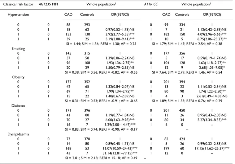
Table 5: Synergistic effect of AGT235 MM and AT1R CC genotypes and classical risk factors in CAD patients and controls
Classical risk factor AGT235 MM Whole population1 AT1R CC Whole population1
Hypertension CAD Controls OR(95%CI) CAD Controls OR(95%CI)
0 0 88 293 1 0 99 334 1
0 1 18 62 0.97(0.52–1.78)NS 1 7 21 1.12(0.42–2.89)NS
1 0 153 130 3.92(2.77–5.55)*** 0 182 150 4.09(2.96–5.66)***
1 1 39 25 5.19(2.88–9.41)*** 1 10 5 6.75(2.06–23.27)**
SI = 1.44; SIM = 1.36; RERI = 1.30; AP = 0.25 SI = 1.79; SIM = 1.47; RERI = 2.54; AP = 0.38 Smoking
0 0 145 315 1 0 177 356 1
0 1 37 58 1.39(0.86–2.24)NS 1 5 17 0.59(0.19–1.74)NS
1 0 96 108 1.93(1.36–2.75)** 0 104 128 1.63(1.18–2.27)**
1 1 20 29 1.50(0.79–2.85)NS 1 12 9 2.68(1.03–7.05)*
SI = 0.38; SIM = 0.56; RERI = -0.82; AP = -0.55 SI = 7.64; SIM = 2.79; RERI = 1.46; AP = 0.54 Obesity
0 0 172 352 1 0 201 394 1
0 1 42 65 1.32(0.84–2.07)NS 1 13 23 1.11(0.52–2.34)NS
1 0 69 71 1.99(1.34–2.95)** 0 80 90 1.74(1.22–2.50)**
1 1 15 22 1.40(0.67–2.89)NS 1 4 3 2.61(0.49–14.83)NS
SI = 0.31; SIM = 0.53; RERI = -0.91; AP = -0.65 SI = 1.89; SIM = 1.35; RERI = 0.76; AP = 0.29 Diabetes
0 0 171 396 1 0 201 450 1
0 1 41 80 1.19(0.77–1.84)NS 1 11 26 0.95(0.43–2.05)NS
1 0 70 27 6.00(3.63–9.98)*** 0 80 34 5.27(3.34–8.33)***
1 1 16 7 5.29(2.00–14.47)*** 1 6 0
--SI = 0.83; --SIM = 0.74; RERI = -0.90; AP = -0.17 --Dyslipidaemia
0 0 73 370 1 0 82 424 1
0 1 14 80 0.89(0.45–1.71)NS 1 5 26 0.99(0.32–2.83)NS
1 0 168 53 16.07(10.59–24.43)*** 0 199 60 17.15(11.62–25.37)***
1 1 43 7 31.14(12.81–79.15)*** 1 12 0
--SI = 2.01; --SIM = 2.18; RERI = 15.18; AP = 0.49
--SI, Rothman's synergy index for interaction; RERI, relative excess risk due to interaction; AP, proportion of disease attributable to interaction; *P < 0.05, **P < 0.005, ***P < 0.0001; 1We performed no further subdivision when analysing these polymorphisms because several classes resulted
BMC Cardiovascular Disorders 2008, 8:15 http://www.biomedcentral.com/1471-2261/8/15
Page 9 of 12
(page number not for citation purposes)
visible in females. The association of ACE I/D DD
geno-type with CAD in men but not in women, has been reported previously in several studies [27,28]. While our patient subpopulation was in Hardy-Weinberg
equilib-rium for I/D polymorphism of ACE gene, the control
sub-population was not, due to an excess of heterozygotes ID. Similar results were previously reported [5,29] and may be interpreted as a case of heterosis, or even confirm the higher risk homozygotes DD have to develop CAD.
The deletion polymorphism of the ACE gene has been
shown to be associated with both CAD and MI [5,30,31].
The mechanism by which the ACE I/D or ACE11860
gen-otypes may predispose an individual to the development of MI remains unclear. ACE is responsible for the conver-sion of angiotensin I to the peptide precursor angiotensin II, which has been implicated in the pathogenesis of atherosclerosis [32,33]. In contrast, other studies
con-cluded that ACE polymorphisms did not influence the
development of MI or other manifestations of CAD [34,35]. There are several possible reasons for these dis-crepancies: besides the different genetic backgrounds of the study populations, some of the studies cited have only
minor statistical power or the associations between ACE
gene polymorphisms and CAD or MI have been restricted to relatively small subgroups. Even more, in some studies, the presence or absence of CAD was not determined by angiography and might even have used false-negative con-trol populations. These findings also stress the necessity of considering ethnic factors in the assessment of genetic risk identifiers.
The total lack of association between the 2 AGTs and AT1R polymorphisms and CAD is in agreement with other stud-ies [14,36] that found no relation between these
polymor-phisms with CAD albeit a strong association between AGT
variants and angiotensinogen levels. Nevertheless, our study revealed a significant influence of the A allele and
AA genotype of AT1R upon an increase in carotid-femoral
PWV values, which may be regarded as a risk factor for CAD (results not shown).
The presence of the ACE I/D I and ACE11860 A alleles in ACE haplotypes corresponds to the lowest risk of
develop-ing CAD in the whole population, male and female sub-groups, expressed by ORs lower than 1. When analysing the influence of RAS haplotypes in the development of CAD, it is clear that the major influence comes from both
ACE polymorphisms. Yet again, the simultaneous
pres-ence of ACE I/D I and ACE11860 A alleles, corresponds to a significant trend towards a decrease in CAD, both in male subgroup and overall analysis. The combined set of
RAS alleles ACE11860 G/ACE I/D D/AGT174 T/AGT235
T/AT1R A was the only one found to significantly increase CAD risk in the whole population analysis. No significant
association was found in females, showing once again the interest of separating this type of data between sexes.
Premature CAD is known to have a particularly strong genetic component. Previous data have suggested that genetic factors are more likely to affect young rather than old people [37]. We conducted a population subdivision, analysing separately individuals who developed CAD before the age of 45. We found no significant allele or gen-otype association between any of the 5 polymorphisms and early onset CAD. The only significant results were found when associating the RAS combined set of alleles with the disease, where individuals under 45 have a 2.47 relative risk of CAD occurrence when in the join presence of ACE11860 G/ACE I/D D/AGT174 T/AGT235 T/AT1R A. Here we must point the impossibility to further subdivide this group according to sex, due to small sample size, as a limitation to perform a more thorough analysis. One should be aware that in almost all case-control studies, particularly those involving haplotype analysis, problems related to multiple comparisons (even when statistically corrected), the potential influence of genetic and environ-mental factors not considered, limited sample size for subgroup analysis and the possible inclusion of patients with silent CAD in the control group should be taken into account. The fact that the control and CAD patients groups are not sex and age matched may also be regarded as a constraint in the study design.
The complex aetiology around cardiovascular disorders and the multiple environmental conditionings are most of the times not evaluated together. In most cases, CAD has a multifactorial genetic basis, involving a number of genes and environmental factors interacting to determine whether or not the disease will develop. Therefore, the inherited genes generally predispose to a greater or lesser extent of CAD, but it is the environmental factors (e.g. cig-arette smoking, obesity, hypertension, sedentarism) inter-acting with the individual's genotype that determine whether or not CAD will develop. We performed further analysis in order to determine whether the simultaneous presence of genetic polymorphisms and well established risk factors – hypertension, smoking habit, obesity, diabe-tes and dyslipidaemia – would enhance the effect in CAD onset of these last. Some interesting results were obtained, particularly in the subgroups analysis. As both ACE poly-morphisms have shown to be in strong linkage disequilib-rium, we chose not to show the results of this analysis for
ACE11860 GG. They are very alike those presented in
table 4 as the exact same associations were found with similar, yet slightly lower values. We found a proportion of CAD attributable to the interaction between
hyperten-sion and ACE I/D DD genotype around 30% for the whole
vascu-BMC Cardiovascular Disorders 2008, 8:15 http://www.biomedcentral.com/1471-2261/8/15
Page 10 of 12
(page number not for citation purposes)
lar risk factors that might be related to serum ACE activity are not yet fully understood. Nevertheless, a previous study has shown a correlation between male sex and his-tory of hypertension with serum ACE activity [38], which may partially support our findings. This effect was also
found when considering AGT235 TT or AT1R CC as the
risk genotypes (SI = 1.44, SIM = 1.36, AP = 0.25; SI = 1.79, SIM = 1.47, AP = 0.38, respectively). Smoking may increase ACE levels by means of a nicotine enhancement
of ACE gene expression [39]. A large population based
study [40] found a positive association between the D allele of the I/D polymorphism and carotid artery thick-ness among smokers: individuals carrying only one of the risk factors did not show significant differences in artery thickness when compared to non-smokers with II geno-type, while carriers of both risk factors had significantly higher artery thickness. In our study we found a
synergis-tic effect due to smoking and ACE I/D DD join presence,
which is in accordance with a previous study [41]. More-over, these authors found a positive association between total high cholesterol, high LDL or overweight/obesity and the DD genotype. In the present report we also found this association, concerning dyslipidaemia and obesity. This synergy effect is particularly striking when consider-ing the early onset CAD subgroup analysis (obesity: SI = 4.13, SIM = 2.36, AP = 0.65; dyslipidaemia: SI = 9.19, SIM = 9.46, AP = 0.89). Obesity and hypercholesterolemia
have been shown to play a role in ACE gene expression,
but also in other RAS intervenients, such as AGT or AT1R [42]. Nevertheless, we found no association between
obesity and AGT235 TT or dyslipidaemia and AT1R CC,
even though all 12 dyslipidaemic individuals carrying AT1R CC were CAD patients. A similar study, analysing
AGT235 T allele synergistic effect with several risk factors,
found a positive association with hypercholesterolemia but neither with hypertension nor smoking [43]. The
association between ACE I/D polymorphism and Type 2
diabetes has been rather controversial. Some studies have affirmed a clear association between DD genotype and the disease in Caucasians [44] while others have excluded this hypothesis [45]. We found a female driven synergistic effect of DD genotype together with diabetes (SI = 5.04, SIM = 4.11, AP = 0.79).
Conclusion
The most relevant findings of this research are based on the influence of RAS gene polymorphisms evaluated together with the presence of the main classical risk fac-tors for CAD, showing how at least two polymorphisms – ACE I/D and ACE11860 A/G – interact synergistically with
them in CAD onset. Taking into account that CAD is a multifactorial disorder driven by numerous environmen-tal, behavioural and genetic components interacting together, our results seem to corroborate the hypothesis that RAS gene polymorphisms may indeed enhance the
influence of traditional risk factors in CAD development. The sample used in this study is also a novelty because it is based on the population of Madeira Island that until the beginning of the 20th century was a relatively isolated island. To our knowledge this analysis on such a popula-tion had not yet been performed. Madeiran inhabitants with a history of a small founder population, long lasting isolation and population bottleneck represent an excep-tional resource in the identification of genes involved in the pathogenesis of multifactorial diseases.
Competing interests
The authors declare that they have no competing interests.
Authors' contributions
AIF performed this work as part of her PhD thesis, under guidance of AB and AC. AB, IM and RPR conceived the idea for the study. Lab and clinical data were performed under AB, IM and AC guidance. All authors performed data analysis, interpretation and discussion of results and also read and approved the final manuscript.
Acknowledgements
This study was supported by contract POCTI/38697/MGI/2001 (co-financed by POCI 2010 and FSE) from Foundation for Science and Technol-ogy, Ministry of Science, Technology and Higher Education (Portugal). AIF is recipient of a PhD research fellowship under contract SFRH/BD/8592/ 2002. The authors are indebted to Dr. José Jesus for valuable criticisms over the data analysis.
References
1. van Berlo J, Pinto Y: Polymorphisms in the RAS and cardiac function. Int J Biochem Cell Biol 2003, 35:932-943.
2. Munroe PB, Caulfield MJ: Genetics of hypertension. Curr Opin
Genetics Dev 2000, 10:325-329.
3. Kamitani A, Rakugi H, Higaki J, Yi Z, Mikami H, Miki T, Ogihara T:
Association analysis of a polymorphism of the angiotensino-gen angiotensino-gene with essential hypertension in Japanese. J Hum
Hypertens 1994, 8:521-524.
4. Kamitani A, Rakugi H, Higaki J, Ohishi M, Shi SJ, Takami S, Nakata Y, Higashino Y, Fujii K, Mikami H, Miki T, Ogihara T: Enhanced pre-dictability of myocardial infarction in Japanese by combined genotype analysis. Hypertension 1995, 25:950-953.
5. Cambien F, Poirer O, Lecerf L, Evans A, Cambou JP, Arveiler D, Luc G, Bard JM, Bara L, Ricard S, Tiret L, Amouyel P, Alhenc-Gelas F, Sou-brier F: Deletion polymorphism in the gene for angiotensin-converting enzyme is a potential risk factor for myocardial infarction. Nature 1992, 359:641-644.
6. Sakuma T, Hirata R, Hirata M: Five polymorphisms in genes can-didates for cardiovascular disease in afro-Brazilian
individu-als. J Clin Lab Anal 2004, 18:309-316.
7. Keavney B, McKenzie C, Parish S, Palmer A, Clark S, Youngman L, Delépine M, Lathrop M, Peto R, Collins R: Large-scale test of hypothesised associations between the angiotensin-convert-ing-enzyme insertion/deletion polymorphism and myocar-dial infarction in about 5000 cases and 6000 controls: International Studies of Infarct Survival (ISIS) Collaborators.
Lancet 2000, 355:434-442.
8. Hamon M, Fradin S, Denizet A, Filippi-Codaccioni E, Grollier G, Morello R: Prospective evaluation of the effect of an angi-otensin I converting enzyme gene polymorphism on the long term risk of major adverse cardiac events after percutane-ous coronary intervention. Heart 2003, 89:321-325.
BMC Cardiovascular Disorders 2008, 8:15 http://www.biomedcentral.com/1471-2261/8/15
Page 11 of 12
(page number not for citation purposes)
the individual coronary risk profile. Eur Heart J 2000,
21:633-638.
10. Tiret L, Bonnardeaux A, Poirier O, Ricard S, Marques-Vidal P, Evans A, Arveiler D, Luc G, Kee F, Ducimetière P, Soubrier F, Cambien F:
Synergistic effects of angiotensin-converting enzyme and angiotensin II type 1 receptor gene polymorphisms on risk of myocardial infarction. Lancet 1994, 344:910-913.
11. Nakauchi Y, Suehiro T, Yamamoto M, Yasuoka N, Arii K, Kumon Y, Hamashige N, Hashimoto K: Signficance of angiotensin I-con-verting enzyme and angiotensin II type I receptor gene pol-ymorphism as risk factors for coronary heart disease.
Atherosclerosis 1996, 125:161-169.
12. Sato N, Katsuya T, Nakagawa T, Ishikawa K, Fu Y, Asai T, Fuduka M, Suzuki F, Nakamura Y, Higaki J, Oqihara T: Nine polymorphisms of angiotensinogen gene in the susceptibility to essential hypertension. Life Sci 2000, 68:259-272.
13. Hernández Ortega E, Medina Fernández-Aceituno A, Rodríguez-Esparragón FJ, Hernández Perera O, Melián Nuez F, Delgado Espinosa A, Fíuza Pérez D, Anabitarte Prieto A, Rodríguez Pérez JC: The involvement of the renin-angiotension system gene poly-morphisms in coronary heart disease. Rev Esp Cardiol 2002,
55:92-99.
14. Renner W, Nauck M, Winkelmann BR, Hoffmann MM, Scharnagl H, Mayer V, Boehm BO, Marz W: Association of angiotensinogen haplotypes with angiotensinogen levels but not with blood pressure or coronary artery disease: the Ludwigshafen Risk and Cardiovascular Health Study. J Mol Med 2005, 83:235-239. 15. Hernández D, Lacalzada J, Salido E, Linares J, Barragán A, Lorenzo V, Higueras L, Martín B, Rodríguez A, Laynez I, González-Posada JM, Torres A: Regression of left ventricular hypertrophy by lisino-pril after renal transplantation: Role of ACE gene polymor-phism. Kidney Int 2000, 58:889-897.
16. Doria A, Ji L, Warram JH, Krolewski AS: DdeI polymorphism in the AGTR1 gene. Hum Mol Gen 1994, 3:1444.
17. Russ AP, Maerz W, Ruzicka V, Stein U, Gross W: Rapid detection of the hypertension-associated Met235→ Thr allele of the human angiotensinogen gene. Hum Mol Gen 1993, 2:609-610. 18. Niu T, Yang J, Wang B, Chen W, Wang Z, Laird N, Wie E, Fang Z,
Lindpaintner K, Rogus JJ, Xu X: Angiotensin Gene Polymor-phism M235T/T174M. No Excess Transmission to Hyperten-sive Chinese. Hypertension 1999, 33:698-702.
19. Zhu X, Bouzekri N, Southam L, Cooper RS, Adeyemo A, McKenzie CA, Luke A, Chen G, Elston RC, Ward R: Linkage and Association Analysis of Angiotensin I-Converting Enzyme (ACE)-Gene Polymorphisms with ACE Concentration and Blood Pres-sure. Am J Hum Genet 2001, 68:1139-1148.
20. Raymont M, Rousset F: GENEPOP (V.1.2) A population genet-ics software for exact tests and ecumenicism. J Hered 1995,
95:248-249.
21. Schneider S, Kueffer J-M, Roessli D, Excoffier L: Arlequin: a software for
population genetic data analysis Genetics and Biometry Laboratory,
University of Geneva, Switzerland; 2000.
22. Stephens M, Smith NJ, Donnelly P: A new statistical method for haplotype reconstruction from population data. Am J Hum
Genet 2001, 68:978-989.
23. Goudet J: FSTAT, a program to estimate and test gene diver-sities and fixation indices (version 2.9.3). 2001 [http:// www2.unil.ch/popgen/softwares/fstat.htm]. Updated from Goudet (1995)
24. Rothman KJ: Synergy and antagonism in cause-effect relation-ship. Am J Epidemiol 1974, 99:385-388.
25. Assman SF, Hosmer DW, Lemeshow S, Mundt KA: Confidence intervals for measures of interaction. Epidemiology 1996,
7:286-290.
26. Yang Q, Khoury MJ: Evolving methods in genetic epidemiology III. Gene-Environment interaction in epidemiologic research. Epidemiol Rev 1997, 19:33-43.
27. Kee F, Morrison C, Poirier O, McCrum E, Mallet C, Nicaud V, McMas-ter D, Dallongeville J, Fruchart JC, Evans AE: Angiotensin II type-I receptor and ACE polymorphisms and risk of myocardial inf-arction in men and women. Eur J Clin Invest 2000, 30:1076-1082. 28. Petrovic D, Bregar D, Guzic-Salobir B, Skof E, Span M, Terziæ R, Petrovic MG, Keber I, Letonjia M, Zorc M, Podbregar M, Peterlin B:
Sex difference in the effect of ACE-DD genotype on the risk of premature myocardial infarction. Angiology 2004,
55:155-158.
29. Mata-Balaguer T, de la Herrana R, Ruiz-Rejon C, Ruiz-Rejon M, Gar-rido-Ramos MA, Ruiz-Rejon F: Angiotensin-converting enzyme and p22phox polymorphisms and the risk of coronary heart disease in a low-risk Spanish population. Int J Cardiol 2004,
95:145-151.
30. Ruiz J, Blanché H, Cohen N, Velho G, Cambien F, Cohen D, Passa P, Froguel P: Insertion/deletion polymorphism of the angi-otensin-converting enzyme gene is strongly associated with coronary artery disease in non-insulin-dependent diabetes mellitus. Proc Natl Acad Sci USA 1994, 91:3662-3665.
31. Mattu RK, Needham EW, Galton DJ, Frangos E, Clark AJ, Caulfield M:
A DNA variant at the angiotensin-converting enzyme gene locus associates with coronary artery disease in the Caer-philly Heart Study. Circulation 1995, 91:270-274.
32. Campbell-Boswell M, Robertson AL: Effects of angiotensin II and vasopressin on human smooth muscle cells in vitro. Exp Mol
Pathol 1981, 35:265-376.
33. Naftilan AJ, Pratt RE, Dzau VJ: Induction of platelet-derived growth factor A-chain and c-myc gene expressions by angi-otensin II in cultured rat vascular smooth muscle cells. J Clin
Invest 1989, 83:1419-1424.
34. Samani NJ, Thompson JR, O'Toole L, Channer K, Woods KL: A meta-analysis of the association of the deletion allele of the angiotensin-converting enzyme gene with myocardial infarc-tion. Circulation 1996, 94:708-712.
35. Lindpaintner K, Pfeffer MA, Kreutz R, Stampfer MJ, Grodstein F, LaMotte F, Buring J, Hennekens CH: A prospective evaluation of an angiotensin-converting enzyme gene polymorphism and the risk of ischemic heart disease. N Engl J Med 1995,
332:706-711.
36. Alvarez R, Reguero JR, Batalla A, Iglesias-Cubero G, Cortina A, Alva-rez V, Coto E: Angiotensin-converting enzyme and angi-otensin II receptor 1 polymorphisms: association with early coronary disease. Cardiovasc Res 1998, 40:375-379.
37. Roncaglioni MC, Santoro L, D'Avanzo B, Negri E, Nobili A, Ledda A, Pietropaolo F, Franzosi MG, La Vecchia C, Feruglio GA: Role of fam-ily history in patients with myocardial infarction: an Italian Case-Control study. GISSIEFRIM Investigators. Circulation
1992, 85:2065-2072.
38. Hung J, McQuillan BM, Nidorf M, Thompson PL, Beilby JP: Angi-otensin converting enzyme polymorphism and carotid wall thickening in a community population. Arterioscler Thromb Vasc Biol 1999, 19:1969-1974.
39. Zhang S, Day I, Ye S: Nicotine induced changes in gene expres-sion by human coronary artery endothelial cells. Atherosclero-sis 2001, 154:277-283.
40. Sayed-Tabatabaei FA, Schut AF, Hofman A, Bertoli-Avella AM, Ver-geer J, Witterman JC, van Duijn CM: A study of gene-environ-ment interaction on the gene for angiotensin converting enzyme: a combined functional and population based approach. J Med Genet 2004, 41:99-103.
41. Niemec P, Zak I, Wita K: Modification of the coronary artery disease risk associated with the presence of traditional risk factors by insertion/deletion polymorphism of the ACE gene. Genet Test 2007, 11:353-359.
42. Gorzelniak K, Engeli S, Janke J, Luft FC, Sharma : Hormonal regula-tion of the human adipose-tissue renin-angiotensin system: relationship to obesity and hypertension. J Hypertens 2002,
20:965-973.
43. Niemec P, Zak I, Wita K: The M235T polymorphism of the AGT gene modifies the risk of coronary artery disease associated with the presence of hypercholesterolemia. Eur J Epidemiol
2008, 23:349-354.
44. Stephens JW, Dhamrait SS, Cooper JA, Acharya J, Miller GJ, Hurel SJ, Humphries SE: The D allele of the ACE I/D common gene var-iant is associated with Type 2 diabetes mellitus in Caucasian subjects. Mol Genet Metab 2005, 84:83-89.
45. Grammer TB, Renner W, von Karger S, Boehm BO, Winkelmann BR, Maerz W: The angiotensin-I converting enzyme I/D polymor-phism is not associated with type 2 diabetes in individuals undergoing coronary angiography. (The Ludwigshafen Risk and Cardiovascular Health Study). Mol Genet Metab 2006,
Publish with BioMed Central and every scientist can read your work free of charge
"BioMed Central will be the most significant development for disseminating the results of biomedical researc h in our lifetime."
Sir Paul Nurse, Cancer Research UK
Your research papers will be:
available free of charge to the entire biomedical community
peer reviewed and published immediately upon acceptance
cited in PubMed and archived on PubMed Central
yours — you keep the copyright
Submit your manuscript here:
http://www.biomedcentral.com/info/publishing_adv.asp
BioMedcentral
BMC Cardiovascular Disorders 2008, 8:15 http://www.biomedcentral.com/1471-2261/8/15
Page 12 of 12
(page number not for citation purposes)
Pre-publication history
The pre-publication history for this paper can be accessed here: