ANTIMYCOBACTERIAL ACTIVITY OF THE ANTIINFLAMMATORY AGENT DICLOFENAC SODIUM, AND ITS SYNERGISM WITH STREPTOMYCIN
Noton K. Dutta1; Sujata G. Dastidar1*; Asok Kumar1; Kaushiki Mazumdar1; Raja Ray2; Atindra N. Chakrabarty2
1Department of Pharmaceutical Technology, Jadavpur University, Calcutta, India; 2Department of Medical Microbiology and
Parasitology, Calcutta University College of Medicine, Calcutta, India
Submitted: October 05, 2003; Returned to authors for corrections: April 26, 2004; Approved: December 20, 2004
ABSTRACT
Diclofenac sodium, an antiinflammatory agent, exhibited remarkable inhibitory action against both drug sensitive and drug resistant clinical isolates of Mycobacterium tuberculosis, as well as other mycobacteria. This drug was tested in vitro against 45 different strains of mycobacteria, most of which were inhibited by the drug at 10-25 µg/ml concentration. When tested in vivo, diclofenac, injected at 10 µg/g body weight of a
Swiss strain of white mice, could significantly protect them when challenged with 50 median lethal dose of M. tuberculosis H37 Rv 102. According to χ2 test, the in vivo data were highly significant (p<0.01). Diclofenac
was further tested for synergism with the conventional antimycobacterial drug streptomycin against M. smegmatis 798. When compared with their individual effects, synergism was found to be statistically significant (p<0.05). By the checkerboard assessment procedure, the fractional inhibitory concentration index of this combination was found to be 0.37, confirming synergism.
Key words: antiinflammatory drug, diclofenac sodium, antimycobacterial activity, streptomycin, synergism, non-antibiotic
INTRODUCTION
Mycobacteriosis, particularly tuberculosis, has become a global problem. The occurrence of multi-drug resistance among Mycobacterium tuberculosis in particular and mycobacteria in general needs surveillance and control. Failure to cure multi-drug resistant tuberculosis (MDR-TB) with the currently available antitubercular drugs leads to a search for newer and potent drugs to treat such cases, and thereby prevent an emerging multidimensional problem. Different studies aimed at discovering newer antimycobacterial agents have revealed moderate to powerful action in several compounds belonging to various pharmacological groups, e.g., promethazine (22), chlorpromazine (8), trifluoperazine (24),methdilazine (7), thioridazine (1) and other phenothiazines (17). Further studies have revealed the enhancement of antibiotic activity against MDR-TB by phenothiazines (25). Many of these agents have exhibited powerful inhibitory action against Gram positive and
Gram negative bacteria as well (18,20,21). Such compounds having antimicrobial properties in addition to their predesignated pharmacological action are entitled as “Non-antibiotics”. The antiinflammatory drug diclofenac sodium was seen to possess powerful antibacterial activity against Gram positive and Gram negative bacteria (3,9). It also exhibited significant synergism with an antibiotic streptomycin (4) and a non-antibiotic trifluoperazine (11). The present paper describes the antimycobacterial action of diclofenac both through in vitro and in vivo tests, and potentiation of its activity by combination with known antitubercular drugs.
MATERIALS AND METHODS Drugs
The drugs were obtained as pure dry powder from their respective manufacturers in India. Diclofenac sodium (Dc) and rifampicin (Rf) were obtained from Hindustan Ciba Geigy,
streptomycin (Sm) from Sarabhai Chemicals, ethambutol (Eb) from Lyka Laboratories and isonicotinic acid hydrazide (INH) from Glaxo Laboratories. They were preserved at 4ºC.
Bacteria
Forty-five strains of mycobacteria were tested. The strains and their sources are given in Table 1. The strains were identified by Radiometric method (BACTEC 460) and biochemical tests (Niacin, Nitrate, Urease, Catalase, Tween80, Tellurite and 5% NaCl tests).
Media
Liquid medium used was Kirchner’s Liquid medium (KLM) (16), which was used to grow and suspend the organisms.
Solid medium was Lowenstein Jensen Medium (LJM), prepared as described by the International Union Against Tuberculosis and Lung Diseases (IUT, 1955) (15).
Preparation of inocula for susceptibility tests
The bacterium was first grown in KLM. The inoculum was prepared by homogenizing the KLM culture with glass beads, spinning down the larger particles, and matching the supernate against Mc Farland’s standard (23).
Determination of minimum inhibitory concentration (MIC) of antibiotics / non- antibiotics against different strains of mycobacteria
While determining MIC by tube dilution method (12), Sm, Rf, INH and Eb each were used in the following concentrations (µg/ml) in KLM: 0 (Control), 0.25, 0.5, 1, 2, 4 and 8. Dc was used
in KLM in concentrations of 0 (Control), 5, 10, 15, 20, 25 and 50
µg/ml. For some selected strains, the drug was tested in
concentrations ± 2 of its MIC value, in order to find out its mean ± standard deviation values with respect to those organisms. Amount of inoculum used to inoculate each tube above was 0.01 ml. Incubation was done at 37ºC for 10-20 days as required. The MIC of each organism was defined as the lowest concentration of antibiotic where the growth obtained was reduced to 1% or less when compared to the control slopes. All the tests were run in duplicate. The resistant strains were further processed for determining the resistant break point by using 8, 16, 32, 64 and 128 µg/ml of the drug in LJM. The following were the MIC levels of primary antitubercular drugs indicating resistance - streptomycin (Sm) > 32 mg/l, isoniazid (INH) > 1 mg/l, rifampicin (Rf) > 128 mg/l and ethambutol (Eb) > 8 mg/l.
Determination of synergism between Dc and Sm by disc diffusion tests (5)
0.5 ml of inoculum was applied on LJM, poured in plates. Filter paper discs (Whatman No.1) containing 50 µg of Dc and
10 µg of Sm were placed on the LJM, and incubation was done
at 37ºC. The clear zones of inhibition around each disc individually or in combination were measured in three different directions to obtain the mean values of each test. The increase or decrease of surface area (πr2) due to a particular combination
as well as those due to single effects was statistically evaluated by χ2 test (6) for the level of significance of alteration. The
occurrence of mutual influence /interference when drugs were used in combination was assessed as (i) indifference, when both tangential circles of inhibition were unaffected, (ii) antagonism, when the circles receded and assumed kidney shape, (iii) synergism (24), when the circles enlarged. The
Table 1. Source of mycobacterial strains tested.
Mycobacteria Source
Reference strains Tuberculosis Research Center, Chennai,
M. tuberculosis H37 Rv102, ICMR, Govt. of India
H37Ra16
M. marinum 50, M. scrofulaceum 1323, M. gordonae 1324,
M. flavescens 1541, M. xenopi 160, M. avium 724, Central JALMA Institute for Leprosy, ICMR, M. intracellulare 1406, M. terrae 1450, M. trivate 1453, Agra, India
M. smegmatis 798, M. smegmatis 1546, M. fortuitum 1529, M. phlei L1
Clinical strains
M. tuberculosis Bajaj, J15, N23, 912042, 911928, 905574,
911454,910708,911831,905358,911447,911884,912234,91 Tuberculosis Research Centre, Chennai, India 1677,90657,912359,911337,912073,912447,912056,91105
3,906909
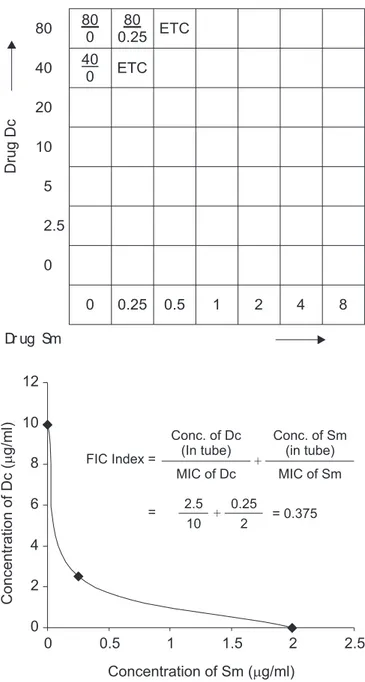
combined effects of two drugs were further determined by the checkerboard dilution technique for derivation of the Fractional Inhibitory Concentration (FIC) indices (19). The checkerboard was such arranged that in the first horizontal row, all the tubes had 80 µg/ml of Dc in KLM against 0, 0.25, 0.5, 1, 2, 4 and 8 µg/
ml of Sm in a final volume of 2 ml. The same was followed in the next 6 rows with 40, 20, 10, 5, 2.5 and 0 µg/ml of Dc, respectively.
The index was calculated as
-Agent A Agent B
————— + ————— = FIC A + FIC B MIC of A MIC of B
Bactericidal activity
It was measured as the average reduction in log10 colony
forming units (CFU)/ml/day when exposed to the respective concentration of the drug. In this test, MIC of an antibiotic or an antimicrobic agent was taken to be the lowest concentration that inhibited visible bacterial growth in vitro after incubation upto 7 days at 37ºC, using an initial inoculum of ca.105 CFU/ml.
The minimum bactericidal concentration (MBC) of these agents was determined by subculturing from the tube of MIC dilution to antibiotic free solid medium (LJM) and determining the % kill [(CFU survivors / 105) x 100]and incubating at 37ºC for 3 weeks
for colonies to develop (6). Similar culture inoculum from the drug free medium provided the control.
Animal Experiments
Swiss albino mice maintained in our animal house were used in this study; the animals were maintained at standard conditions at 21 ± 1ºC and 50-60% relative humidity with a photoperiod of 14:10 h of light-darkness. Water and a dry pellet diet were given ad libitum. M. tuberculosis H37 Rv 102 was the test bacterium
as it was naturally virulent to mice. The median lethal dose (MLD/LD50) of the strain (after repeated passage through mice)
was determined by using graded challenges in batches of mice and recording the mortality upto 30 days. The LD50 was not
affected by freeze-drying and reconstitution. Reproducibility of the challenge dose was ensured by standardizing its optical density at 640nm in a Klett-Summerson colorimeter to obtain the desired CFU on KLM.
Systemic infections were produced in groups of 20 inbred Swiss Albino male mice (ca. 18-20 g). Each mouse was administered intraperitoneally 0.05 ml of a suspension (containing 0.5 mg homogenized KLM culture deposit, representing c < 9 x 10⁹ CFU)(13); of these, 10 were protected
by Dc (dose 10 µg/g body weight/day x 6 weeks) while the
other 10 did not receive any drug and served as the control. The viscera from the animals autopsied 6 weeks after infection were obtained, taking strict precaution respecting sterility and examined for macroscopic lesions of systemic infections, e.g., tubercles and caseation, both for the treated and untreated
groups (27). Portions of each organ were processed for histological study of the lesions, while the remainder were homogenized aseptically in sterile glass homogenisers in saline, examined under the microscope as stained smears (Hematoxylin and Eosin, as well as, Ziehl-Neelsen stains) for presence of acid fast bacilli (AFB)/ and contaminants, and inoculated onto nutrient/blood agar plates to determine rapid growth, if any. Sterile specimens (as well as contaminated specimens after adequate decontamination by Petroff’s method) were plated out on LJM in 0.1 ml amounts and examined for growth of the infecting M. tuberculosis. The growth was confirmed by Radiometric method.
RESULTS
Minimum inhibitory concentration (MIC) of Sm/Rf and Dc against different strains of mycobacteria
The MIC of different agents with respect to 45 strains of mycobacteria tested is given in Table 2. The MIC of Dc is much higher (5-6 times) than the MIC of the conventional antimycobacterial drugs (Sm/Rf). It can also be seen that the MIC of Dc as well as Sm and Rf is higher against the MDR strains as compared to the sensitive strains.
Out of 45 strains of mycobacteria tested, 5 strains (M. tuberculosis Bajaj, J15, N23, H37Rv102 and H37Ra16) were
inhibited by diclofenac at 10 µg/ml, while 13 strains (M. marinum
50, M. scrofulaceum 1323, M. gordonae 1324, M. flavescens 1541, M. xenopi 160, M. avium 724, M. intracellulare 1406, M. terrae 1450, M. trivate 1453, M. fortuitum 1529, M. phlei L1,M. smegmatis 798, M. smegmatis 1546) were inhibited at 15 µg/ml of
Dc. These 18 strains were highly to moderately sensitive with respect to conventional antitubercular drugs. Eight strains (M. tuberculosis BTA1, BTA2, BTA3, BTA4, BTA5, BTA6, BTA7, BTA8) were found to be multidrug resistant. They were inhibited by Dc at 20 µg/ml. Finally, M. tuberculosis 912042, 911928, 905574,
911454, 910708, 911831, 905358, 911447, 911884, 912234, 911677, 90657, 912359, 911337, 912073, 912447, 912056, 911053, 906909 were inhibited by Dc at 25 µg/ml. These strains were polydrug
resistant. The susceptible strains like M. tuberculosis H37Rv102
were inhibited at lower doses of conventional antitubercular agents (0.5 to 2 µg/ml), while the single-, poly- and multidrug
resistant clinical isolates (like M. smegmatis 798, M. tuberculosis 912042and M. tuberculosis BTA8 and so on) were inhibited at much higher concentrations, and some were even resistant. MIC of Dc against M. tuberculosis H37Rv102 was 10 µg/ml, while it
was 25 µg/ml for the drug-resistant strains. The MIC values of
Activity of Dc against M. tuberculosis H37Rv 102
The MIC and MBC of Dc against M. tuberculosis H37Rv 102
were 10 and 40 µg/ml respectively, i.e., the MBC value was 4
times higher than the MIC value for a complete killing of the population in the initial inoculum. The bactericidal activity was 0.33 with 40 µg/ml of Dc on day 3; it was 0.27 with 40 µg/ml and
0.16 with 20 µg/ml on day 7 (Table 3, Fig. 1).
Synergism between Dc and Sm by disc diffusion tests The synergism between Dc and Sm with respect to M. smegmatis 798 is shown in Fig. 2. The individual average inhibition zone for Sm was 17.2 mm and for Dc was 16.6 mm, while their combined activity was synergistic; the inhibition zone for Sm increased by 0.8 mm while that of Dc increased by 0.5 mm. The percentage increase of surface area of inhibition zones was 9.52 and 6.12 for Sm and Dc, respectively (Table 4). Statistical analysis of these values by Student’s ‘t’ test showed the result was significant (p<0.05). The FIC index for M. smegmatis 798 was 0.37, thus confirming synergism between Dc and Sm. (Fig. 3).
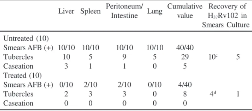
In vivo assessment
Table 5 shows that of the 10 animals in the untreated group, all developed minute tubercles in the liver, 5 in the spleen, 5 in the lungs and 9 in the peritoneum and intestines; microscopic necrosis suggestive of caseation was found in the liver of 3 animals and in the spleen, peritoneum and intestines each in one animal. Smears for acid-fast bacilli by Z-N stain, from centrifuged deposits (for 100 fields) of tissue homogenates,
Table 2. Minimum inhibitory concentration (MIC) of diclofenac, rifampicin and streptomycin.
Strains MIC (µg/ml)
Dc Sm Rf
Highly sensitive
M. tuberculosis Bajaj, J15, 10 0.25-2 0.5-2
N23, H37 Rv102, H37Ra16 Moderately sensitive
M. marinum 50, M. scrofulaceum 1323, M. gordonae 1324, M. flavescens 1541,
M. xenopi 160, M. avium 724, M. intracellulare 1406, M. terrae 1450, M. trivate 1453, 15 0.5- 2 1-2 M. fortuitum 1529, M. phlei L1
M. smegmatis 798, M. smegmatis 1546 15 2 2
Multidrug Resistant
M. tuberculosis BTA1, BTA2, BTA3, BTA4, BTA5, BTA6, BTA7, BTA8 20 4-8 4-8
Polydrug resistant
M. tuberculosis 912042, 911928, 905574, 911454, 910708, 911831, 905358, 911447, 25 >8 >8 911884, 912234, 911677, 90657, 912359, 911337, 912073, 912447, 912056, 911053, 906909
Table 3. Activity of Dc against M. tuberculosis H 37 Rv102.
Conc. Viable counts (log10 CFU/ml) on day
(µg/ml) 0 3 7
Nil 4.60 6.16 8.39 40 4.60 3.60(0.3)* 2.66(0.27) 20 4.60 4.94 4.23(0.16) 10 4.60 5.58 6.03
*Bactericidal activity.
effects of Dc and Sm, as well as Dc and the non-antibiotic trifluoperazine on Gram positive and Gram negative bacteria provided significant synergism both through in vitro and in vivo experiments (4,11).
In the present study, Dc has shown a remarkable antitubercular activity against a number of mycobacteria. Streptomycin and rifampicin are known conventional antitubercular drugs, and their MIC ranged from 0.5 to 8 µg/ml
with respect to most of the strains tested. The susceptible strains like M. tuberculosis H37Rv102 were inhibited at lower doses Figure 2. Synergism between diclofenac (50 µg/ml) and
streptomycin (10 µg/ml) against Mycobacterium smegmatis 798.
showed all 10 animals to be smear positive at the time of autopsy, which suggested successful infections in these animals. In contrast, macroscopic examination of the treated group (10 animals) showed tiny tubercles to be present in some of the liver specimens (2) and in the spleen, peritoneum, as well as in the intestine (3 each), but in the lungs, Z-N stained smears showed presence of AFB only in 4 cases (Table 5). In 5 animals of the untreated group, M. tuberculosis H37Rv 102 could actually
be recovered on subculture (as confirmed by BACTEC test) in comparison with only one of the treated groups, which appeared to be significant (p<0.01). The failure to recover the bacterium in other untreated animals was probably due to a non-viability of these bacilli, although these could readily be detected in smears in all cases. The histopathological sections of liver also revealed a considerable decrease in number of infiltrations in infected mice treated with Dc as compared to the untreated ones.
DISCUSSION
The non-steroidal antiinflammatory drug Dc had proved to be a powerful bactericidal antimicrobic agent, when tested against a large number of Gram positive and Gram negative bacteria, the MIC ranging from 25-100 µg/ml in most of the
instances, and even lower in some cases. This bactericidal agent could also offer significant protection to mice, when challenged with a virulent bacterium (3,9). Moreover, the antibacterial activity of Dc was found to be due to its inhibition of bacterial DNA synthesis, which was demonstrated using 2 µ Ci (3H)
deoxythymidine uptake (10). Further studies on the combined
(0.5 to 2 µg/ml), while the drug-resistant varieties were inhibited
at much higher concentrations, and some were totally resistant. A similar pattern was noticed in the in vitro activity of Dc as well – while its MIC against M. tuberculosis H37Rv102 was 10 ±
0.4 µg/ml, it was 25 ± 0.4 µg/ml for the drug resistant strains. It
was noticed that even the multidrug resistant strains like those obtained from BTA and Tuberculosis Research Centre were susceptible to Dc, although at higher concentrations (20-25 µg/
ml). The MIC of Dc seems to be high against mycobacteria in the in vitro studies. However, the antimycobacterial chemotherapeutics like INH and pyrazinamide also have quite high MIC values against mycobacteria; such high doses are often toxic to liver and other organs.
Dc was found to be bactericidal in action against M. tuberculosis H37 Rv 102.
Table 5. Effects of Dca on M. tuberculosis H
37 Rv 102b infection in mice.
Liver Spleen Peritoneum/ Lung Cumulative Recovery of Intestine value H37Rv102 in
Smears Culture
Untreated (10)
Smears AFB (+) 10/10 10/10 10/10 10/10 40/40
Tubercles 10 5 9 5 29 10c 5
Caseation 3 1 1 0 5
Treated (10)
Smears AFB (+) 0/10 2/10 2/10 0/10 4/40
Tubercles 2 3 3 0 8 4d 1
Caseation 0 0 0 0 0
Untreated, did not receive Dc; Treated, received Dc; a 10
µg/g body wt./day; b 4.5 x 10⁹ CFU/mouse i.p.; c from at least one viscera of 10 animals, all viscera did not yield
(+) culture; the viable counts (CFU) varied:103-106/ml; d only from 4 animals, recovery
from even single organ being counted as positive, other organs did not yield any growth; CFU101-103/ml in the positive samples.
Table 4. Individual and combined (synergistic) effects of Sm and Dc on M. smegmatis 798.
Diameter of the inhibition zone in mm2
Strain Single (A) Combined (B) Percentage increase Drug Effect Drug Effect on the basis of πr2
. Sm 10* Dc 50 Sm 10 + Dc 50 Sm Dc
M. smegmatis 798 17.2 16.6 18 17.1 9.52 6.12
*Amount (µg) of the drug /disc; Mean surface area of the inhibition zone (mm2) was calculated as πr2 on the basis of their mean diameter (2r) and % increase was calculated as (B-A)/A x 100, which was statistically significant (p<0.05).
The activity of Dc against M. smegmatis 798 was enhanced in the presence of an antitubercular drug Sm (Fig. 2).
In the animal experiments with M. tuberculosis H37 Rv 102 in mice, several minute tubercles were
observed in the liver, spleen, lungs, peritoneum and intestines of infected mice. However, there was a definite reduction in these macroscopic lesions in Dc-treated animals. The tubercle bacilli could not be recovered from all the untreated animals (Table 5), possibly because of the relatively few bacilli that mouse lesions had during autopsy, with even fewer survivors some weeks after infection, since it is known that compared to mice, guinea pig is a better animal model for producing experimental tuberculosis infection.
Although Dc is reported to be a rather toxic agent for human consumption, this drug could be tolerated by mice for the entire period of 6 weeks when this was administered intraperitoneally everyday in the dose of 10 µg/g body weight.
Protection at such a low concentration could be achieved possibly due to the fact that Dc is rapidly and completely absorbed after oral administration. There is a substantial first pass effect, such that only about 50% of the drug is available systematically. Its half-life in plasma is 1 to 2 hours. Dc produces side effects in only 20% of patients when used as an antiinflammatory agent, and only 2% of them discontinue therapy as a result (14). This depends upon genetic factors, nutritional factors and physiological state of the patient.
Earlier studies by Amaral and Kristiansen (2) had proved the efficacy of chlorpromazine in combating tuberculosis in vivo, along with a significant in vitro action. Apparently, the drug Dc has remarkable structural correlation with chlorpromazine in having two complete benzene rings attached to each other as phenyl acetic acid derivative through an NH group, and two halogen (Cl) atoms.
Most antimycobacterial non-antibiotics reported so far have shown in vitro MIC values ranging from 10 to 25 µg/ml, which
This suggests that such drugs, including Dc, might be used as adjuvants to current regimens used for the management of freshly diagnosed tuberculosis.
An elaborate study on the effects of combination of Dc plus Sm proved synergism, which could be further substantiated by carrying out tests for FIC index. Both these drugs have been in use satisfactorily in clinical medicine with known toxicity limits. The combination of Sm-Dc may prove to be a breakthrough in the treatment of tuberculosis. Furthermore, in course of time, it may be possible to obtain compounds with much greater synergistic effect with the help of suitable structural modification, thereby making a new generation of potential non-antibiotic antitubercular drugs. The actual factors responsible for attributing antimycobacterial activity to Dc are yet to be ascertained. QSAR studies may reveal the actual moieties responsible for conferring antimycobacterial activity to Dc.
ACKNOWLEDGEMENTS
We are grateful to All India Council for Technical Education (AICTE) for providing financial support for carrying out the work.
RESUMO
Atividade antimicobacteriana do agente antiinflamatório diclofenac sódico e seu sinergismo
com estreptomicina
Diclofenac sódico, um agente antiinflamatório, mostrou ação inibitória marcante contra isolados clínicos de Mycobacterium tuberculosis sensíveis e resistentes à drogas, bem como contra outras micobactérias. A droga foi testada in vitro contra 45 cepas diferentes de micobactérias, sendo que a maioria foi inibida pela droga na concentração de 10-25 µg/ml. Quando testado in vitro, diclofenac injetado em ratos albinos da linhagem Swiss, na proporção de 10 µg/g de peso corporal, provocou proteção
significativa dos animais desafiados com metade da dose letal de M. tuberculosis H37 Rv 102. De acordo com o teste χ2, os
dados in vivo foram altamente significativos (p < 0,01). Diclofenac foi posteriormente testado quanto ao sinergismo com a droga antimicobacteriana convencional estreptomicina, frente a M. smegmatis 798. Quando comparado aos seus efeitos individuais, o sinergismo foi estatisticamente significativo (p < 0,05). Através da análise checkerboard, o índice fracional de concentração inibitória para essa combinação foi 0,37, confirmando o sinergismo.
Palavras-chave: droga antiinflamatória, diclofenac sódico, atividade antimicobacteriana, estreptomicina, sinergismo, não-antibiótico
REFERENCES
1 . Amaral, L.; Kristiansen, J.E.; Abebe, L.S.; Millet, W. Inhibition of the respiration of multi-drug resistant clinical isolates of
Mycobacterium tuberculosis by thioridazine: potential use for initial therapy of freshly diagnosed tuberculosis. J. Antimicrob. Chemother., 38:1049-1053, 1996.
2 . Amaral, L.; Kristiansen, J.E. Phenothiazines. An alternative to conventional management of suspect multi-drug resistant tuberculosis. A call for studies. Int. J. Antimicrob. Agents, 14:173-176, 2000.
3 . Annadurai, S.; Basu, S.; Ray, S.; Dastidar, S.G.; Chakrabarty, A.N. Antimicrobial activity of the antiinflammatory agent diclofenac sodium. Indian. J. Exp. Biol., 36:86-90, 1998.
4 . Annadurai, S.; Guha-Thakurta, A.; Sa, B.; Dastidar, S.G.; Ray, R.; Chakrabarty, A.N. Experimental studies on synergism between aminoglycosides and the antimicrobial antiinflammatory agent diclofenac sodium. J. Chemother., 14(1):47-53, 2002.
5 . Antimicrobial Susceptibility Testing for Mycobacterium tuberculosis: Tentative standard document M24-T, Vol. 15, No.-16. National Committee for Clinical Laboratory Standards (NCCLS). December 1995.
6 . Bhattacharya, G.K.; Johnson, R.A. Statistical Concept and Methods.
John Wiley and Sons, New York, London, 1977, p.425.
7 . Chakrabarty, A.N.; Bhattacharya, C.P.; Dastidar, S.G. Antimycobacterial activity of methdilazine (Md), an antimicrobic phenothiazine. Acta Path. Microbiol. Immun. Scand., 101:449-454, 1993.
8 . Crowle, A.J.; Douvas, G.S.; May, M.H. Chlorpromazine: a drug potentially useful for treating mycobacterial infections. Chemother., 38:410-419, 1992.
9 . Dastidar, S.G.; Basu, S.; Annadurai, S.; Chakrabarty, A.N. In vitro and
in vivo antibacterial activity of the antiinflammatory agent diclofenac sodium. In: Recent advances in chemotherapy. Proceedings of the Eighteenth International Congress of Chemotherapy. Stockholm, Sweden, 1994, pp.339-340.
10. Dastidar, S.G.; Ganguly, K.; Chaudhuri, K.; Chakrabarty, A.N. The antibacterial action of diclofenac shown by inhibition of DNA synthesis. Int. J. Antimicrob. Agents, 14:249-251, 2000. 11. Dastidar, S.G.; Annadurai, S.; Kumar, A.K.; Dutta, N.K.; Chakrabarty,
A.N. Evaluation of a synergistic combination between the non-antibiotic microbicides diclofenac and trifluoperazine. Int. J. Antimicrob. Agents, 21(6):601-603, 2003.
12. Dickinsion, J.M.; Mitchison, D.A. In vitro property of rifapentine (MDL 473) relevant to its use in the intermittent chemotherapy of tuberculosis. Tubercule, 113-118, 1987.
13. Fenner, F. The pathogenic behavior of Mycobacterium ulcerans and
Mycobacterium balnei in the mouse and the developing chick embryo.
Amer. Rev. Tubercul., 73:650-660, 1956.
14. Goodman Gilman, A. In: Hardman, J.G.; Limbird, L.E. and Goodman Gilman, A. (Eds.). Goodman and Gilman’s The Pharmacological Basis of Therapeutics. McGraw- Hill, New York, 2001, Vols. I and II. 15. International Union against Tuberculosis 1955 Year Book 1955.
International Union against Tuberculosis, Paris, p.89.
16. Kirchner, O. Die Leistungsfahigkeit der Tiefenkultur des Tuberkelbazillus bei Verwendung besonders geeigneter flussiger Nahrboden. Zentralblat fur Bakt., 124:403-413, 1932.
17. Kristiansen, J.E.; Vergman, B. Antibacterial effect of selected phenothiazines and thioxanthines on slow growing mycobacteria.
Acta Path. Microbiol. Immun. Scand., 94:393-398, 1986. 18. Kristiansen, J.E. The antimicrobial activity of non-antibiotics. Acta
Path. Microbiol. Scand., 100(Suppl.):7-19, 1992.
19. Krogstad, D.J.; Moellering, R.C. Jr. Combinations of antibiotics, mechanism of interaction against bacteria. In: Lorian, V. (ed).
20. Mazumdar, R.; Ganguly, K.; Dastidar, S.G.; Chakrabarty, A.N. Trifluoperazine: a broad-spectrum bactericide specially active on staphylococci and vibrios. Int. J. Antimicrob. Agents, 18:403-406, 2001. 21. Molnár, J.; Mandi, Y.; Király, J. Antibacterial effect of some phenothiazine compounds and the R-factor elimination by chlorpromazine. Acta Microbiol. Acad. Sci. Hung., 23:45-54, 1976. 22. Molnár, J.; Beladi, I.; Foldes, I. Studies on antitubercular action of some phenothiazine derivatives in vitro. Zbl. Bakt. Hyg. I. Abt. Orig. A., 239:521-526, 1977.
23. Oberhofer, T.R. Manual of Practical Medical Microbiology and Parasitology. John Wiley and Sons, Toronto, 1985, p.352.
24. Ratnakar, P.; Murthy, P.S. Antitubercular activity of trifluoperazine, a calmodulin antagonist. FEMS Microbiol. Lett., 76:73-76, 1992. 25. Viveiros, M.; Amaral, L. Enhancement of antibiotic activity against
poly-drug resistant Mycobacterium tuberculosis by phenothiazines.
Int. J. Antimicrob. Agents, 17:225-228, 2001.
26. Williams, J.D.; Hedges, A.J. Antimicrobial substances. In: Linton, A.H.; Dick, H.M. (eds.). Topley and Wilson’s Principles of Bacteriology, Virology and Immunity. Edward Arnold, London, 1990, pp.111-143.