Reneé Pieterse1, Svetoslav D. Todorov1,2,*
1
Department of Microbiology, University of Stellenbosch, 7600 Stellenbosch, South Africa; 2Universidade de São Paulo,
Faculdade de Ciências Farmacêuticas, Departamento de Alimentos e Nutrição Experimental, Laboratório de Microbiologia de
Alimentos, São Paulo, SP, Brasil.
Submitted: May 29, 2009; Approved: March 16, 2010.
ABSTRACT
Mastitis is considered to be the most costly disease affecting the dairy industry. Management strategies
involve the extensive use of antibiotics to treat and prevent this disease. Prophylactic dosages of
antibiotics used in mastitis control programmes could select for strains with resistance to antibiotics. In
addition, a strong drive towards reducing antibiotic residues in animal food products has lead to research in
finding alternative antimicrobial agents.
In this review we have focus on the pathogenesis of the mastitis in dairy cows, existing antibiotic
treatments and possible alternative for application of bacteriocins from lactic acid bacteria in the treatment
and prevention of this disease.
Key words: mastitis, antibiotic, milk, bacteriocin, food safety
MASTITIS
The general health and well being of individuals depends
largely on meeting basic nutritional needs. Milk and fermented
milk products such as cheese, cultured milks and yoghurt have
formed an important part of daily nutrition, and the variety of
products produced from milk has increased dramatically over
the years, as modern food processing technologies have
improved. An increase in global population coupled with the
increasing demands for milk as an economic food and as an
industrial raw food product has necessitated an increase in
production by dairy farmers.
Current statistics indicate that the annual milk production
in South Africa has increased steadily over the last 20 years
from approximately 1700 million litres in 1985 to an estimated
3400 million litres in 2009. Consumption of dairy products has
also increased at similar levels with a sharper increase in recent
years, due primarily to a larger personal income base for
individuals (46).
In a commercial milking environment, dairy cattle need to
be in perfect physical condition to maintain a high level of milk
production. The risk of lesions and infections that develop in
modern dairy farming has consequently increased. Low milk
production has been attributed to a large extent to the control of
diseases in dairy cattle, of which mastitis accounts for the
largest economic losses on dairy farms in many countries in the
world, including the USA, United Kingdom, Europe, Australia
and South Africa (29, 63).
Improving udder health and decreasing the incidence of
udder infection and inflammation in dairy herds, will result in
increased milk production as huge losses are directly or
______________
Pieterse, R. and Todorov, S.D. Bacteriocins alternatives in mastitis treatment
indirectly incurred through loss of milk during treatment
periods, culling of cows and death of clinically infected cattle.
Mastitis control programmes addressing various aspects of
dairy farming such as feeding practices, animal husbandry,
hygiene and general health care can contribute towards
reducing the incidence of udder infections. Treating infection
with antimicrobials can, in conjunction with good farming
practices, assist in this endeavour to eliminate, or at least
decrease, the incidence of mastitis infection within a dairy
herd.
“Mastitis” describes an inflammatory reaction in the
mammary gland. The term comes from the Greek derived
word elements masto- referring to the mammary gland and -itis
meaning – “inflammation” (6). Although “mastitis” could
technically be used to describe any udder injury that may result
in inflammation, it is generally accepted that the causative
agents for the inflammatory reaction are microorganisms that
have gained entry into the teat canal and mammary tissue (65).
The extent of the infection that occurs as microorganisms
multiply and proliferate within the mammary tissue determines
the type of mastitis affecting the cow udder.
Mastitis-causing pathogens
The main etiological agents responsible for mastitis
infections can be divided into different groups of organisms
depending on the source of the organism involved. These
include contagious pathogens, environmental bacteria,
opportunistic bacteria and other organisms that less frequently
cause mastitis less frequently (65).
Contagious organisms
Contagious microorganisms are usually found on the
udder or teat surface of infected cows and are the primary
source of infection between uninfected and infected udder
quarters, usually during milking. The organisms that fit into
this category include: Staphylococcus aureus
(coagulase-positive staphylococci), Streptococcus agalactiae and the less
common sources of infection caused by Corynebacterium bovis
and Mycoplasma bovis (65, 67).
Environmental organisms
Environmental pathogens are found in the immediate
surroundings of the cow, such as the sawdust and bedding of
housed cows, the manure of cattle and the soil. Bacteria
include streptococcal strains other than S. agalactiae, such as
Streptococcus dysgalactiae, Streptococcus uberis and
Streptoccous bavis, Enterococcus faecium and Enterococcus
faecalis and coliforms such as Escherichia coli, Klebsiella
pneumonia and Enterobacter aerogenes (67,79). Mastitis
caused by environmental organisms is essentially opportunistic
in nature and becomes established if the immune system of the
host is compromised or if sanitation and hygiene is not
adequately practiced (80).
Opportunistic organisms
Opportunistic pathogens result in mild forms of mastitis
and include coagulase-negative staphylococci. The coagulase
test correlates well with pathogenicity and strains that are
coagulase-negative are generally regarded as non-pathogenic
(67). These staphylococci occur commensally and may be
isolated from milk but usually illicit a minor immune response
in cattle and infections caused are slight. They include S.
epidermidis, S. saprophyticus (23,67), S. chromogenes (20) and
S. simulans (23).
Other organisms
Many other bacteria and even yeasts may be responsible
for causing mastitis, but are less common and occur if
conditions in the environment change to increase exposure to
these organisms. A condition known as “summer mastitis”
occurs mostly in European countries in the summer months
when wet, rainy conditions prevail. The source of infection is
usually traced to an increase in exposure of the cows to flies in
pastures that transmit infecting Arcanobacterium pyogenes and
Peptostreptococcus indolicus strains and is more common in
non-lactating cows (67, 84).
Mastitis caused by Pseudomonas aeruginosa is often
traced to contaminated water sources and will result in a
endotoxemia occurs (65, 67).
Nocardia asteroides causes severe cases of mastitis
resulting in fibrosis and permanent damage to mammary
tissues (67). Treatment is usually ineffective and a high
mortality rate occurs. The source of the infection caused by
Nocardia asteroides is usually from the soil and could be
prevented by ensuring that effective sanitation measures are
enforced before treatment with intramammary infusions (65).
Less common causes of bovine mastitis include Bacillus
cereus, resulting in peracute and acute mastitis and also the
human pathogens Streptococcus pyogenes and S. pneumonia
that causes acute mastitis and is accompanied by fever
symptoms in the host (67).
Current aetiology of mastitis
Contagious organisms have usually been responsible for
the highest incidence of both clinical and sub-clinical cases of
mastitis. Bradley (8) sites the changes that have occurred in
the United Kingdom from 1967, where S. aureus and S.
agalactiae were primarily responsible for the highest number
of clinical mastitis cases in dairy herds. Three decades later in
1998, after the implementation of control strategies in the late
sixties, the number of incidences of contagious pathogens
responsible for clinical mastitis decreased significantly,
accounting for only 10 % of cases. E. coli and
Enterobacteriacae, however, were responsible for 34.7 % and
40.9 %, respectively, of all cases (9).
Adequate mastitis control strategies have thus played a
key role in reducing contagious cases of mastitis. It would
appear however, that as contagious pathogens were reduced,
opportunistic and environmental pathogens seemed to play a
greater role in causing persistent infections (8). The importance
of the correct diagnosis and identification of the aetiological
agent causing inflammation in the udder tissue is essential in
determining the treatment strategies. It is also important to
understand the history of mastitis incidence within a herd over
a period of time and to understand the different periods when a
cow may be at higher risk for infection. For example, cows are
especially susceptible to mastitis during the periparturient
period (just before and after calving) and at drying off - due to
structural changes occurring in the mammary gland. A
decrease in the number and functionality of white blood cells
caused by interactions with specific hormones during these
periods results in a compromised defence system (61,95).
Infection
Mammary structure is composed of the milk-producing
tissue or alveoli that lead into the lactiferous ducts, gland
cistern, teat canal and finally the teat opening or duct. The
alveoli are lined with epithelial cells that become specialised
during the gestation period, before calving, and after calving.
These specialised cells produce colostral and lacteal secretions
and finally, milk. Connective tissue and muscle cells support
the alveoli glands and contract and squeeze milk from the
alveoli during milking (29, 65).
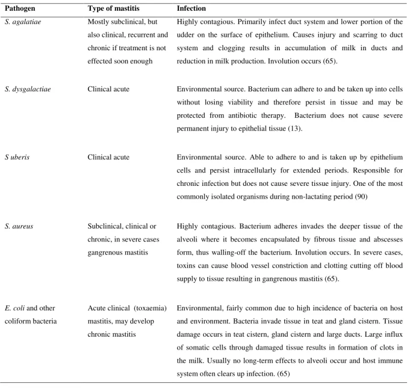
Table 1 summarises the type of mastitis infection that
occurs when pathogens invade the teat canal and mammary
tissue. Some pathogens are well adapted for the udder tissue
environment and are the primary source for recurrent
intramammary infections, especially contagious mastitis caused
by S. aureus and S. agalactiae. Most microorganisms,
including S. uberis (2), S. dysgalactiae (3) and E. coli (21,22)
adhere to and internalise into epithelium cells. Persistence of
the pathogen in the tissue may vary, some are easily destroyed
by the host immune system while others such as S. aureus are
well-adapted and cause serious injury within the mammary
tissue, producing virulence factors that disarm the host immune
systems cells (2, 36).
E. coli and other coliform pathogens are not only able to
adhere to and invade epithelium (22) but are also able to
multiply rapidly in the gland cistern, which elicits a rapid
inflammatory response that destroys a large number of the
invading pathogens. However, upon cell lyses endotoxins are
released causing severe toxaemia in the blood stream of the
cow (65, 67).
Mastitis control strategies
Pieterse, R. and Todorov, S.D. Bacteriocins alternatives in mastitis treatment
standard for control strategies for many years (29), and has
been successful in reducing the incidence of mastitis. The
strategy addresses areas where the risk of infection is the
greatest and promotes the use of treatment at specific times.
The five points listed by Giesecke et al. (29) include: (A) Teat
disinfection after milking; (B) Proper hygiene and milking
procedures and adequate milking equipment; (C) Culling of
chronically mastitis cows; (D) Antibiotic dry-cow therapy; (E)
Prompt treatment of clinical mastitis during dry period and
during lactation.
Table 1. Characteristics of common mastitis-causing pathogens, invasiveness and infection
Pathogen Type of mastitis Infection
S. agalatiae Mostly subclinical, but
also clinical, recurrent and
chronic if treatment is not
effected soon enough
Highly contagious. Primarily infect duct system and lower portion of the
udder on the surface of epithelium. Causes injury and scarring to duct
system and clogging results in accumulation of milk in ducts and
reduction in milk production. Involution occurs (65).
S. dysgalactiae Clinical acute Environmental source. Bacterium can adhere to and be taken up into cells
without losing viability and therefore persist in tissue and may be
protected from antibiotic therapy. Bacterium does not cause severe
permanent injury to epithelial tissue (13).
S uberis Clinical acute Environmental source. Able to adhere to and is taken up by epithelium
cells and persist intracellularly for extended periods. Responsible for
chronic infection but does not cause severe tissue injury. One of the most
commonly isolated organisms during non-lactating period (90)
S. aureus Subclinical, clinical or
chronic, in severe cases
gangrenous mastitis
Highly contagious. Bacterium adheres invades the deeper tissue of the
alveoli where it becomes encapsulated by fibrous tissue and abscesses
form, thus walling-off the bacterium. Involution occurs. In severe cases,
toxins can cause blood vessel constriction and clotting cutting off blood
supply to tissue resulting in gangrenous mastitis (65).
E. coli and other
coliform bacteria
Acute clinical (toxaemia)
mastitis, may develop
chronic mastitis
Environmental, fairly common due to high incidence of bacteria on host
and environment. Bacteria invade tissue in teat and gland cistern. Tissue
damage occurs in teat cistern, gland cistern and large ducts. Large influx
of somatic cells through damaged tissue results in formation of clots in
the milk. Usually no long-term effects to alveoli occur and host immune
Farm management
A strategy to control mastitis must be practical and
economical. The primary goal would be to reduce the rate of
new infections and the duration of current infections within a
herd. It would also be essentially important to maintain normal
udder health ensuring that the natural immune response in the
cow can resist and fight disease while still producing the
required level of milk (65).
Control strategies need to target every facet and process of
dairy farming and can begin with maintaining good hygiene
practices in the environment. The holding yards or stalls
should be kept clean and dry. The water supply should be
adequate and free of coliform bacteria and equipment should
be maintained and sanitised between milking (29). The welfare
of animals is becoming increasingly important in modern dairy
production as consumers become more concerned about the
manner in which farm animals are treated. The Farm Animal
Welfare Council in the UK has defined “the five freedoms” of
animals, which highlight issues relating to the treatment and
management of animals. The advantage of implementing such
quality control measures within the herd would ensure that
dairy cows are free of a stressful environment, injury, pain,
hunger and discomfort, which in turn would promote a healthy
immune system and udder health in general (77).
The milking practice is of paramount importance as this is
most often the route of infection. The udder should be
prepared before milking by washing the teats, followed by
disinfection and drying with clean paper towels. If the teat area
is dripping with water from run-off of areas that were heavily
soiled it could lead to pathogens gaining access to the teat
canal. Milker’s hands should also be disinfected to prevent the
transfer of pathogens. Post milking treatment is also important
and all cows should be treated with a teat dip disinfectant to
reduce the risk of infection (29, 65).
Monitoring SCC on a regular basis and follow-up
investigations give an indication of the success of good animal
husbandry and hygiene practices. It therefore forms an integral
part of mastitis control strategies and assists in diagnosis and
treatment.
The elimination of mastitis in a herd may require the
culling of cows that are incurable or are so severely infected
that the mammary tissue has been scarred and damaged to the
extent that the tissue no longer functions (29).
Treatment
A cow may spontaneously recover from mastitis, but this
will usually occur in mild cases of subclinical mastitis.
Theoretically, the mechanism by which a cow recovers from
infection without treatment can be capitalised upon to produce
a vaccine (65). Research in this area continues and some
vaccines such as E. coli J5 can reduce the number and severity
of coliform mastitis cases by 70 – 80 % (17). Recent
technology has focused on a DNA vaccine that expresses
virulence factors in vivo and is primarily targeted against S.
aureus mastitis, as antibiotic therapy is usually less effective
against this pathogen (89,103).
Antimicrobial agents can be administered either during
lactation or during the dry period. Treatment during lactation
will be necessary if clinical mastitis is present, whereas dry
cow therapy can be used to treat existing infections and can
also be administered in a prophylactic manner to prevent new
infections from developing during this period. A cow will
usually lactate for a period of approximately 300 days per year
and have a dry period of between 50 to 60 days. The most
vulnerable period when new mastitis infections occur is at the
end of the lactation period and again just before the start of the
next lactation period (29). This can be attributed to hormonal
and structural changes occurring in the mammary tissue which
affects the immune system as the cow prepares for calving or
for the drying-off stage (61, 95).
Dry cow therapy
Dry cow therapy is as much a management issue as it is a
treatment issue. The manner in which the cows enter this
period is important and the way in which the housing
conditions and nutrition is handled impacts on the success of
the treatment itself. The energy intake of the cows should be
Pieterse, R. and Todorov, S.D. Bacteriocins alternatives in mastitis treatment
and then, as soon as drying-off occurs, they need to be treated
immediately with either antimicrobial infusions (containing
slow release antibiotic preparations) or with internal teat
sealant products (60). Antimicrobials will be required if an
existing infection is present, whereas an internal teat sealant
can be used alone if no infection is present. Commercially
available teat sealants such as Orbeseal® (Pfizer Animal
Health) are approved for use in North America and Europe.
The teat sealant is composed of an inert salt (bismuth
subnitrate) in a paraffin base. The paste is infused into the teat
of each quarter using a sterile syringe. After drying-off, the
product is stripped out at first milking (64). To ensure that
other pathogens are not introduced into the teat along with the
teat sealant, trained personnel should perform the
administration of the product.
The teat sealant forms an impermeable plug as it lines the
teat canal and results in a physical barrier against invading
microorganisms through the teat opening, thereby preventing
new infections during the dry period. Research has shown that
the internal teat sealant (Orbeseal®, Pfizer Animal Health) is
effective in reducing the infection rate when compared to
untreated cows (4). A recent study also demonstrated the
benefit of administering Orbeseal® (Pfizer Animal Health)
along with an antibiotic infusion (Orbenin® Extra Dry Cow,
Pfizer Animal Health) containing cloxacillin. The use of the
teat sealant and the antibiotic infusion performed slightly better
in preventing clinical mastitis in the dry period compared with
using only the antibiotic infusion (10).
Lactation therapy
The use of antimicrobials during lactation must be
carefully considered. Only cases of clinical mastitis and some
specific cases of subclinical mastitis, where the quality and
production of the milk is severely affected, are treated.
Mastitis caused by S. agalactiae can be treated most readily
during lactation and has a high cure rate (90-95 %). Mastitis
caused by S. aureus has the lowest cure rate and along with
environmental streptococci should be treated during the dry
period (65).
An important consideration for treatment during lactation
is the presence of antibiotic residues in the milk. A waiting
period is required for the duration of the treatment and for a
given period after treatment where milk and meat products
need to be withheld to ensure that the level of antibiotics
present in the product meets the legislative requirements. The
withdrawal period and the type of product that is administered
vary in different countries (34). The cost of treatment and the
loss of milk during the withdrawal period are important in
determining the type of product used and the manner in which
it is administered. The withdrawal period for milk products
marketed during lactation varies between 1 and 4 days (Table
3). A product is considered excellent if it has a high cure rate
and a minimum withdrawal period (34).
Efficacy of drug delivery
The administration of drugs can be done either directly
into the teat canal, as previously described for dry cow therapy,
in the form of intramammary infusions, but can also be given
parenterally by intravenous or intramuscular injection (65).
The route of choice for subclinical mastitis is usually by
intramammary infusion; and in the case of severe acute clinical
mastitis, a combination of parenteral and intramammary
treatment is usually necessary (104).
To be effective, the drug has to exert specific
antimicrobial activity at the site of infection (34) and must have
certain characteristics to be an effective agent in the mammary
tissue. The pH of blood plasma is 7.4. The pH of milk varies
between 6.4 and 6.6, but increases to 7.4 in the case of an
infection. Most antibiotics are weak organic acids or bases and
exist in both an ionised and non-ionised form in varying
proportions in blood and milk, depending on the change in pH
of the environment. Drugs that are administered parenterally
must pass from the circulatory blood system and into the milk
and milk tissue via lipid membranes. The active fraction of the
drug must be in a non-ionised, non-protein bound, lipid-soluble
form to pass this blood-to-milk barrier (104).
Antibiotics that are administered via the teat opening must
often the distribution is uneven and diffusion through the
mammary ducts where severe inflammation and swelling is
present may block the movement of the therapeutic agent (24).
Added to this, most pathogens have the ability to invade the
epithelium tissue. In the case of S. aureus infection, interaction
with antibiotics is prevented by the formation of fibrous scar
tissue. The scar tissue may also have no blood supply,
rendering intramuscular or intravenous drug therapy less
effective (65). Some bacteria may also evade interactions with
antibiotics once engulfed by macrophages, where they remain
active within the leukocyte and can cause recurrent infections
once the antibiotic has been eliminated from the area (65). The
formation of biofilms within the teat canal as bacteria adhere to
bacteria on the epithelium surface may also contribute to the
ineffectiveness of local intramammary infusions (52).
The type of drug used to treat an infection can be
determined once an accurate diagnosis has been made and the
pathogens identified. The minimum inhibitory concentration
(MIC) is defined as the lowest concentration of a drug that
prevents the growth of a specific pathogen (59). Antimicrobial
disk diffusion tests are performed on the pathogens isolated
from mastitic milk samples to determine the drug sensitivity
profile of the pathogens. The veterinarian is then able to select
the most effective drug for treatment (65). The ideal drug
should have the lowest MIC against the majority of udder
pathogens. No single drug can, however, be effective against
all pathogens and most need to be used in combinations and in
different formulations to increase efficacy and bioavailability
within the udder tissue (34,104).
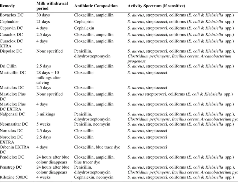
Types of antimicrobial agents
Commonly used remedies available for dry cow and
lactation therapy, the recommended withdrawal period and the
possible activity spectrum of mastitis pathogens (24) are shown
in Table 2 and 3. The antibiotic groups and antimicrobials
used in these remedies have different mechanisms of action
and many new semi-synthetic compounds have been developed
to counter the threat of antimicrobial resistance. The majority
of antibiotics used are broad-spectrum antibiotics acting
against Gram-positive and Gram-negative bacteria (59).
-lactam Penicillins (penicillins, ampicillin, cloxacillin,
amoxycillin, nafcillin, methicillin) and -lactam
Cephalosporins (cephalexin, cefuroxime, cephapirin) inhibit
cell wall synthesis by preventing the formation of cross-links
between polysaccharide chains in the cell wall. Many
staphylococcal strains produce the enzyme penicillinase, which
acts by breaking the -lactam ring structure of the antibiotic
and are therefore resistant. Penicillinase-resistant penicillins
such as cloxacillin are specifically used to treat the
penicillinase-producing, methicillin-susceptible staphylococci
(59).
Clavulanic acid inhibits the activity of penicillinase
produced by staphylococcal strains. Combined with -lactam
antibiotics such as amoxicillin it can eliminate -lactamase
activity by pathogens and improve susceptibility to the
antibiotic (83).
Tetracyclines such as oxytetracycline inhibit protein
synthesis by binding to the 30S ribosomal sub-unit and
interfere with amino-acyl-tRNA binding. Tetracycline is
bacteriostatic and usually more active against Gram-positive
organisms (59). Oxytetracycline is an irritant and should
therefore not be administered as an infusion, but rather
intravenously (24).
Aminoglycosides (streptomycin, neomycin) inhibit protein
synthesis by binding to the 50S ribosomal sub-unit and inhibits
peptide chain elongation. Aminoglycosides are mostly active
against Gram-negative bacteria and are often formulated
together with -lactam penicillins (59).
Polymixin B is an antimicrobial compound that binds to
the cell membrane and disrupts its structure and permeability
properties. It is the antimicrobial drug of choice for infections
caused by P. aeruginosa (24).
Macrolide antibiotics (tylosin, lincomysin, erythromycin)
are effective in treating Gram-positive udder infections both by
parenteral and intramammary administration (24). They are
bacteriostatic and thus act in conjunction with the host immune
system to fight infection. The mechanism of action is to inhibit
Pieterse, R. and Todorov, S.D. Bacteriocins alternatives in mastitis treatment
prevent peptide elongation (66).
What are the alternatives?
The risks involved in the treatment of mastitis has been
discussed in terms of the development of antibiotic resistance,
but from a commercial standpoint, milk products containing
specific levels of antibiotic residues cannot be sold for human
consumption. Processing of milk for cheese and yoghurt
manufacture is also affected as bacterial starter cultures are
inhibited and the quality of the product produced is generally
compromised (54). Completely eliminating the use of
antibiotics for the treatment of mastitis is unlikely, as modern
intensive farming practices and high demand dictate rapid and
intensive treatment strategies, which involve the use of
antibiotic therapy in both lactation and dry periods. The
ultimate goal would be to reduce the use of antibiotics. This
could primarily be achieved through better management and
hygiene practices and legislation enforcing a reduction in the
indiscriminate use of antibiotics for treatment and for growth
promotion, as was done in Nordic countries in 1980’s (25).
Improving host defences can result in rapid elimination of new
infections. Supplementing of selenium and vitamin E and
improving general nutrition during high-risk periods such as
periparturient and drying-off periods can increase host defence
mechanisms (58).
Table 2. Recommended remedies for dry cow treatment, withdrawal period and activity spectrum (24).
Remedy Milk withdrawal
period Antibiotic Composition Activity Spectrum (if sensitive)
Bovaclox DC 30 days Cloxacillin, ampicillin S. aureus, streptococci, coliforms (E. coli & Klebsiella spp.)
Cephudder 21 days Cephapirin S. aureus, streptococci, coliforms (E. coli & Klebsiella spp.)
Cepravin DC 4 days Cephalexin S. aureus, streptococci, coliforms (E. coli & Klebsiella spp.)
Curaclox DC 2.5 days Cloxacillin, ampicillin S. aureus, streptococci, coliforms (E. coli & Klebsiella spp.)
Curaclox DC XTRA
4 days Cloxacillin, ampicillin S. aureus, streptococci, coliforms (E. coli & Klebsiella spp.)
Dispolac DC None specified Penicillin,
dihydrostreptomycin
S. aureus, streptococci, coliforms (E. coli & Klebsiella spp.),
Clostridium perfringens, Bacillus cereus, Arcanobacterium pyogeness
Dri Cillin 2.5 days Cloxacillin, ampicillin S. aureus, streptococci, coliforms (E. coli & Klebsiella spp.)
Masticillin DC 28 days + 10
milkings after calving
Cloxacillin S. aureus, streptococci
Masticlox DC 2.5 days Cloxacillin S. aureus, streptococci
Masticlox Plus DC
None specified Cloxacillin, ampicillin S. aureus streptococci, coliforms (E. coli & Klebsiella spp.)
Masticlox Plus DC EXTRA
4 days Cloxacillin, ampicillin S. aureus, streptococci, coliforms (E. coli & Klebsiella spp.)
Nafpenzal DC 3 milkings Penicillin,
dihydrostreptomycin
S. aureus, streptococci, coliforms (E. coli & Klebsiella spp.),
Clostridium perfringens, Bacillus cereus, Arcanobacterium pyogenes
Neomastitar DC 5 weeks Penicillin, neomycin S. aureus, streptococci, coliforms (E. coli & Klebsiella spp.)
Noroclox DC 2.5 days Cloxacillin S. aureus, streptococci
Noroclox DC EXTRA
2.5 days Cloxacillin S. aureus, streptococci
Orbenin EXTRA DC
4 days Cloxacillin, blue trace dye S. aureus, streptococci
Pendiclox DC 24 hours after blue
colour disappears
Cloxacillin, ampicillin, blue tracer dye
S. aureus, streptococci, coliforms (E. coli & Klebsiella spp.)
Penstrep DC 24 hours after blue
colour disappears
Penicillin,
dihydrostreptomycin
S. aureus, streptococci, coliforms (E. coli & Klebsiella spp.),
Clostridium perfringens, Bacillus cereus, Arcanobacterium pyogenes
Table 3. Recommended remedies for lactating cow treatment, withdrawal period and activity spectrum (24, 42).
Remedy
Milk withdrawal period
Antibiotic Composition Activity Spectrum (if sensitive)
Cloxamast LC 3 days Cloxacillin, ampicillin Septic mastitis. S. aureus, streptococci, coliforms (E. coli
& Klebsiella spp.)
Curalox LC 3 days Cloxacillin, ampicillin S. aureus, streptococci, coliforms (E. coli & Klebsiella
spp.)
Dispolac RX 4 24 hours after
blue colour has
disappeared
Penicillin,
dihyrostreptomycin
S. aureus, streptococci, coliforms (E. coli & Klebsiella
spp.), Clostridium perfringens, Bacillus cereus
Lactaclox 2.5 days Cloxacillin S. aureus, streptococci
Lactaciliin 3 days Ampicillin S. aureus, streptococci, coliforms (E. coli & Klebsiella
spp.)
Lincocin Forte 2.5 days Lincomycin, neomycin Staphylococcus aureus, streptococci
Mastijet Forte 4 days Oxytetracycline, neomycin,
bacitracin, cortisone
S. aureus, streptococci, coliforms (E. coli & Klebsiella
spp.)
Nafpenzal MC 6 milkings in
treatment + 3 milkings after treatment
Penicillin,
dyhrostreptomycin, nafcillin
S. aureus, streptococci, coliforms (E. coli & Klebsiella
spp.), Clostridium perfringens, Bacillus cereus,
Arcanobacterium pyogenes
Noroclox QR 24 hours after
blue colour has
disappeared
Cloxicillin, blue tracer dye S. aureus, streptococci
Pendiclox Blue 24 hours after
blue colour has
disappeared
Cloxicillin, ampicillin, blue tracer dye
S. aureus, streptococci, coliforms (E. coli & Klebsiella
spp.)
Penstrep 300 D 24 hours after
blue colour has
disappeared
Penicillin,
dihydrostreptomycin, blue tracer dye
Acute mastitis. S. aureus, streptococci, soliforms (E. coli
& Klebsiella spp.), Clostridium perfringens, Bacillus
cereus, Arcanobacterium pyogenes
Rilexine LC 4 days Cephalexin, neomycin,
cortisone
Acute & chronic mastitis
Spec Form Forte 3 days Penicillin,
dihydrostreptomycin, novobiocin, polymyxin B, cortisone
Acute or chronic mastitis. S. aureus, streptococci,
coliforms (E. coli & Klebsiella spp.), Clostridium
perfringens, Bacillus cereus, Pseudomonas aeruginosa, Arcanobacterium pyogenes
Streptocillin 24 hours after
blue colour has
disappeared
Penicillin,
dihyrostreptomycine, blue tracer dye
S. aureus, streptococci, coliforms (E. coli & Klebsiella
spp.), Clostridium perfringens, Bacillus cereus,
Arcanobacterium pyogenes
BACTERIOCINS – EXPLORING ALTERNATIVES TO
ANTIBIOTIC TREATEMNT
INTRODUCTION
The study of the antibacterial properties of peptides that
became known as colicins began in 1925 when one strain of E.
coli produced an antagonistic effect against another E. coli
culture (33). The antibiotic effect between other enteric
bacteria was also reported by Fredericq and Levine (27) and
further research into these proteinaceous molecules centred on
Pieterse, R. and Todorov, S.D. Bacteriocins alternatives in mastitis treatment
members of the familyEnterbacteriaceae.
Colicin-like molecules produced by Gram-positive
bacteria have also been studied extensively since the first
report of nisin produced by L. lactis subsp. lactis (71). The
term “bacteriocin” was used to describe these antibiotic
substances as not all were produced by coliform bacteria (42)
and according to Tagg et al. (87), were defined as ribosomally
synthesized polypeptides that usually possess a narrow
spectrum of antibacterial activity against bacteria of the same
or closely related species. Jack et al. (41) however noted some
discrepancies in this definition in that some bacteriocins (or
bacteriocin-like substances) have a broader spectrum of
activity and some are even active against Gram-negative
species.
Klaenhammer (45) classified bacteriocins on the structure
and mode of action of the peptide and predominantly included
those produced by lactic acid bacteria (LAB). Four distinct
classes were identified: class I, small lantibiotics (<5 kDa), that
contained the amino acids lanthionine, -methyllanthionine,
dehydroalanine and dehydrobutyrine; class II, small (<10 kDa),
heat-stable, non-lanthionine containing peptides; class III, large
(>30 kDa), heat-labile proteins and class IV, consisting of
complex bacteriocins containing carbohydrate or lipid moieties
that were required for bacteriocin activity.
Applications of bacteriocins
The antibacterial activity of bacteriocins has resulted in
research into the practical applications thereof and can be
broadly divided into two focus areas: food production and
preservation, by preventing the growth of unwanted or
disease-causing organisms and secondly, medical and veterinary
applications. Traditionally, antibiotics have been administered
to prevent and treat disease. However, with the widespread
development of antibiotic drug-resistant strains, the importance
of alternative antimicrobials is becoming increasingly urgent
and bacteriocin-producing organisms could be considered as an
important source of antimicrobial agents in the medical and
veterinary fields. The important role that bacteriocins continue
to play in food production and clinical applications will be
discussed.
Application in medical and veterinary fields
Bacteriocins, by definition usually only target closely
related species; they could offer an advantage over antibiotics
in that treatment could be targeted against specific pathogenic
organisms. Bacteriocins, identified for potential use as
antimicrobials include lantibiotics produced by Gram-positive
lactic acid bacteria, and colicins and microcins, produced by
Gram-negative bacteria (30). Applications are widespread,
ranging from topical applications in the treatment of skin
infections to the treatment of inflammation and ulcers.
Commercial products are currently available for the treatment
of mastitis in dairy cattle and will be discussed in more detail.
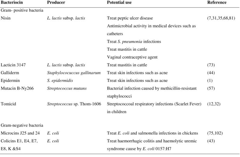
Table 4 summarises some of the potential applications of some
bacteriocins in the medical and veterinary field. Most testing
for clinical applications have been carried out in animal
models, however the bacteriocin nisin has already undergone
human clinical trials for the treatment of peptic ulcers caused
by Helicobacter pylori (35). Bacteriocins produced by
Gram-negative bacteria can be advantageous in that they can be used
to target other pathogenic Gram-negative strains. Bacteriocins
produced by positive LAB are not active against
Gram-negative strains without pre-treatment strategies to compromise
the integrity of the outer membrane (15). For example, nisin,
after treatment with ETDA, citrate and lactate, was shown to be
effective against Salmonella typhimurium and E. coli 0157:H7
(18). In contrast, colicins produced by Gram-negative E. coli
are naturally active against other E. coli strains as well as some
Salmonella strains (11). Microcins produced by enteric
bacteria, usually target strains in the family Enterobacteriaceae
(55).
Bacteriocins produced by Gram-positive strains can
substitute antibiotics such as ionophores routinely applied as
feed additives for livestock animals, such as cattle. The
ruminal bacterial populations of Gram-positive bacteria that
produce excessive fermentation products, such as methane and
ammonia, can be inhibited, without the dangers and perceived
Table 4. Potential medical and veterinary applications of some bacteriocins
Bacteriocin Producer Potential use Reference
Gram- positive bacteria
Nisin L. lactis subsp. lactis Treat peptic ulcer disease
Antimicrobial activity in medical devices such as
catheters
Treat S. pneumonia infections
Treat mastitis in cattle
Vaginal contraceptive agent
(7,31,35,68,81)
Lacticin 3147 L. lactis subsp. lactis Treat mastitis in cattle (73)
Galliderm Staphylococuccus gallinarum Treat skin infections such as acne (44)
Epidermin S. epidermidis Treat skin infections such as acne (1)
Mutacin B-Ny266 Streptococcus mutans Bacterial infection caused by methicillin-resistant
staphylococci
(57)
Tomicid Streptococcus sp. Thom-1606 Streptococcoal respiratory infections (Scarlet Fever)
in children
(12,32)
Gram-negative bacteria
Microcins J25 and 24 E. coli Treat E. coli and salmonella infections in chickens (75,102)
Colicins E1, E4, E7,
E8, K &S4
E. coli Treat haemorrhagic colitis and haemolytic uremic
syndrome cause by E. coli 0157:H7
(43)
Bacteriocins used in the treatment of mastitis
The most economically costly disease in cattle is mastitis.
As a result the dairy industry could benefit greatly from the
development of safe antimicrobial agents and bacteriocins
could be an attractive alternative to antibiotics. The treatment
of mastitis has been a target of research since the inception of
scientific research into the applications of bacteriocins (91).
To date, only the Lactococcal bacteriocin, nisin, has been
developed for commercial application and the lantibiotic,
lacticin 3147, has been extensively researched for dry cow
therapy. Applications for prevention and treatment using these
lactococcal bacteriocins will be discussed in detail below.
Other bacteriocins that are active against mastitis
pathogens have also been investigated. Researchers have
targeted staphylococci and streptococci isolated from the
normal flora of the teat canal and other areas as these could be
a source for bacteriocins to treat mastitis pathogens. The
potential applications for these bacteriocins will also be
discussed.
Lactococcal bacteriocins
Nisin: was the first bacteriocin applied to the preservation
of food products and was approved for use in pasteurised
processed cheese spreads in 1988 by the FDA (19). Nisin is
classified as a class Ia lantibiotic (45) and is a 34 amino acid
peptide (3488 Da). Nisin has a dual mode of action, which
essentially involves the prevention of cell wall synthesis and
pore formation, leading to cell death. The precise mechanism
involves binding to lipid II molecules
(Undecaprenyl-pyrophosphate-MurNAc(pentapeptide)-GlcNAc) located in the
cell membrane of the target cells. Lipid II is the main
Pieterse, R. and Todorov, S.D. Bacteriocins alternatives in mastitis treatment
cell wall and when nisin binds to lipid II, it prevents the
transfer of the peptidoglycan across to the cell wall (15). The
process of pore formation is initiated in the membrane of the
target cell after docking at lipid II occurs and results in the
efflux of cytoplasmic compounds that are required to maintain
ion gradients, thereby affecting trans-membrane potential and
the pH gradient across the membrane. Biosynthetic processes
such as ATP synthesis driven by proton motive force cease and
cell death occurs (69,76).
Nisin has a wide spectrum of activity against
Gram-positive bacteria, including species of Enterococcus,
Lactobacillus, Lactococcus, Leuconostoc and Pediococcus
(14). Nisin is also active against L. monocytogenes and its
efficacy against this food pathogen in raw meat products have
been evaluated by Pawar et al. (62), as well as in dairy
products (5). Nisin has also been applied to cheese products to
control the growth of spores produced by Clostridium
tyrobutyricum (70, 78).
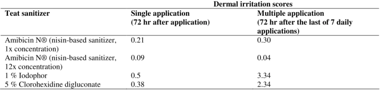
Sears et al. (81) investigated the use of a nisin-containing
germicidal formulation in preventing mastitis in cattle. Teat
sanitisers are routinely used before and after milking cows to
prevent the introduction of pathogens into the teat canal, which
could lead to intramammary infections. The study compared
the nisin-based formulation (Ambicin® N, Applied
Microbiology, Inc., New York, NY) with that of conventional
chemical treatments such as iodines and chlorohexidines.
Initial performance data for a nisin-based teat sanitizer
(Amibicin N®) showed a significant reduction in pathogen in
experimentally challenged teat surfaces after 1-minute
exposure to the germicidal formulation (Table 5). The
formulation also showed little potential for skin irritation after
repeated exposure in contrast to 1 % iodophore and 5 %
chlorohexidine digluconate preparations. Table 6 shows the
skin irritation data reported by Sears et al. (81). Dermal
irritation scores indicated the degree of redness or scab
formation, with a score of <1.0 indicating a product with little
or no potential for irritation. Products with a score of ranging
from 3.0-4.9 would have the potential to cause severe irritation.
Table 5. Performance data for nisin-based germicidal teat sanitizer (81).
Mastitis-causing organisms Reduction using Ambicin N®
S.aureus 61.8 %
S. agalactiae 98.6 %
E. coli 85.5 %
S. uberis 67.1 %
K. pneumonia 76.5 %
Table 6. Comparative skin irritation to rabbit skin after exposure to teat sanitizer.
Dermal irritation scores
Teat sanitizer Single application
(72 hr after application)
Multiple application
(72 hr after the last of 7 daily applications)
Amibicin N® (nisin-based sanitizer, 1x concentration)
0.21 0.30
Amibicin N® (nisin-based sanitizer, 12x concentration)
0.09 0.04
1 % Iodophor 0.5 3.34
Contamination of milk with a sanitizer chemical based
product is a concern if it is not completely removed before
milking. Using bacteriocin-based sanitizers or products would
be advantageous in that complete removal of the product would
not necessarily be required.
In addition to Ambicin®, two other nisin-based products,
namely Wipe-Out® Dairy Wipes and Mast Out® were
developed by Immucell Corporation (15). Mast Out® was used
in January 2004 in initial field trials involving 139 cows with
subclinical mastitis. Significant cure rates were reported and
the product was subsequently licensed to Pfizer Animal Health
for further development and distribution (39). The product has
however not been made available by Pfizer Animal Health and
no further trial results have been reported.
Lacticin 3147: is produced by L. lactis subsp. lactis
DPC3147 and was first isolated from Irish Keffir grain (74).
As with nisin, it is also classified as a Class 1a lantibiotic, but it
differs from nisin in that it is a two-peptide lantibiotic,
requiring both the LtnA1 and LtnA2 peptides for full activity.
The mode of action of lacticin 3147 is similar to that of nisin in
that it results in the inhibition of cell synthesis and pore
formation in the target cell (98).
The primary structure of the lacticin A1-peptide, LtnA1,
consists of 30 amino acids (3306 Da) and has a
lanthionine-bridging pattern resulting in a globular structure similar to class
Ib lantibiotics such as mersacidin. The LtnA2 peptide consists
of 28 amino acids (2847 Da) and is an elongated peptide.
Wiedeman et al. (98) proposed a three-step model to describe
how both peptides are involved for antibacterial activity of
lacticin 3147. LtnA1 first binds to lipid II (i), thereby inducing
a conformation that facilitates the interaction with LtnA2. This
enables the formation of a two-peptide-lipid II complex (ii).
When bound to the complex, LtnA2 is able to adopt a
transmembrane conformation that results in the formation of a
defined pore and the release of ions across the membrane (iii).
In an earlier study, McAuliffe et al. (53) reported that the pore
formation resulted in the efflux of potassium ions and
inorganic phosphate, resulting in the dissipation of the
membrane potential and hydrolysis of internal ATP, the
collapse of the pH gradient and cell death.
Lacticin 3147 has a broad spectrum of antimicrobial
activity and inhibits the growth of Bacillus sp., Enterococcus
sp., Lactobacillus sp., Pediococcus pentriceans, S. aureus, S.
thermophilus and most mastitis-causing streptococci.
Food-borne spoilage bacteria, including L. monocytogenes and C.
tyrobutyricum, are sensitive to lacticin 3147 and the peptide
could be used to prevent food spoilage and disease (74).
Lacticin 3147 was investigated for use as an antimicrobial
agent as it inhibited common mastitis-causing pathogens,
including S. aureus, S. dysgalactiae, S. uberis and S. agalactiae
(73). The producing organism is GRAS and is active at both
low and physiological pH and was heat stable (73,74).
Teat seal formulations such as Orbeseal® (64) are
recommended for use during the dry period as a prophylactic
measure to reduce the number of new mastitis infections (4).
The inert property of the teat seal formulation has no
antimicrobial effect and therefore relies on good udder hygiene
practices for effective treatment. Antibiotics such as
cloxacillin have been added to the formulations (Orbenin®
Extra Dry Cow, Pfizer Animal Health) to prevent new
infections during this period. However, prolonged exposure to
antibiotics at low levels could increase the risk of antibiotic
resistance by pathogenic bacteria. Bacteriocins, such as
lacticin 3147 could replace antibiotics in these formulations
(73, 74, 93). Studies to date have shown that resistance by
mastitis pathogens S. dysgalactiae and S. aureus to the
bacteriocin lacticin 3147 were not significant (73).
In separate studies, the bismuth subnitrate-based teat seal
(Osmonds Teat Seal 2, Cross Vetpharm Group Ltd., Dublin,
Ireland) combined with lacticin 3147 was evaluated against the
mastitis-causing pathogens S. dysgalactiae (73) and S. aureus
(16,93). Irritancy to the teat area and the somatic cell response
were evaluated.
The protection given by the teat seal plus lacticin 3147 and
the teat seal only were compared after experimental challenge
with S. dysgalactiae. The results showed significant
improvements in the level of protection afforded by the teat
Pieterse, R. and Todorov, S.D. Bacteriocins alternatives in mastitis treatment
percent of quarters treated with the teat seal plus lacticin 3147
remained free of new infections compared with only 33.3 % of
quarters treated with the teat seal alone (73, 74).
Tissue tolerance studies were done comparing the SCC in
the milk from quarters treated with the teat seal alone, teat seal
plus lacticin 3147 and with a commercially available antibiotic
infusion containing sodium cloxacillin. The SCC over 5
consecutive days after infusion was 7.22 x 10⁵ and 5.71 x 10⁵
SCC.mL-1 for the teat seal and the teat seal plus lacticin 3147
respectively. The highest SCC of 1.01 x 10⁶ SCC.mL-1 was for
the quarter infused with the antibiotic cloxacillin, while the
untreated quarter had a SCC of 6.27 x 10⁵ SCC.mL-1. This data
indicated that the lacticin 3147 was tolerated within the udder
tissue and no visible sign of irritation or abnormality was
reported (73, 74).
Twomey et al. (93) evaluated the effect of the teat seal
plus lacticin 3147 with untreated quarters as controls, against
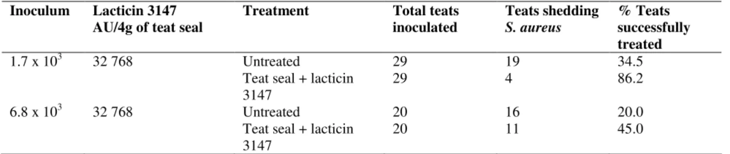
experimental challenge by S. aureus. The concentration of the
bacteriocin and inoculum of the S. aureus challenge was varied
to optimise treatment conditions. The presence of the teat seal
plus lacticin 3147 using a concentration of 32 768 AU/4g of
teat seal, resulted in a significant decrease in the number of
teats shedding S. aureus (Table 8). The antagonistic effect of
the bacteriocin at the same concentration was however reduced
when the inoculum of the S. aureus challenge introduced into
the teats was increased. The concentration of the bacteriocin
used was found to be significant factor for the teat seal to be
effective in reducing S. aureus in the teats.
Table 7. Clinical mastitis and recovery of S. dysgalactiae in non-clinical mastitis in quarters after treatment with the teat seal only
and the teat seal plus lacticin 3147 (73).
Treatment Total no of quarters
treated
New clinical infections by
S. dygalactiae
New non-clinical isolations of S. dysgalactiae
Teat seal 33 16 (48.5 %) 6 (18.2 %)
Teat seal plus lacticin 3147
35 3 (8.6 %) 0 (0 %)
Table 8. The effect of teat seal plus lacticin 3147 in eliminating S. aureus in artificially infected cows. Shedding evaluated after
18h (93).
Inoculum Lacticin 3147 AU/4g of teat seal
Treatment Total teats
inoculated
Teats shedding
S. aureus
% Teats successfully treated
1.7 x 10³ 32 768 Untreated
Teat seal + lacticin 3147
29 29
19 4
34.5 86.2
6.8 x 10³ 32 768 Untreated
Teat seal + lacticin 3147
20 20
16 11
20.0 45.0
The initial evaluation of lactitin 3147 by Ryan et al. (73,
74) indicated that bacteriocin produced in a synthetic growth
medium was not adequately released from the teat seal
formulation without the addition of a surfactant (Tween 80).
Later research improved the efficacy of the teat seal
formulation by producing lacticin 3147 in milk-based (whey)
medium which resulted in an increase in activity from ~320
AU.mL-1 to ~500 AU.mL-1 in the fermentate after 24 hr
incubation. The increase in activity of the bacteriocin
preparation resulted in a significant release of the peptide in the
teat seal formulation without the addition of Tween 80, thereby
quantities of the bacteriocin (16).
The lacticin 3147 produced in the milk-based (whey)
medium reduced the number of S. aureus recovered after
experimental challenge. The average recovery of S. aureus
from teats infused with teat seal plus lacticin 3147 was 7.3 x
102 cfu.mL-1 compared with 1.6 x 10⁴ cfu.mL-1 for those
treated with the teat seal alone. The bacteriocin-teat seal
preparation also appeared to eliminate S. aureus cells already
present in the teat canal prior to the infusion of the product
compared to the teat seal alone. No viable S. aureus cells were
recovered from the teats where the bacteriocin was present in
the teat seal, compared to four of the teats where only the teat
seal was used (n = 8) (16).
The stability of the product for the dry period of 50-60
days would still need to be assessed adequately as the teat
seal-bacteriocin product evaluated by Twomey et al. (93) and
Crispie et al. (16) was only infused for a period of 18 hours.
Ryan et al. (73) however showed that in an 8-day period,
lacticin 3147 retained activity in the teat environment.
To summarise, research has shown that the bacteriocin
lacticin 3147 has the potential for use in a teat seal preparation
to effectively prevent new infections by streptococci and offer
some protection to S. aureus infection. The bacteriocin could
potentially be produced on large scale using a milk-based
(whey) medium at concentrations that are active against target
organisms. The bacteriocin is also active and insoluble at
physiological pH and thus remains effective in the teat canal
environment.
Other bacteriocins that could have potential use in mastitis
treatment
Staphylococcal bacteriocins: Bacteriocins from
Gram-positive bacteria have, to a large extent, been limited to
applications in the food industry. Potential applications of
other bacteriocins in mastitis treatment have been limited to
that of lacticin 3147 (16) and nisin (81).
Growth inhibition studies of mastitis pathogens by normal
bovine teat skin flora (20,101) have been attempted to evaluate
the antagonistic or other effect that these less pathogenic
bacteria could have on major mastitis-causing pathogens such
as S. aureus, E.coli and streptococci. Staphylococcal strains
associated with mastitis were investigated and it was found that
bacteriocins active against mastitis-causing Streptococcus
agalactiae isolates were primarily produced by S. epidermidis,
S. saprophyticus and S. arlettae (23).
Streptococcal bacteriocins: Many streptococci have been
found to produce bacteriocins and the potential applications of
these bacteriocins range from those produced by the
thermophilic lactic acid bacteria, for their potential application
in cheese production to the oral streptococci for use in the
treatment of dental carries.
No potential streptococcal bacteriocins have as yet been
isolated for use in the treatment of mastitis. However, the
natural ecological niche of a particular bacteriocin producer is
often the specific area that is targeted for practical application.
The mastitis pathogen S. uberis is commonly found in the
natural environment of dairy cattle and thus could also be
competing with other bacteria in this ecological niche.
Wirawan et al. (99,100) screened more than 200 S. uberis
strains from their culture collection to determine whether any
of these strains produced bacteriocin-like inhibitory substances.
Strain 42 was found to produce two bacteriocins, a natural
nisin variant, nisin U and a circular peptide, uberlysin (100)
The bacteriocin nisin U had activity spectra against S.
agalactiae, S. dysgalactiae and E. faecalis that are considered
to be potential mastitis-causing pathogens (99). The discovery
of this natural nisin variant, which is active against
mastitis-causing pathogens, could offer a potential alternative to nisin
A, especially in cases where nisin A resistance may occur in
pathogenic strains. A combination of antimicrobials, such as a
nisin variant with other bacteriocins could potentially be more
effective in treatment strategies (100).
Other streptococcal bacteriocin producers occur in the oral
cavity where the normal flora such as S. salivarius, S. pyogenes
and S. mutans are readily found. These produce bacteriocins or
uncharacterised bacteriocin-like inhibitory substances (86, 88).
Normal flora of the nasopharynx also consists of bacteriocin
Pieterse, R. and Todorov, S.D. Bacteriocins alternatives in mastitis treatment
investigated for the prevention of streptococcal pharyngitis and
otitis media (86, 96). The type of treatment used is known as
bacteriotherapy or bacterial interference, where bacteriocin
producing, non-pathogenic strains are introduced into the
nasopharynx to protect against recurrent streptococcal
infections (96). The bacteriocin salvaricin A2 (SalA2),
produced by S. salivarius K12 has been developed as an oral
probiotic (BLIS K12 Throat Guard, BLIS Technologies, New
Zealand) to treat streptococcal infections especially by S.
pyogenes in children (88).
Streptococcal bacteriocins produced by Streptococcus
thermophilus strains are often investigated for use in yoghurt
starter cultures, including thermophilin 81 (40) and
thermophilin 13 (50), while thermophilin 580, produced by S.
thermophilus 580 has been studied for possible application in
cheese production as starter cultures with the added benefit of
bacteriocin inhibition of C. tyrobutyricum in the cheese
ripening process (51).
Larger bacteriocins (>10kDa) also produced by some
streptococci are characterised as non-lytic inhibitory agents or
as bacteriolytic enzymes. Examples include dysgalactin
produced by S. dysgalactiae subsp. equisimilis and
streptococcin A-M57 produced by S. pyogenes M-57.
Stellalysin is an example of a large 29-kDa bacteriocin
produced by S. constellatus subsp. constellatus. The activity
spectra of stellalysin includes S. pyogenes, S. gordonii and S.
mutans (37).
The mutacins B-Ny266, J-T8 and B-JH1140, produced by
S. mutacin have been characterised as belonging to the
lantibiotic class of bacteriocins. Potential practical applications
of mutacins include the treatment of dental carries (38).
Mutacin B-Ny266 has been of particular interest due to its
wide-spectrum of activity against many pathogenic
Gram-positive and Gram-negative bacteria, including staphylococcal
and streptococcal strains resistant to antibiotics. It could
therefore find application for therapeutic use (56, 57).
Rumen streptococci have also been investigated as a
source of bacteriocins, with S. bovis as the predominant strain
isolated (97). Bovacin 255 produced by S. gallolyticus 255, a
class II bacteriocin and bovicin HC5 from S. bovis HC5 could
find application in cattle farming (49, 97). Bacteriocins that
inhibit Gram-positive LAB found in rumen can be
advantageous as these bacterial species, through fermentation
produce large quantities of methane and ammonia waste
products. Bacteriocins could be applied as feed additives to
alter ruminal fermentation, and as a substitute to conventional
antibiotics, such as monesin (72).
The first report of a bacteriocin, namely macedocin
produced by the thermophilic S. macedonicus ACA-DC 198,
was characterised by Geogalaki et al. (28). The bacterium was
first isolated from Greek Kasseri cheese from Macedonia in
Northern Greece and was subsequently named as S.
macedonicus (92). Flint et al. (26) also isolated S. waius from
biofilms on stainless steel structures exposed to milk, but S.
waius was subsequently found to be synonymous to S.
macedonicus isolated by Tsakalidou et al. (92) and reclassified
as such (48). The species forms part of the larger S. bovis / S.
equinus complex but remains as a separate species, as low level
of DNA homology (less than 70 %) exists with other closely
related species such as S. gallolyticus (International Committee
on Systematics of Prokaryotes Subcommittee on the taxonomy
of staphylococci and streptococci, 2003). More recently, S.
macedonicus strains isolated from Italian raw milk cheeses
were characterised (47).
Macedocin ACA-DC 198 is a bacteriocin that has been
assessed as a food grade bacteriocin for use in cheese
manufacturing as a starter culture, because it is able to produce
the lantibiotic at pH and temperature conditions that prevail
during cheese manufacturing, and it also inhibits the food
spoilage bacteria C. tyrobutyricum (94). It has a molecular
mass of 2,794 Da, as determined by electrospray mass
spectrometry. Partial N-terminal amino acid sequence analysis
revealed some homology to other streptococcal bacteriocins,
SA-F22 and SA-M49, both produced by S. pyogenes (28). No
therapeutic applications have as yet been investigated for
macedocin ACA-DC 198 and its activity spectrum has been