67 6767 6767 Mem Inst Oswaldo Cruz, Rio de Janeiro, Vol. 99(Suppl. I): 67-71, 2004
H ypertensive Portal Colopathy in Schistosomiasis M ansoni
-Proposal for a Classification
M aria Angelina C M iranda+, Ana Lúcia C D omingues, H eloisa S D ias, Renata C M iranda, Norma T Jucá, M aria Fátima M Albuquerque, Fernando T Cordeiro
Faculdade de Medicina, Universidade Federal de Pernambuco, Av. Moraes Rego s/no, Cidade Universitária,
50640-900 Recife, PE, Brasil
Portal hypertension is a frequent complication of chronic liver disease, detected not only in schistosomiasis, but also in cirrhosis of any etiology. Vascular alterations in the colonic mucosa are a potential source for acute or chronic bleeding and have been observed in patients with portal hypertension. The purpose of this prospective study was to describe and propose a classification for the vascular alterations of portal hypertension in the colonic mucosa among patients with hepatosplenic schistosomiasis mansoni. One or more alterations of portal colopathy were observed in all patients and they were classified according to their intensity, obeying the classification proposed by the authors. Portal colopathy is an important finding in hepatosplenic schistosomiasis and might be the cause of lower gastrointestinal bleeding in patients with severe portal hypertension.
Key words: Schistosoma mansoni - portal colopathy vascular alterations telangiectasia angiodysplasia erythema -rectal varices
Schistosomiasis mansoni is a public health problem of major importance in Brazil. The infection reaches exten-sive areas in the Northeast affecting a large number of people, particularly youngsters and adults in their most productive phases of life in which 4 to 10% present seri-ous forms of the disease (Coutinho & Domingues 1993). Portal hypertension is a frequent complication of chronic liver disease, detected not only in schistosomia-sis, but also in cirrhosis of any etiology.
Endoscopic abnormalities in the colonic mucosa of patients with portal hypertension due to hepatic cirrhosis with risk of intermittent hemorrhage are well described (Kosareck et al. 1991, Viggiano & Gostout 1992, Rabino-vitz et al. 1995, Ganguly et al. 1995, Chen et al. 1996, Misra et al. 1996, Bresci et al. 1998, Bini et al. 2000). The most frequent alterations are defined as telangiectasias or vas-cular ectasias, angiodysplasia-like lesions, red spots, and erythema.
The initial histologic findings on these lesions showed dilated capillaries, submucosal edema, and non-specific inflammatory infiltrate (Kosareck et al. 1991). The term ectasia should be used for the combination of dilatation and structural alterations of the blood vessels (wall thick-ening and tortuousity), and not for dilatation only (Vig-giano & Gostout 1992).
Alterations in the colonic mucosa caused by Schisto-soma mansoni eggs are described as increased vascular bed, edema, congestion, scattered petechial spots with the aspect of “flea-bites”, ulceration, and polyps (Pereira 1962, Mohamed et al. 1990, Sanguino et al. 1993).
Consid-erable controversy still exists regarding the colonic ab-normalities in patients with portal hypertension due to schistosomiasis mansoni (Mohamed et al. 1990, Sanguino et al. 1993, Geboes et al. 1995).
The aim of this study was to better define the colonic alterations that are present in patients with portal hyper-tension (portal colopathy) due to schistosomiasis mansoni, to classify the severity of these abnormalities and to associate them with esophageal varices and portal gastropathy.
MATERIALS AND METHODS
After approval from the Research Ethics Committee of the Federal University of Pernambuco, 31 patients over 18 year of age, presenting the hepatosplenic form of schis-tosomiasis mansoni participated in this study. All patients had a history of specific treatment for S. mansoni and antecedents of upper or lower gastrointestinal bleeding. Other parasites eventually identified were treated before the inclusion of the patients in the study, in order to pre-vent possible lesions in the colon that might alter the results (Pereira 1962). Patients with alcohol ingestion up to 60 g/day were excluded, as were those with other he-patic disease, positive HbsAg and anti-HCV, or other dis-ease that could cause vascular alterations, such as chronic nephropathy, stenosis of the abdominal aorta, abdominal surgery (ileostomy and colostomy), and inflammatory bowel disease. After the feces, hematologic and biochem-istry exams, an abdominal ultrasonography was performed to confirm the diagnosis of hepatosplenic schistosomia-sis mansoni, as well as to characterize the portal hyper-tension and to exclude other hepatopathies. The ultra-sonography classifications of peripheral fibrosis used in this study were proposed by WHO (Niamey 2000). An upper gastrointestinal endoscopy was carried out to char-acterize esophageal (Japanese Research 1980), gastric varices (Hosking & Johnson 1988) and the portal hyper-tensive gastropathy. The entire colon was visualized us-+Corresponding author. Fax: +55-813325.3315. E-mail:
6 8 6 8 6 8 6 8
6 8 Schistosomal Colopathy • M aria Angelina C M iranda et al.
ing an Olympus CF-VI colonoscope with conventional resolution. The patients had their colon prepared by us-ing manitol at 10% (Habar-Gama et al. 1981). Durus-ing the examination, they received an intravenous sedation of meperidine associated with midazolan, monitored through pulse oximetry. In accordance to criteria described by Tam et al. (1995), the lesions were characterized as: erythemas (diffuse hyperemia in the mucosa), red spots (hyperemic focus in the mucosa), telangiectasia (dilated and tortuous small vessels), angiodysplasia (coiled vessels measuring about 1 cm), rectal varices (dilated, crooked vessel). The current authors elaborated a classification for the colopathy in patients with schistosomiasis and portal hypertension, based on both literature concerning cir-rhotic patients and personal experience. It was classified into three levels: degree I (erythemas and telangiectasias); degree II (erythemas, telangiectasias, and angiodyspla-sias); and degree III (erythemas telangiectasias, angio-dysplasias, red spots, and rectal varices). During the
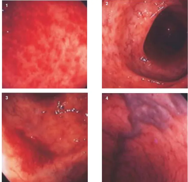
colonoscopy, mucosal biopsy specimens were obtained, by way of the FB-24U-1 Olympus forceps, in normal ap-pearing mucosa of the rectum, sigmoid and in areas with lesions, avoiding to perform biopsy in areas with angiodysplasia-like lesions. Histological sections were stained with hematoxilina and eosine (HE) (Figs 1-4).
RESULTS
The 31 hepatosplenic schistosomotic patients stud-ied were between 20 to 73 years old. Fifteen patients were male and 16 female. All were from endemic schistosomia-sis region. Twenty-six patients (83%) reported having one or more episodes of upper gastrointestinal bleeding and 8 (25.8%) had had lower gastroentestinal bleeding.
The stool examinations were negative for S. mansoni
eggs in 30 patients (96.7%). The HbsAg and anti-HCV serology were negative in all cases.
The upper gastrointestinal endoscopy findings are displayed in Table I.
Fig. 1: rectal erythemas. Fig. 2: colonic telangiectasias. Fig. 3: colonic angiodysplasia. Fig. 4: rectal varices.
1 2
6 9 6 96 9 6 9 6 9 Mem Inst Oswaldo Cruz, Rio de Janeiro, Vol. 99(Suppl. I), 2004
All patients presented portal hypertensive colopathy. One or more alterations were identified simultaneously, in the same patient. Erythemas were observed in 10 (32.2%), telangiectasias in 31 (100%), angiodysplasia-like lesions in 17 (54.8%), and red spots in 3 patients (9.6%). Rectal varices were identified in 9 cases (29%). Erythemas pre-dominated in the distal segments (70%). Telangiectasias were distributed throughout the entire colon in 93.5% of the patients. Angiodysplasia-like lesions also had a pan-colonic presentation in 58.8% of the cases. The frequen-cies of the vascular alterations in portal colopathy are displayed in Table II.
After grouping the cases according to the classifica-tions proposed by the authors, mild colopathy was found in 10 patients (32.2%), moderate colopathy was found in 10 patients (32.2%), and severe colopathy was found in 11 (35.5%).
The histological examination identified mononuclear inflammatory infiltrated (lymphocytes and plasma cells) of mild intensity in 28 cases (90.3%), moderate intensity in 3 (9.7%); mild hyperemia in 12 cases (38.7%), and mod-erate in 1 (3.3%); modmod-erate edema in 13 cases (41.9%) and severe in 3 (9.7%), moderate capillary ectasia in 12 cases (38.6%) and severe in 2 (6.5%). S. mansoni eggs were observed in three cases: viable eggs in the sigmoid with and without schistosomotic granuloma in one; viable egg without granuloma in the distal ileo in another; and calci-fied eggs in the sigmoid in the last one.
DISCUSSION
While vascular lesions in the colonic mucosa of cir-rhotic patients with portal hypertension have been re-ported (Kosareck et al. 1991, Viggiano & Gostout 1992, Rabinovitz et al. 1995, Chen et al. 1996, Ganguly et al. 1995, Misra et al. 1996, Bresci et al. 1998, Bini et al. 2000), studies on hepatosplenic schistosomiasis mansoni are scarce (Mohamed et al. 1990, Sanguino et al. 1993, Geboes et al. 1995).
For ethical reasons, we did not study patients without prior episode of upper or lower gastrointestinal bleeding and the patients selected had already been treated for schistosomiasis to avoid the alterations due to the para-sitose per si (Pereira 1962, Mohamed et al. 1990, Sanguino et al. 1993, Geboes et al. 1995).
The characteristic alterations of the portal colopathy in schistosomiasis are similar to those of portal colopathy in cirrhotics (erythemas, telangiectasias, angiodysplasias, red spots, and rectal varices), but they tend to be more frequent and accentuated. It has been suggested that the schistosomotic portal colopathy happens due to the in-tense and permanent rise of the portal venous pressure (Lamps et al. 1998). This is associated to the neovascu-larization process stimulated during the active phase of the disease, through the blockage of the vessels caused by the schistosome eggs, as well as the secretions re-leased by them (Wyler et al. 1987). Telangiectasias are the most frequent finding in this series and in Justo’s work (2003). These vascular changes, which are similar to the injuries of the upper gastrointestinal tract, can be the cause of lower gastrointestinal bleeding, a process that is often obscure and co-responsible for the chronic anemia found in these patients. This may have occurred in 8 (25.8%) cases of this present study. The patients in this series had advanced portal hypertension, as determined by endoscopy and ultrasonography. All of them had vas-cular alterations of portal colopathy, 32.2% mild, 32.2% moderate, and 35.5% severe. A retrospective study by Bini et al. (2000) on cirrhotic patients with portal hyper-tension identified both medium and large esophageal va-rices in 51% of the cases, gastropathy in 47%, and at least one characteristic alteration of the colopathy in just 57%. However, in a study by Eleftheriadis et al. (1993) on cir-rhotic patients with accentuated portal hypertension who had undergone sclerotherapy for the erradication of esophageal varices and had no prior history of lower gas-trointestinal bleeding, a high frequency of portal colopathy (93%) was observed, with lesion-like telangiectasias pre-dominating (70%). In the present series, colitis-like (ery-themas) alterations in the mucosa were seen in 32.2% of the cases in relation to what was observed in cirrhotic patients, 27% (Misra et al. 1996), 32% (Rabinovitz et al. 1995) and 38% (Bini et al. 2000). Rectal varices are collat-eral portosystemic dilated veins in patients with portal hypertension and communicate the venous flow of the superior hemorrhoid veins to the medium and inferior hem-orrhoid veins (Jacobs et al. 1989, Hosking et al. 1989). Therefore, they may occasionally be subjected to mas-sive bleeding (Weinshall & Cheer 1986). The prevalence of the rectal varices has varied from 0% to 92% in cirrhotic
TABLE I
Upper gastrointestinal endoscopy in patients with hepatosplenic schistosomiasis mansoni
Degree of esophageal varices a/b (n = 27) n %
Degree I 1 3.7
Degree II 9 33.3
Degree III 17 63
Gastric varices b/c (n = 27)
Cardia 20 64.5
Fundus 4 12.9
Body 3 9.7
Portal gastropathy d (n = 31)
Mild 29 93.5
Severe 2 6.5
a: Japanese Society Classification for Portal Hypertension Research; b: 4 patients did not present esophageal and gastric varices; c: Hosking and Johnson classification; d: McCormack et al. classification
TABLE II
Vascular alterations of portal colopathy in patients with hepatosplenic schistosomiasis
Vascular alterations n %
Erythemas 10 32.2
Red spots 3 9.6
Telangiectasias 31 100
Angiodysplasias 17 54.8
7 0 7 0 7 0 7 0
7 0 Schistosomal Colopathy • M aria Angelina C M iranda et al.
patients ( Hosking et al. 1989, Rabinovitz et al. 1995, Misra et al. 1996). Our findings are in agreement with Hosking et al. (1989), who concluded that rectal varices reflect a later stage in the development of the portal hypertension, be-ing observed more frequently in patients with large esoph-ageal and gastric varices. This fact was demonstrated in this series, in which 6 of the 9 cases with rectal varices (66.6%) had large esophageal varices. In the histological studies, a mild mononuclear inflammatory infiltrate was evidenced, particularly lymphocytes and plasma cells in 90.3% of the cases, excluding inflammatory bowel dis-ease. Edema and the loss of the villous architecture of the lamina (propria?) of the colonic mucosa are secondary to the vessel wall thickness present in this study with mod-erate to accentuated intensity in 51.6% of the cases. This can be explained by the chronic increase of the intralumi-nal pressure (Misra et al. 1997). Endoscopic biopsies of the vascular lesions in the colonic mucosa were avoided due to the high risk of bleeding (Tam et al. 1995). How-ever, when it is obtained, the descriptions include dilata-tion and tortuosity of the mucosa vessels, as well as an increase in the number of small vessels and the wall thick-ness in the lamina propria (Viggiano & Gostout 1992). In this study, we followed the criteria established by Viggiano and Gostout (1992) and observed moderate to severe cap-illary ectasia in only 45.1% of the cases. In these two cases, who had viable eggs in the biopsia, probability due to re-infection, an increase in the degree of colopathy was not verified when compared to the other patients.
There was no correlation between the severity of por-tal gastropathy and the presence of hypertensive colo-pathy. Meanwhile, it was observed a predominance of severe portal colopathy (87.5%) in patients with large esophageal varices.
We conclude that in a well-characterized portal hyper-tension in hepatosplenic schistosomiasis mansoni, hy-pertensive colopathy is an important entity, as it was present in all cases. The identification and classification of these vascular lesions in the colonic mucosa is of great significance for the diagnosis and control of the evolu-tion of the disease, as these alteraevolu-tions can be the cause of anemia, hypoproteinemia, and obscure lower bleeding, contributing to a higher morbidity from the disease. We suggest that colonoscopy should be done in every pa-tient with hepatosplenic schistosomiasis who has anemia without a prior episode of upper gastrointestinal hemor-rhage. The classification proposed in this work would be useful to standardize the categorized alterations accord-ing to intensity.
REFERENCES
Bini EJ, Lascarides CE, Micale PL, Weinshel EH 2000. Mu-cosal abnormalities of the colon in patients with portal hypertension: an endoscopic study. Gastrointest Endosc 52: 511-516.
Bresci G, Gambardella L, Parisi G, Federici G, Bertini M, Rindi G 1998. Disease in cirrhotic patients with portal hyperten-sion. J Clin Gastroenterol 26: 222-227.
Chen LS, Lin HC, Lee FY, Hou MC, Lee SD 1996. Portal hy-pertensive colopathy in patients with cirrhosis. Scand J Gastroenterol 31: 490-494.
Coutinho A, Domingues AL 1993. Esquistossomose mansoni.
In R Dani, LP Castro (eds), Gastroenterologia Clínica, 2nd ed., Guanabara Koogan, Rio de Janeiro, p. 1697-1728. Eleftheriadis E, Kotzampassi K, Karkavelas G 1993. Portal
hypertensive colopathy: endoscopic, hemodynamic and morphometric study. Dig Endosc 5: 224-230.
Ganguly S, Sarin SK, Bhatia V, Lahoti D 1995. The prevalence and spectrum of colonic lesions in patients with cirrhotic and noncirrhotic portal hypertension. Hepatology 21: 1226-1231.
Geboes K, El-Deeb G, El-Haddad S, Amer G, El-Zayadi AR 1995. Vascular alterations of the colonic mucosa in schisto-somiasis and portal colopathy. Hepato-Gastroenterol 42: 343-347.
Habr-Gama A, Gama-Rodrigues JJ, Teixeira MG, Alves PRA, Ventura TCM, Quintanilha AG 1981. Preparo intestinal pela ingestão de manitol a 10%. Rev Bras Colo-Proct 4: 16-26.
Hosking SW, Smart HL, Johnson AG, Triger DR 1989. Anorec-tal varices, hemorrhoids, and porAnorec-tal hypertension. Lancet 1: 349-52.
Jacobs DM, Bubpick MP, Onstan GR, Hitchcook CR 1980. The relationship of hemorrhoids to portal hypertension.
Dis Colon Rectum 23: 567-569.
Justos CRE 2003. Efeito da Esplenectomia e Ligadura da Veia Gástrica Esquerda na Colopatia da Hipertensão Porta Esquistossomótica, MSc Thesis, Univesidade Federal de Pernambuco, 32 pp.
Kosareck RA, Botoman VA, Bredfeldt JE, Roach JM, Patterson DJ, Ball TJ 1991. Portal colopathy: prospective study of colonoscopy in patients with portal hipertensive. Gastro-enterology 101: 1192-1197.
Lamps LW, Hunt CM, Green A, Gray GF, Washington K 1998. Alterations in colonic mucosal vessels in patients with cir-rhosis and noncirrhotic portal hypertension. Human Pa-thology 29: 527-535.
McComarck TT, Sims J, Eyre-Brook I, Kennedy H, Goepel J, Johnson AG, Triger DR 1985. Gastric lesions in portal hypertension: inflammatory gastrits or congestive gastropathy? Gut 26: 1226-1232.
Misra SP, Dwivedi M, Misra V 1996. Prevalence and factors influencing hemorrhoids, anorectal varices, and colopathy in patients with portal hypertension. Endoscopy 28: 340-345.
Misra V, Misra SP, Dwivedi M, Gupta SC 1997. Histomor-phometric study of portal hypertensive enteropathology.
Am J Clin Pathol 108: 652-657.
Mohamed AE, MA Al Karawi, MI Yasawi 1990. Schistosomal colonic disease. Gut 31: 439-442.
Naveau S, Bedossa P, Poynard T, Mory B, Chaput JC 1991. Portal hypertensive colopathy, a new entity. Dig Dis Sci 36: 1774-1781.
Nebel OT, El Masry NA, Castell DO, Farid Z, Fornes MF, Sparks HA 1974. Schistosomal disease of the colon: a re-versible form of polyposis. Gastroenterology 67: 939-943. Niamey Working Group 2000. Ultrasonography in schistoso-miasis. A practical guide to the standardized use of ultra-sonography for the assessment of schistosomiasis-related morbidity. WHO-World Health Organization TDR/SCH/ ULTRASON document, Geneva, Switzerland, in press. Pereira OA 1962. Aspectos endoscópicos da esquistossomose
mansoni. Rev Brasil Gastroenterol 14: 5-12.
Rabinovitz M, Schade RR, Dindzans VJ, Belle SH, Van Thiel DH, Gavaler JS 1995. Colonic disease in cirrhosis. Gastro-enterology 99: 195-199.
7 1 7 17 1 7 1 7 1 Mem Inst Oswaldo Cruz, Rio de Janeiro, Vol. 99(Suppl. I), 2004
Raso P, Pedroso ERP 1987. Patologia das principais doenças tropicais do Brasil. Esquistossomose mansônica. In L Bogliolo, Patologia, 4th ed., Guanabara Koogan, Rio de Janeiro, p. 1065-1086.
Sanguino J, Peixe R, Guerra J, Rocha C, Quina M 1993. Schis-tosomiais and vascular alterations of the colonic mucosa.
Hepato-Gastroenterol 40: 184-187.
Tam TN, Ng WW, Lee SD 1995. Colonic mucosal changes in patients with liver cirrhosis. Gastrointest Endosc 42: 408-412.
Viggiano TR, Gostout C 1992. Portal hypertensive intestinal vasculopathy: a review of the clinical, endoscopic, and his-topathologic features. Am J Gastroenterol 87: 944-946. Weinshall E, Cheer W 1986. Hemorrhoids or rectal varices:
defining the cause of massive rectal hemorrhage in pa-tients with portal hypertension. Gastroenterology 90: 744-747.