71 71 71 71 71 Mem Inst Oswaldo Cruz, Rio de Janeiro, Vol. 94, Suppl. I: 71-80, 1999
Immunopathology of Chagas Disease
Zilton A Andrade
Laboratório de Patologia Experimental, Centro de Pesquisas Gonçalo Moniz-Fiocruz, Rua Valdemar Falcão 121, 40295-001 Salvador, BA, Brasil
The main clinical forms of Chagas disease (acute, indeterminate and chronic cardiac) present strong evidences for the participation of the immune system on pathogenesis. Although parasite multiplication is evident during acute infection, the intense acute myocarditis of this phase exhibits clear ultrastruc-tural signs of cell-mediated immune damage, inflicted to parasitized and non-parasitized myocardiocytes and to the endothelium of myocardial capillaries (microangiopathy). Inflammation subsides almost completely when immunity decreases parasite load and suppressor factors modulate host reaction, but inflammation does not disappear when the disease enters the indeterminate phase. Inflammation be-comes mild and focal and undergoes cyclic changes leading to complete resolution. However, the pro-cess is maintained because the disappearance of old focal lesions is balanced by the upsurge of new ones. This equilibrium allows for prolonged host survival in the absence of symptoms or signs of dis-ease. The chronic cardiac form is represented by a delayed-type, cell-mediated diffuse myocarditis, that probably ensues when the suppressive mechanisms, operative during the indeterminate phase, become defaulted. The mechanism responsible for the transition from the indeterminate to the cardiac form, is poorly understood.
Key words: Trypanosoma cruzi - immunopathology - myocarditis
The major aspects of the pathogenesis of Chagas disease remain poorly understood. Some crucial pathological findings strongly suggest the participation of immunological fators. Inflamma-tory lesions in the myocardium show no direct cor-relation with the presence of parasites, regardless the clinical stage of the disease. The acute myo-carditis that follows primary infection with Trypa-nosoma cruzi, spontaneously subsides, after a pe-riod of two-three months in most cases, but does not disappear. In its place a mild focal myocarditis remains and may last for years without signs of cumulative damage. Suddenly or slowly, severe, diffuse, progressive and fatal chronic myocarditis ensues after a prolonged period of silent infection, in a small proportion of cases (about 30%). This complex chain of events is unusual to be seen in other parasitic diseases. It has stimulated research on immunology and immunopathology of Chagas disease, but efforts are often hampered by lack of an adequate experimental model. Almost any mam-mal can be infected with T. cruzi. Acute infection (positive parasitemia in peripheral blood) can variably be documented a week or more after in-oculation. In some hosts infection will eventually
Fax: +55-71-359-4292 Received 9 June 1999 Accepted 9 August 1999
evolve toward spontaneous cure. Others will re-main infected, without signs of disease (indeter-minate form), but, different from the situation in man, none will develop progressive chronic myo-carditis, with cardiomegaly, arrhytmias and signs of chronic cardiac failure following a prolonged time of silent infection. The dog is an exception. Young dogs usually develop severe acute disease when naturally or experimentally infected with T. cruzi (Andrade 1984). When acute manifestations abate, a period of latent infection follows and may last for life. Eventually, chronic myocarditis, accompanied by cardiomegaly, arrhytmias, dispnea, peripheral edema will appear, without any evidence of rapid resumption of parasite multipli-cation (Anselmi et al. 1966, Laranja & Andrade 1980). Unfortunately, immunological data are scanty for this model, although a good amount of clinical and morphological data are avaliable. Most of the subject to be discussed in this chapter will be based on experimental studies made in dogs. This report will also be limited to heart involve-ment in Chagas disease. Lesions in other organs, especially involving the digestive tract, which may result in megaesophagus and megacolon, have been little explored as far as immunopathology is con-cerned.
ACUTE PHASE MYOCARDITIS
72 72 72 72
72 Immunopathology of Chagas Disease Zilton A Andrade
tissue forms of T. cruzi multiplied within cardiomyocytes and caused their rupture, liberat-ing inflammatory mediators of several kinds. This was sufficient to explain the whole picture, since parasitism was easily observed in tissue sections. This contrasted to the situation with the chronic cardiac form, when parasites are rarely demon-strated by the usual techniques or even by a com-bination of them. Therefore, the pathogenesis of acute myocarditis was considered radically dif-ferent from that of the chronic cardiac form. How-ever, closer observations of acute cases revealed that, although parasites were present, the intensity of the inflammatory process did not always corre-lated with the overall presence of tissue forms of the parasites. Furthermore, destruction of non-parasitized cardiomyocytes was found to be an outstanding feature, and that seemed to require a more elaborated explanation.
Sequential examination of experimental mate-rial shows that the first changes related to T. cruzi infection appear around ruptured parasitized fibers. Presence of immuno-globulin and complement can
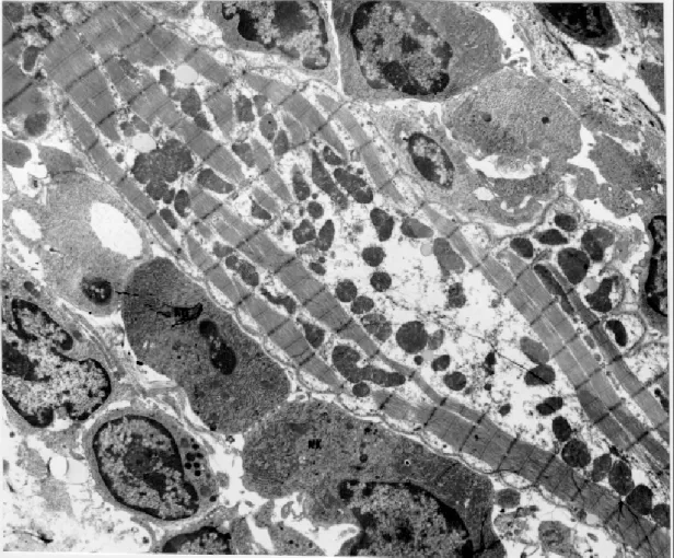
be demonstrated in these focal lesions by immun-ofluorescence, revealing the early participation of humoral immune factors in pathogenesis (Silva et al. 1985). Initially focal reactions are seen scat-tered distributed throughout the myocardium a few days after experimental inoculation. This is soon followed by diffuse and more intense inflamma-tion. When this happens, isolated necrosis of non-parasitized cardiomyocytes becomes more or less prominent. From then on, signs of cell-mediated immune damage can be ultrastructurally disclosed (Andrade et al. 1994). There is close adhesion of mononuclear cells (small and large granular lym-phocytes, probably NK-cells and also macroph-ages) to cardiomyocyte membrane, with fusion of membrane at focal points, accompanied by focal disruption of T tubules, sarcoplasmic reticulum, myofibrils, mitochondria and even intercallated disks (Fig. 1). Damage to the cardiocyte some-times is multifocal, and leads to lytic or coagula-tive necrosis of large segments of the car-diomyocytes or even of their entire length. Cells in close proximity to the damaged ones usually
73 73 73 73 73 Mem Inst Oswaldo Cruz, Rio de Janeiro, Vol. 94, Suppl. I, 1999
appear entirely preserved. Possible causes for these changes have been suggested: (a) immunological injury after adsorption of T. cruzi antigens on non-parasitized cells (Ribeiro dos Santos & Hudson 1980); (b) ischemic injury due to platelet aggrega-tion and obstrucaggrega-tion of myocardial capillaries (Rossi et al. 1984, Tanowitz et al. 1990); (c) di-rect or antibody-mediated cytotoxic damage by inflammatory and immune effector cells, includ-ing lymphocytes, neutrophils, eosinophils, mac-rophages and mast cells (Lopes et al. 1977, Tafuri et al. 1983, Cabral 1988, Molina & Kierszenbuam 1989, Rossi 1990).
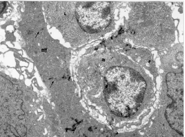
Besides these destructive changes of myocytes, a microangiopathy is also observed. It consists of myocardial capillary involvement, following close contact between lymphocytes or macrophages to the endothelial cells (Andrade et al. 1994). In the contact points there occur membrane fusion and in-creased pinocytosis, tumefaction, vacuolation and cytoplasmic disruption of endothelial cells, associ-ated or not with platelet aggregation and fibrin microthrombi (Fig. 2). The role of a vascular factor
in acute Chagas myocarditis has been emphasized, especially by investigators who observed platelet thrombi within myocardial capillaries of mice (Rossi et al. 1984, Tanowitz et al. 1990). However, the ultrastructural signs of cytotoxic damage to the myocytes seen in the canine model far exceeded those indicative of ischemic injury (Andrade 1984). On the other hand, microangiopathy appears as an important clue suggesting the participation of cytokines in acute Chagas myocarditis. Similar vas-cular changes have been observed in patients dying of the so-called “leaky syndrome” after being treated with large doses of interleukin-2 (Cotran et al. 1987). Experimentally, rats treated either with recombi-nant interleukin-2, interleukin-1 and tumor necro-sis factors also exhibited similar myocardial microangiopathy (Yi & Ulich 1992, Zhang et al. 1993). Recent studies have demonstrated that cytokines associated with CD4 response of the Th-1 type, can be enhanced by the administration of 12, which results in the production of INF-γ, IL-2 and TNF-α, causing reduction in parasitemic lev-els of infected animals (Silva et al. 1995,
74 74 74 74
74 Immunopathology of Chagas Disease Zilton A Andrade
Abrahamsohn & Coffman 1996, Cardillo et at. 1996, Hunter et al. 1996). Probably this mechanism of protection, when overwhelming, would contribute to capillary and other myocardial changes during the course of acute Chagas disease.
IMMUNOLOGICAL MODULATION
There are two basic host reactions to T. cruzi, which are represented by focal and diffuse inflam-mation. Focal reaction is parasite-mediated. It oc-curs anywhere the parasite enters host cells and then multiplies, causing cell-rupture. Focal heart lesions, observed in human tissue in which para-site could not be demonstrated in routine histologi-cal sections, revealed the presence of parasite antigen by immunohistochemistry (Higuchi et al. 1993) or its genomic segments by PCR technique (Jones et al. 1993), which is in keeping with the idea that focal inflammatory reactions are parasite-induced.
The heart may present both types of reactions. Diffuse inflammation is not directly parasite re-lated. It occurs during the acute and chronic car-diac forms, while mild focal inflammation is the hallmark of the indeterminate form of the disease. All other organs exhibit only focal reactions following parasitized-cell rupture, regardless the type of reaction present in the heart. This simple observation reveals that some important pathogenic factors of chagasic myocarditis are related to the heart itself. Also, by observing the cardiac histo-pathology one can follow the changes suggestive of immunological modulation during the several clinical stages of Chagas disease. The complete picture regarding what happen when diffuse in-flammation of the acute phase subsides and turns into a mild focal myocarditis, is poorly understood. The same is true for the transition of focal myo-carditis of the indeterminate form toward the dif-fuse and fibrosing chronic myocarditis of the car-diac form. Of course immunity factors, both hu-moral and cellular, contribute to decreasing para-site load. When cytokines patterns were investi-gated, transition from the acute form to the inde-terminate form was identified in infected children as a CD4-Th1 response changing into a Th0 pat-tern, with expression of both interferon-gamma and IL-4 (Smudio et al. 1998). There are evidences that immunossupression plays a role in diminish-ing and modifydiminish-ing the character of the inflamma-tory reaction ellicited by T. cruzi (Tarleton 1998). Depressed delayed skin reaction to parasite anti-gens and in-vitro demonstration of inhibition of IL-2 lymphocyte production have been reported in mice. Immunossupresed individuals with latent T. cruzi infection, especially those affect by Aids,
are seen to present acute exacerbation of the infec-tion (Tarleton 1993, Rocha et al. 1994).
THE INDETERMINATE FORM
The indeterminate phase of Chagas disease is defined as the prolonged clinically silent period that follows the phase of acute primary infection with T. cruzi. The individuals present serological and/or parasitological evidences of infection, but remain asymptomatic and do not exhibit electro-cardiographic signs of heart involvement, nor X-rays abnormalities of the digestive tract.
Microscopically, infected individuals at the in-determinate form of Chagas disease reveal mild focal myocarditis when their hearts are examined following biopsies (Mady et al. 1984, Palacios-Pru et al. 1989) or autopsies, that latter material being available after suicidal or accidental deaths (Lopes et al. 1975, 1981). However, longitudinal clinical studies have demonstrated that subjects present-ing the indeterminate form tend to remain asymp-tomatic for prolonged time, and thus prognosis can be considered good. This also indicate that lesions of mild focal myocarditis are not cumulative.
75 75 75 75 75 Mem Inst Oswaldo Cruz, Rio de Janeiro, Vol. 94, Suppl. I, 1999
Transition to chronic cardiac form probably oc-curs through disappearance of, or interference with, suppressive factors. Therefore, both parasites and heart tissue appear to be important to explain diffuse and progressive inflammatory lesions of the cardiac form, since parasites outside the heart will continue to induce focal reactions only (Barbosa & Andrade 1984). The factors responsible for the transition from the indeterminate to the chronic, progressive stage of Chagas disease remain to be elucidated. This transition, in which the suppres-sion of cell-mediated immune responses ceases to occur, seems to be related to reactivation of the inflammation. The inflammatory reaction of the chronic phase of Chagas disease in humans has been recently characterized as consisting mainly of cytotoxic CD8+ T-lymphocytes (Tostes et al.
1994). These cells were seen to express the Gramzyme A cytotoxic factor (Reis et al. 1993). It has been now a consensus that CD8+
lympho-cytes are the main T-cell type responsible for im-mune activation in chronic chagasic myocarditis (Kumar & Tarleton 1998). These cells are activated, through Class I MHC molecules, by macrophages containing remnants of T. cruzi. The absence of a CD4+ T-cell response in the presence of T. cruzi antigens suggests that the presentation of these an-tigens through Class II MHC molecules is inhib-ited. However, other evidence suggests that the depletion of CD4+ lymphocytes in the chronic phase of Chagas disease is related to selective apoptosis of these cells (Lopes et al. 1995). In the mouse model of chronic Chagasic cardiomyopathy, stimulation of T cells results in apoptosis of CD4+ cells but not of CD8+ cells (DosReis et al. 1995) Taken together, the morphologic and immunologic observations re-viewed above support the concept that the indeter-minate phase of Chagas disease represents a state of host-parasite equilibrium rather than of progres-sive chronic myocardial damage.
76 76 76 76
76 Immunopathology of Chagas Disease Zilton A Andrade
THE CHRONIC CARDIAC FORM
The phase of chronic, progressive myocarditis and congestive heart failure which are associated with myocardial hypertrophy, myocyte degenera-tion, severe interstitial fibrosis and thickening of the basement membranes characterize the patho-logical picture of the chronic cardiac form of Chagas disease. One can assume that this form originates from the indeterminate stage of latent infection, in 30% of the cases in man, and much less for the dog. Some important points usually pass unnoticed. A progressive, destructive and repara-tive process appears in the heart, previously af-fected during a long time by mild diffuse myocardi-tis. Although rarely, parasites can also be found in several other organs always accompanied by focal inflammation, a situation entirely similar to that seen in the heart during the indeterminate stage. Except for such focal and rare microscopic para-site-related changes, no other abnormalities are apparent outside of the heart during the chronic cardiac form, except those derived from chronic passive congestion and thrombo-embolic phenom-ena. No evident modification, from the status of the indeterminate phase, occurs regarding
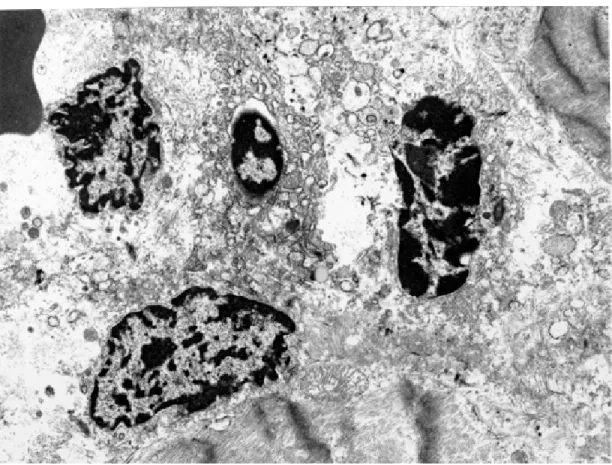
para-Fig. 4: heart of dog with indeterminated Chagas disease. Chromatic nuclear materials (DNA) belonging to four disintegrating cells, probably cardiomyocytes, show different degree of condensation and fragmentation (apoptosis). Electron micrograph, X5,000.
77 77 77 77 77 Mem Inst Oswaldo Cruz, Rio de Janeiro, Vol. 94, Suppl. I, 1999
sitism. Also, there are no definite changes regard-ing serum antibodies. There have been some at-tempts to investigate the behavior of mononuclear cells in peripheral blood, with the result that some suppressor cells can be differentially stimulated with anti-idiotypic antibodies (Ouaissi et al. 1988, Gazzineli et al. 1988). Also, lymphocytes directly isolated from the heart of patients with either the indeterminate or the cardiac forms of Chagas dis-ease have exhibited different patterns of reactiv-ity when in the presence of a particular myocar-dial epitope (Cunha Neto et al. 1995).
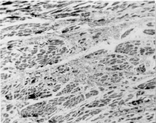
Experimental reproduction of the transition from the period of silent infection to chronic pro-gressive myocarditis has been achieved with the canine model (Andrade et al. 1987). Animals with known acute infection at early age, and with pro-longed silent period of infection, with no electro-cardiographic alterations, were treated with low doses of cyclophosphamide. This procedure has been assumed to exacerbate cell-mediated lesions, probably by selectively destroying suppressor T-lymphocytes, their precursor cells, or other ele-ments in the host-immune supressor network (Turk et al. 1972, Schwartz et al. 1978, Colley et al. 1979, Liew & Russel 1983).
In contrast to non-treated dogs, that remained with mild focal myocarditis, all treated animals
exhibited intense and diffuse myocarditis (Fig. 6). Destruction of isolated myocells was extensive throughout the contractile myocardium and the conduction system. Mononuclear cells were seen at the periphery and in the interior of disintegrat-ing myocardiocytes. Tissue forms of T. cruzi were not detected in histological section although ex-haustively searched. Parasitemia in peripheral blood was always negative.
The mechanism of cell-mediated myocardial damage remains to be elucidated. The suggestion for an auto-immune process has been frequently presented, but has not been proved (Kierszembaum 1986). A strong support in favor of an auto-im-mune mechanism for Chagas myocarditis came from experiments with transplanted neonatal hearts into the external ear of isogenic mice (Ribeiro dos Santos et al. 1992). While the transplants remained viable for a long time, when into the ear of normal receptors, they were rejected if the receptor was chronically infected with T. cruzi. Effector cells were identified as CD4 lymphocytes. However, recent data from Tarleton et al. (1997) contradict these findings. They failed to observe rejection of transplanted neonatal hearts placed into chroni-cally infected mice. Severe inflammation occurred only when T. cruzi was directly injected into the transplant, showing that the parasite, and not an
78 78 78 78
78 Immunopathology of Chagas Disease Zilton A Andrade
auto-immune process, was responsible for the ef-fect observed in experiments with transplanted neonatal hearts.
The auto-immune theory is supported by the lack of correlation between parasitism and chronic myocarditis. However, the great age variation of the patients at the onset of clinical manifestations, the lack of exacerbations and remissions, the ab-sence of typical auto-antibodies at high and con-stant levels, the lack of involvement of other or-gans, are not in keeping with an auto-immune pro-cess. Some investigators have documented the identity of epitopes present in the parasite and myocardium (Sadigursky et al. 1982). These cross-reacting molecules could attract the destructive power of the sensitized immune system. Similarly, the same could occur if parasite antigens happen to be expressed in the myocyte membrane, or be adsorbed onto it (Ribeiro dos Santos & Hudson 1909). There are several other suggestions to ex-plain myocardial damage in T. cruzi infection, all of them in needing of further research. Some data refer to the idiotype/anti-idiotype network. When generating antibodies against anti-T. cruzi globu-lins, the host organism would be in effect produc-ing antibodies that would have the same configu-ration as that of the parasite. These antibodies could therefore, either directly react with or acti-vate immune cells against receptors present in the cardiomyocyte membranes. One such cross react-ing antigen found in man was represented by an enzyme, acetyl-cholinesterase. The anti-idiotype antibody against this epitope was found to be more frequent in the sera of patients with the cardiac form of Chagas disease than in those of the inde-terminate form (Quaissi et al. 1988). Other differ-ences regarding anti-idiotypic antibodies have been registered for the latent and active chronic forms of the disease. These findings are not only inter-esting for the supporters of the theory of auto-im-munity, but points toward the existence of a pro-cess of modulation or repression/des-repression in Chagas disease pathogenesis.
REFERENCES
Abrahamsohn IA, Coffman RL 1996. Trypanosoma cruzi: Il-10, TNF, INF-γ, and Il-12 regulate innate and acquired immunity to infection. Exp Parasitol 84: 231-244.
Andrade ZA 1984 The canine model of Chagas disease. Mem Inst Oswaldo Cruz 79: 77-83.
Andrade ZA, Andrade SG, Correa, R, Sadigursky M, Ferrans VJ 1994. Myocardial changes in acute Try-panosoma cruzi infection. Ultrastructural evidence of immune damage and the role of microangiopathy. Am J Pathol 144: 1403-1411.
Andrade ZA, Andrade SG, Sadigursky M 1987. En-hancement of chronic Trypanosomacruzi
myocardi-tis in dogs treated with low doses of cyclophospha-mide. Am J Pathol127: 467-473.
Andrade ZA, Andrade SG, Sadigursky M, Wenthold Jr RJ, Hilbert SL, Ferrans VJ 1997. The indetermi-nate phase of Chagas disease: ultrastructural char-acterization of cardiac changes in the canine model. Am J Trop Med Hyg 57: 328-336.
Anselmi A, Pifano F, Suarez JA, Gurdiel O 1966. Myo-cardiopathy in Chagas disease. I. Comparative study of pathologic findings in chronic human and experi-mental Chagas myocarditis. Am Heart J 72: 469-481.
Barbosa Jr AA, Andrade ZA 1984. Identificação do Trypanososoma cruzi nos tecidos extracardíacos de portadores de miocardite crônica chagásica. Rev Soc Bras Med Trop17: 123-126.
Cabral HRA 1988. Mastocitos en contacto com fibras musculares cardíacas en miocardio de pacientes com cardiopatia de Chagas severa. Prensa Med Argent 75: 490-496.
Cardillo F, Voltarelli JC, Reed SG, Silva JS 1996. Regu-lation of Trypanosoma cruzi infection in mice by gamma interferon and interleukin 10: role of NK cells. Infect Immun64: 128-134.
Colley DG, Lewis FA, Todd CW 1979. Adoptive sup-pression of granuloma formation by T lymphocytes and by lymphoid cells sensitive to cyclophospha-mide. Cell Immunol 46: 192-200.
Cotran RS, Pober JS, Giambrone Jr MA, Springer TA, Wiebke EA, Gaspari AA, Rosenberg SA, Lotze MT 1987. Endothelial activation during interleukin 2 immunotherapy. A possible mechanism for the vas-cular leaky syndrome. J Immunol139: 1883-1888 Cunha-Neto E, Duranti M, Gruber A, Zingales B,
Messias I, Stolf N, Bellotti G, Patarroyo ME, Pilleggi F, Kalil J 1995. Autoimmunity in Chagas disease cardiopathy: biological relevance of a car-diac myosin-specific epitope crossreactive to an immunodominant Trypanosoma cruzi antigen. Proc Natl Acad Sci USA92: 3541-3545.
DosReis GA, Fonseca MEF, Lopes MF 1995. Pro-grammed T-cell death in experimental Chagas dis-ease. Parasitol Today11: 390-394.
Gazzinelli RT, Morato MJ, Nunes RM, Cançado JR, Brener Z, Gazzinelli G 1988. Idiotype stimulation of T Iymphocytes from Trypanosoma cruzi-infected patients. J Immunol140: 3167-3172.
Higuchi ML, Brito T, Reis M, Barbosa A, Bellotti G, Pereira Barretto AC, Pileggi F 1993. Correlation between T. cruzi parasitism and myocardial inflam-matory infiltrate in human chronic chagasic myo-carditis. Light microscopy and immunohistochemi-cal findings. Cardiovasc Pathol 2: 101-106. Higuchi M, Reis M, Gutierrez P, Aiello V 1995.
Myo-cardial T. cruzi antigens induces increases of CD8+ but not of CD4+ T cell numbers in human chronic Chagas cardiopathy. Circulation 92 (Suppl. 1): 1-470.
79 79 79 79 79 Mem Inst Oswaldo Cruz, Rio de Janeiro, Vol. 94, Suppl. I, 1999
Jones EM, Colley DG, Tostes S, Lopes ER, Vnencak-Jones CL, McCurley TL 1993. Amplifi-cation of a Trypanosoma cruzi DNA sequence from inflammatory lesions in human chagasic cardiomy-opathy. Am J Trop Med Hyg48: 348-357.
Kierszembaum F 1986. Autoimmunity in Chagas dis-ease. J Parasitol72: 201-211.
Kumar S, Tarleton RL 1998. The relative contribution of antibody production and CD8+T cell function to immune control of Trypanosoma cruzi. Parasite Immunol 20: 207-216.
Laranja FS, Andrade ZA 1980. Forma crônica cardíaca da doença de Chagas no cão. Arq Bras Cardiol35: 377-380.
Liew FY, Russel SM 1983. Inhibition of pathogenic ef-fect of efef-fector T cells by specific suppressor T cells during influenza virus infection in mice. Nature304: 541-543.
Lopes ER, Chapadeiro E, Almeida HO, Rocha A 1975. Contribuição ao estudo da anatomia patológica dos corações de chagásicos falecidos subitamente. Rev Soc Bras Med Trop9: 269-282.
Lopes ER, Chapadeiro E, Andrade ZA 1981. Anatomia patológica do coração em chagásicos assintomáticos falecidos de modo violento. Mem Inst Oswaldo Cruz 76: 189-197.
Lopes ER, Tafuri WL, Bogliolo L, Almeida HO Chapadeiro E 1977. Miocardite chagásica aguda humana (ganglionite subpicárdica; agressão a fibra cardiaca por linfócitos; relação entre amastigotas e a fibra muscular). Rev Inst Med Trop São Paulo19: 301-309.
Lopes MF, Veiga VF, Santos AR, Fonseca ME, DosReis GA 1995. Activation-Induced CD4+ T cell death by apoptosis in experimental Chagas disease. J Immunol154: 744-752.
Mady C, Pereira-Barretto AC, lanni BM, Lopes EA, Pileggi F 1984. Right ventricular endomyocardial biopsy in undetermined form of Chagas disease. Angiology35: 755-759.
Molina HA, Kierszenbaum F 1989. Eosinophil activa-tion in acute and chronic chagasic myocardial le-sions and deposition of toxic eosinophil granule pro-teins on heart myofibers. J Parasitol 75: 129-133. Morgan J, Dias JC, Gontijo ED, Oliveira LB, Oliveira
RC, Colley DG, Powell MR 1996. Anti- Trypano-soma cruzi antibody isotype profiles in patients with different clinical manifestations of Chagas disease. Am J Trop Med Hyg 55: 355-359.
Ouaissi A, Cornette J, Velge P, Capron A 1988. Identifi-cation of anti-acetylcholinesterase and anti-idiotype antibodies in human and experimental Chagas dis-ease: pathological implications. Eur J Immunol 18: 1889-1894.
Palacios-Pru E, Carrasco H, Scorza C, Espinoza R 1989. Ultrastructural characteristics of different stages of human chagasic myocarditis. Am J Trop Med Hyg 41: 29-40.
Reis DA, Jones EM, Tostes Jr S, Lopes ER, Gazzinelli G, Colley DG, McCurley TL 1993. Characteriza-tion of inflammatory infiltrates in chronic chagasic myocardial lesions: presence of tumor necrosis
fac-tor-α+ cells and dominance of granzyme A+, CD8+ lymphocytes. Am J Trop Med Hyg48: 637-644. Ribeiro dos Santos R, Hudson L 1980. Trypanosoma
cruzi: binding of parasite antigen to mammalian cells. Parasite Immunol2: 1-10.
Ribeiro dos Santos RR, Rossi M, Laus JL, Silva JS, Savino W, Mengel J 1992. Anti-CD4 abrogates re-jection and reestablishes long-term tolerance to syn-geneic newborn heartys grafted in mice chronically infected with Trypanosoma cruzi. J Exp Med175: 29-39.
Rocha A, Meneses ACO, Silva AM, Ferreira MS, Nishioka AS, Burgarelli MKN, Almeida E, Turcato Jr G, Metze K, Lopes ER 1994. Pathology of pa-tients with Chagas disease and acquired immunode-ficiency syndrome. Am J Trop Med Hyg50: 261-268.
Rossi MA 1990. Myocardial damage in Trypanosoma cruzi myocarditis: a role for macrophages. Can J Cardiol 6: 293-298.
Rossi MA, Gonçalves S, Ribeiro dos Santos R 1984. Experimental Trypanosoma cruzi myocardiopathy in Balb/c mice: the potential role of intravascular platelet aggregation in its genesis. Am J Pathol114: 209-216.
Sadigursky M, Acosta AM, Santos Buch CA 1982. Muscle sarcoplasmic reticulum antigen shared by a Trypanosoma cruzi clone. Am J Trop Med Hyg31: 934-941.
Schwartz A. Askenase PW, Gershon RK 1978. Regula-tion of delayed-type hypersensitivity reacRegula-tions by cyclophosphamide-sensitive T cells. J Immunol121: 1573-1577.
Silva JC, Pirmez C, Morgado MG, GalvãoB 1985. Im-munopathological aspects of experimental Trypano-soma cruzi infection: correlation of immune com-plexes and other serological features with muscle lesions during the infection. Parasite Immunol7: 457-466.
Silva JS, Vespa GNR, Cardoso MAG, Aliberti JCS, Cunha FO 1995. Tumor necrosis factor alpha medi-ate resistance to Trypanosoma cruzi infection in mice by inducing nitric oxide production in infected gamma interferon-activated macrophages. Infect Immun 63: 4862-4867.
Smudio M, James SM, Cabral M, Martinez J, Arias AR, James MA 1998. Cytokine responses in Trypano-soma cruzi -infected children in Paraguay. Am J Trop Med Hyg58: 119-121.
Tafuri WL, Lopes ER, Chapadeiro E, Miziara HL, Santos BG, Raso P 1993. Miocardite chagásica humana: provável agressão à célula cardíaca pelo granulócito eosinófilo. Rev Soc Bras Med Trop 16: 122-124. Tanowitz HB, Burns ER, Shina AK, Khan NN, Morris
AS, Factor SM, Hatcher VB, Bilezikian JP, Baum SG, Wittner M 1990. Enhanced platelet adherence and aggregation in Chagas disease: a potential patho-genic mechanism to cardiomyopathy. Am J Trop Med Hyg 43: 274-281.
Edi-80 80 80 80
80 Immunopathology of Chagas Disease Zilton A Andrade
tion, Blackwell Scientific Publications, Boston. Tarleton RL 1998. Trypanosoma cruzi-induced
suppres-sion of Il-2 production. II Evidence for a role for suppressor cells. J Immunol140: 2769-2773. Tarleton RL, Zhang L, Downs MO 1997. Rejection of
neonatal heart transplants in experimental Chagas disease is a parasite-specific response to infected host tissue. Proc Natl Acad Sci USA199: 3932-3937. Tostes Jr S, Lopes ER, Pereira FEL, Chapadeiro E 1994.
Miocardite chagásica crônica humana: estudo quantitativo dos linfócitos CD4+ e dos CD8+ no exsudato inflamatório. Rev Soc Bras Med Trop27:
127-134.
Turk JL, Parker D, Poulter LW, 1972. Functional as-pects of the selective depletion of Iymphoid tissue by cyclophosphamide. Immunology23: 493-501. Yi ES, Ulich TR 1992. Endotoxin, interleukin-1, and
tumor necrosis factor cause neutrophil-dependent microvascular leakage in post capillary venules. Am J Pathol 140: 656-663.