Isolation of
Dickeya dadantii
strains from potato disease and biocontrol
by their bacteriophages
Abbas Soleimani-Delfan, Zahra Etemadifar, Giti Emtiazi, Majid Bouzari
Department of Biology, Faculty of Sciences, University of Isfahan, Islamic Republic of Iran, Iran.
Submitted: June 11, 2014; Approved: December 28, 2014.
Abstract
One of the most economically important bacterial pathogens of plants and plant products isDickeya dadantii. This bacterium causes soft rot disease in tubers and other parts of the potato and other plants of the Solanaceae family. The application of restricted host range bacteriophages as biocontrol agents has recently gained widespread interest. This study purposed to isolate the infectious agent of the potato and evaluate its biocontrol by bacteriophages. Two phytopathogenic strains were isolated from infected potatoes, identified based on biochemical and 16S rRNA gene sequencing, and submit-ted to GenBank asD. dadantiistrain pis3 (accession no. HQ423668) andD. dadantiistrain sip4 (ac-cession no. HQ423669). Their bacteriophages were isolated from Caspian Sea water by enriching the water filtrate with D. dadantii strains as hosts using spot or overlay methods. On the basis of morphotypes, the isolated bacteriophages were identified as members of the Myoviridae and
Siphoviridaefamilies and could inhibit the growth of antibiotic resistantD. dadantiistrains in culture medium. Moreover, inDickeyainfected plants treated with bacteriophage, no disease progression was detected. No significant difference was seen between phage-treated and control plants. Thus, iso-lated bacteriophages can be suggested for the biocontrol of plant disease caused byDickeyastrains.
Key words:Dickeya dadantii, soft rot, bacteriophage, antibiotic resistance, biocontrol.
Introduction
Each year, bacterial diseases cause significant loss of plant productions during cultivation and storage. Some members of the Enterobacteriaceae family, such as
Dickeya dadantii sp. (Samson et al., 2005), formerly known asErwinia chrysanthemi(Haubenet al., 1998), and
Pectobacterium chrysanthemi,cause soft rot and black age disease in a wide range of crop plants such as sorghum, urn plant, sugarcane, rice, poinsettia, taro, nephthytis, carna-tion, African violet, pineapple, Aglaonema, philodendron, Dieffenbachia, orchid, cyclamen, tomato,Geranium, bego-nia, dahlia, corn, sweet potato, banana, gladiolus, potato, and carrot (Dickey, 1979; McFadden, 1958). These are Gram-negative, oxidase negative bacteria which grow at 39 °C. Samsonet al.(2005) placedP. chrysanthemiin the genus Dickeya spp. and reclassified Erwinia
(Pectobacterium) into the following sixDickeya species:
D. chrysanthemi, D. paradisiacal, D. dadantii, D.
dianthicola,D. dieffenbachiae,andD. zeae(Samsonet al., 2005). In Finland,PectobacteriumandDickeya sp. were isolated from infected potato stems and tubers and identi-fied by sequencing asP. atrosepticum,P. carotovorum,and pathogens in the genus Dickeya. Furthermore, Dickeya
strains were isolated from river water samples (Laurilaet al., 2008).E. chrysanthemidisease on potato was reported in the 1980s, andD. dadantiisp. disease on potato was re-ported in 2008 (Tsroret al., 2009). Several reports have in-dicated that D. dadantii could cause soft rot disease in potato seeds (Czajkowskiet al., 2009; Ngadzeet al., 2010). Hedjarood first isolatedE. chrysanthemi on cyclamen in Iran in 1967.
The microbial agent of Erwinias soft rot disease is widespread in the environment; thus, controlling it is diffi-cult. Several chemical compounds including copper, form-aldehyde, 8-hydroxyquinoline, anthium dioxide, chlorine dioxide, gluytaraldehyde, benzalkonium chloride, cetalko-nium chloride, and others are used to control Erwinias soft
Send correspondence to Z. Etemadifar. Department of Biology, Faculty of Sciences, University of Isfahan, I.R. Iran. E-mail: z_etemadifar@yahoo.com, z.etemadifar@sci.ui.ac.ir.
rot disease, but they are not very effective on this bacterium (Letal, 1977; Lund and Lyon, 1975; Wyatt and Lund, 1981). Potato diseases have been effectively controlled with antibiotics such as streptomycin, tetracycline, and vancomycin, but the development of antibiotic resistance in soil pathogens has limited their application (Gnanama-nickam, 2007).
Recently, bacteriophages have been used as biocon-trol agents to conbiocon-trol pathogenic bacteria (Górski and Weber-Dabrowska, 2005; Joneset al., 2007; Smithet al., 1987; Verthé et al., 2004). The advantages of bacterio-phages over other biocontrol agents include specificity to their bacterial hosts and non-pathogenicity for humans and animals (Czajkowskiet al., 2013).
Not much information exists aboutD. dadantiiorE. chrysanthemilytic bacteriophages or their use in the bio-control of plant disease. However, some studies have re-ported the use of bacteriophages against Erwinia and
Dickeyaspp. For example, Czajkowskiet al.(2013) iso-lated lytic bacteriophages infectingDickeyaspp. Biovar 3, and used them to protect potato tuber tissue from symptoms of Dickeya infection. Fourteen bacteriophages of E. carotovorasubsp.carotovorawere isolated from samples of fertilizer solutions by Ravensdale et al. (2007), who were able to reduce the incidence of soft rot symptoms.
The current study aimed to isolate and identify new specific native bacteriophages againstD. dadantiias poten-tial agents for the biocontrol of plant disease caused by
Dickeya.
Materials and Methods
D. dadantiiisolation
Potato tuber and stem samples were collected from potato farms in Flavarjan, Isfahan, Iran (GPS coordinates of sampling locations include: 1: latitude: 32°33’33.26” N and longitude: 51°29’11.10” E; 2: latitude: 32°33’33.06” N and longitude: 51°29’10.62” E; 3: latitude: 32°32’29.53” N and longitude: 51°30’6.33” E). Samples were stored at 4 °C immediately after collection. To isolate bacteria, the exter-nal surface of the infected tubers and stems were disin-fected in two steps: first with 70% ethanol, and then with 0.1% HgCl2. Finally, they were rinsed 4-5 times with sterile distilled water. Small amounts of tuber and stem samples were taken from the margin of healthy and diseased tissue and plated on Nutrient Agar (NA, Scharlou, Spain) and Brain Heart Infusion Agar (BHI, Merck, Germany) media (Laurila et al., 2008). After incubation at 28 °C for 24 hours, distinct colonies were purified by repeated sub-culturing on NA and BHI agar media.
Biochemical and molecular identification of isolated bacteria
Primary identification was performed according to Brenneret al.(2005). For molecular identification, the
iso-lated bacteria were cultured on NB medium at 28 °C for 48 hours. Genomic DNA was extracted by a commercial extraction kit (DNA extraction kit, Cinnagen, Iran) and am-plified by two universal primers including RW01 as for-ward primer (5’AAC TGG AGG AAG GTG GGG AT3’) and DG74 as reverse primer (5’AGG AGG TGA TCC AAC CGC A3’) (TAG Copenhagen, Denmark) (Leong and Greisen, 1993). The nucleotides position of RW01 forward primer is 1172-1191 and of DG74 reverse primer is 1540-1522 of the DNA sequence coding for 16S ribosomal RNA of E. coli (GenBank accession number: E05133). PCR mixture for amplification of the 16S rRNA gene con-tained 2.5mL 10X PCR buffer, 0.7mL MgCl2, 0.5mLdNTP, 20 pM of forward and reverse primers, 2mL DNA template, and 0.5mL DNA Taq polymerase (Cinnagen, Iran). PCR conditions were as follows: initial denaturation at 95°C for 5 min (1 cycle), denaturation at 94 °C for 45 seconds, an-nealing at 54 °C for 30 seconds, extension at 72 °C for 45 seconds in 30 cycles, and final extension at 72 °C for 5 min. The PCR products (370 bp length) were sequenced by Macrogen, South Korea.
Antibiotics susceptibility test
The disk diffusion method was used for the antibiotic susceptibility test in Muller Hinton Agar (MHA, Merck, Germany). Zones of inhibition were measured (mm) and susceptibility was determined by standard tables of anti-biogram (CLSI 2010).
Isolation of bacteriophages
cul-ture was then centrifuged at 12,000xgfor 10 min and fil-trated through 0.22mfilters (Gillet al., 2003; Ritchie and Klos, 1977). The soft-agar overlayer method was used for bacteriophage plaque formation (Clokie and Kropinski, 2009). Diluted bacteriophage filtrates were added to a tube of warm overlay NA medium (0.7%), enriched with 100mL ofD. dadantiibacterial culture as host, plated on sublayer (NA medium), and incubated at 28 °C for 12 hours. This procedure was repeated 5-7 times to purify phages isolated from single plaques.
Preparation of high titer bacteriophage
Phages were propagated on isolated D. dadantii
strains. First the phage originating from a single plaque was mass streaked on the lawn of the propagating bacterium on a soft NA (0.7%) with a sterile toothpick. Following over-night incubation at 28 °C, the phages were eluted with 5 mL sterilized glycine buffer (1%), and the resulting phage sus-pensions (1010 PFU/mL) were used to prepare high-titer suspensions as follows: 100 mL Nutrient Broth (NB) me-dium (Gillet al., 2003; Ritchie and Klos, 1977) was inocu-lated by the propagating bacterium to 108 cells/mL final concentrations and 107 PFU/mL from phage. After a 15-min preincubation time, the cultures were placed in a ro-tary shaker (50 rpm) for overnight incubation at 28 °C. Bac-terial debris was eliminated from the lysates by further centrifugation (12000x g, 10 min), then either glycine buffer (1% v/w) or chloroform-treated buffer was added for preservation of the bacteriophages, and they were stored at 4 °C. All controls were stored under the same conditions in sterile double-distilled water without using glycine or chlo-roform. Bacteriophages were titrated using the direct plate plaque assay based on the method introduced by Mazzocco
et al.(2009) and calculated as follows:
Average number of plaques x 5 x reciprocal of counted dilution = PFU/mL
Growth curve of isolatedDickeyawith bacteriophage
The purified colonies of isolated strains were cultured in NB medium (28 °C, 120 rpm, overnight). Two percent (v/v) of the enriched bacterial culture was inoculated to 250 mL NB medium in 1 L flasks and incubated on a rotary shaker (120 rpm) for 30 hours. The optical densities of cul-ture were measured at 2-hour intervals. The experiment was repeated using the same conditions except that the bacteriophage suspension (3x 10⁹PFU/mL) was added to the bacterial culture for analysis of bacterial lysis activity of the isolated phages.
Electron microscopy
High titer bacteriophage suspensions were trans-ferred onto copper-coated grids with formvare for 2 min. The additional suspension was dried using Watman filter paper, and the grade was stained by 1% uranyl acetate for
30 seconds. The grids were viewed using Philips CM10 transmission electron microscopy.
Antibacterial effects of phages onDickeya
dadantii-infectedGeraniumspp. at laboratory scale The two Siphoviridae- and Myoviridae-related phages were used separately to control D. dadantiisip4 pathogenicity in cranesbill (Geraniumspp.). Bacterial sus-pension grown in an NB medium at logarithmic growth phase (A600nm= 0.6) was washed with sterile deionized
wa-ter and resuspended in the same wawa-ter to 5x10⁸cells/mL. Then it was used to create lesions in Geranium being sprayed onto leaves (group 1). Similar to group 1 and two days before spraying the bacterial suspension onto the leaves, 3x10⁹PFU/mL of Siphoviridae-and
Myoviridae-related bacteriophages were sprayed onto leaves in experi-mental group 2. Plants in group 3 were tested without any bacteria or bacteriophages treatments. The 3 groups of plants were stored in greenhouse conditions at 32 °C and 80-90% humidity. This experimental procedure was re-peated nine times under the same conditions, and the results were examined using Mann-Whitney U and Kruskal-Wallis tests. All statistical analyses were carried out at
a= 0.05 significance level.
Results
Dickeya dadantiiisolation and identification
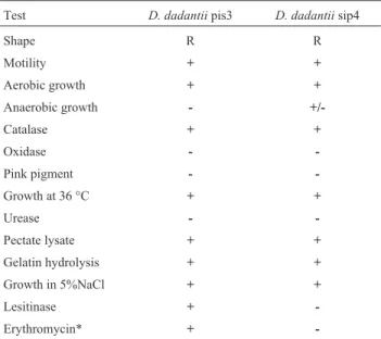
The bacteria isolated from infected potatoes (Solanum tuberosum) were identified by biochemical tests for theErwiniagroup (Table 1). In molecular analyses, the amplified 16S rRNA genes of two isolated bacterial strains
Table 1- The results of morphological and biochemical characteristics of
Dickeyaspp.
Test D. dadantiipis3 D. dadantiisip4
Shape R R
Motility + +
Aerobic growth + +
Anaerobic growth -
+/-Catalase + +
Oxidase -
-Pink pigment -
-Growth at 36 °C + +
Urease -
-Pectate lysate + +
Gelatin hydrolysis + +
Growth in 5%NaCl + +
Lesitinase +
-Erythromycin* +
were identified using the megablast program of NCBI (www.NCBI.nlm.nih.gov). Due to the high homology of the amplified ribosomal DNA sequences withD. dadantii, two bacterial strains isolated in this experiment were sub-mitted to GenBank asD. dadantiipis3 (GenBank accession number HQ423668) andD. dadantiisip4 (GenBank acces-sion number HQ423669).
Antibiotic sensitivity
The results of the antibiotic susceptibility test for iso-latedDickeyaare shown in Table 2. The isolated strain pis3 was sensitive to amikacin, siprofloxacillin, and oxytetra-cyclin. Strain pis3 was also sensitive to erythromycin, but strain sip4 was resistant (Table 2).
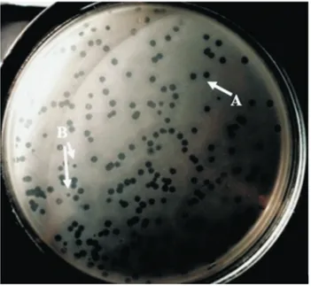
Isolation of bacteriophages and assessment of plaque morphology
The bacteriophage produced two various types of plaque morphology on theD. dadantiilawn culture: large plaques with 5 mm diameters and opaque halos in their margins and small plaques with 1-2 mm diameters and opaque halos (Figure 1).
Growth curve of isolatedDickeyawith bacteriophage The growth rates of the isolated bacteria with and without bacteriophage are shown in Figure 2. In the pres-ence of bacteriophage, bacteria growth was inhibited for 17 hours, after which, it was initiated and increased.
Transmission electron microscopy
Two types of bacteriophage were identified based on their morphology. Bacteriophages with regular hexagonal head of 90-100 nm length, 25-30 nm width, and 120 nm long tails were identified as theSiphoviridaefamily
(Figu-re 3a) and bacteriophages with hexagonal heads (approxi-mately 100 nm length and 50-60 nm width) and 90-100 nm long tails were identified as theMyoviridaefamily (Figu-re 3b).
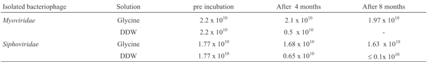
Bacteriophage enumeration in preservative buffer
The obtained phage suspensions had titers of up to 1.9 x 10¹⁰PFU/mL. The bacteriophage was enumerated both pre- and post-incubation at 4 °C for 8 months. All results indicated that glycine could preserve bacterio-phage particles for up to 8 months at 4 °C in good condi-tions with no apparent change in phage titer, while for phages preserved with chloroform-treated buffer, and double distilled water, bacteriophage titration decreased (Table 3).
Table 2- Antibiotic sensitivity test ofDickeya dadantiistrains pis3 and sip4.
Antibiotics g per disc Sensitivity
Strain pis3 Strain sip4
Oxytetracyclin 30 S I
Carbenicillin 100 R R
Tobramycine 10 R R
Erythromycin 15 S R
Nalidixic acid 30 I S
Vankomycin 30 R R
Cloxacillin 5 R R
Chloramphonicle 30 I R
Clindamycin 2 R R
Ceftisoxim 30 R R
Siprofloxacillin 5 S I
Amikacin 30 S S
R = Resistant; I = Intermediate; S = Sensitive. Figure 2- Growth curve ofDickeya dadantii.
Figure 1- Plaques formation in agar medium. The isolatedDickeya propa-gated on soft nutrient yeast agar medium, and the phage suspension spread on the medium. A) plaques up to 4-5 millimeter diameter (Myoviridae
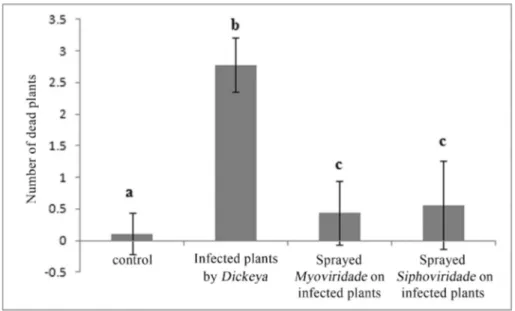
D. dadantiipathogenesis reduced by its bacteriophage
After 5 days of incubation,Geraniumspp. with the bacterial suspension in group 1 showed several symptoms of lesions, such as chlorosis and necrosis on leaves and stems. After 5 days, the leaves had rotted, and plants in-fected withD. dadantiisip4 were completely rotten. In the second group of plants that were treated with bacterial and bacteriophage suspensions, only 3 of the 27 tested plants treated withMyoviridae-related bacteriophage faded com-pletely after five days. Other treated geraniums in this group grew without any symptoms of disease. Data was an-alyzed with the Kruskal-Wallis test, and four groups within the groups were significantly different (p = 0.00). More-over, the differences between bacteria/phages (p = 0.00), bacteria/control (p = 0.00), bacteria/phage siph (p = 0.00) were statistically significant; between phage/control treat-ment, phage myo/control (p = 0.258), phage myo/phage siph (p = 0.863), and control/phage siph (p = 0.222), the data for phages and control treatment showed no statistical differences (Figure 4).
Discussion
D. dadantii causes soft rot disease in plants of the
Solanaceaefamily, including the potato (Czajkowskiet al., 2009; Laurilaet al., 2008; Ngadzeet al., 2010; Tsroret al., 2009). In the current study, two strains ofD. dadantiiwere isolated from infected potato tissue for the first time in Iran. Also isolated were two lytic bacteriophages of the
Myoviridae andSiphoviridae families from water of the Caspian Sea, located in northern Iran, against isolated strains ofD. dadantiifrom infected potatoes from central Iran (Isfahan province). Adriaenssenset al. identified two bacteriophages, LIMEstone 1 and LIMEstone 2, againstD. solani belonging to Myoviridae which had icosahedral heads of 91.4 nm and tail dimensions of 113.8x17 nm (Adriaenssenset al., 2012).
Schenabel and Jones (2001) reported five bacterio-phages ofE. amylovoraisolated from apple orchards that belonged toMyoviridae. Czajkowski et al. isolated nine bacteriophages infectingDickeyaspp. biovar 3 from soil of distinct geographical locations in Poland, which were mor-phologically related to the order Caudovirales, family
Myoviridae(Czajkowskiet al., 2013). Gillet al. isolated 42 bacteriophages againstE. amylovorafrom pear and apple
Figure 3- Electron micrograph ofDickeya dadantiiphages: (a)Myoviridaefamily, (b)Siphoviridaefamily.
Table 3- Titers of two isolated bacteriophages (PFU/mL) in glycine solution and pure water (DDW).
Isolated bacteriophage Solution pre incubation After 4 months After 8 months
Myoviridae Glycine 2.2 x 10¹⁰ 2.1 x 10¹⁰ 1.97 x 10¹⁰
DDW 2.2 x 10¹⁰ 0.5 x 10¹⁰
-Siphoviridae Glycine 1.77 x 10¹⁰ 1.68 x 10¹⁰ 1.63 x 10¹⁰
DDW 1.77 x 10¹⁰ 0.65 x 10¹⁰ £0.1x 10¹⁰
that were from the Myoviridae family and were smaller than ones isolated in the current study (Gillet al., 2003). Only Kishkoet al. seem to have reported the isolation of bacteriophages of Siphoviridae infecting E. carotovora
(Kishkoet al., 1983). This study isolated two members of
Myoviridae and Siphoviridae bacteriophage infecting D. dadantiistrains from rotting potato tubers for the first time in Iran.
In many studies the use of a few drops of chloroform in the buffer is suggested for the preservation of various bacteriophages at 4 °C (Clokie and Kropinski, 2009). The isolated phages that were identified as theSiphoviridaeand
Myoviridaefamilies were unstable in chloroform. There-fore, for bacteriophage preservation, the isolated phages in this study were found to be stable in the solutions made with 1% (w/v) glycine buffer rather than in buffer solutions with chloroform or in double distilled water. Glycine buffer could preserve phages with no significant change in the ti-ter for up to eight months at 4 °C.
Isolated lytic bacteriophages from the Caspian Sea samples were able to significantly reduce population of iso-latedDickeyastrains from central Iran in the culture media. In first phage titration of Caspian Sea water samples with-out phage enrichment, approximately 271 plaque units were observed on Dickeya strains. This massive plaque forming unit (PFU) of bacteriophage indicated that Caspian Sea water has great potential for phage therapy in this case. This observation indicates that the sampling regions (Guilan province and Kiashahr farmlands and rivers) are contaminated and infected withDickeyaspp. or other re-lated phytopathogens.
Erwiniacannot be controlled by specific bactericides (like other phytopathogenic bacteria) (Gnanamanickam,
2007). The incidence of antibiotic resistance may increase among bacteria due to the use of antibiotics in treating plant diseases, and this may be an origin of antibiotic-resistance genes in agroecosystems and the transfer of these genes to human pathogenic bacteria. Other compounds such as cop-per- and sulfur-containing formulations are not very effec-tive and cause environmental pollution (Gnanamanickam, 2007). In this study, the isolated bacteria were resistant to most antibiotics but were lysed completely by isolated bac-teriophages; so, these bacteriophages are capable of bio-controlling plant disease caused by specific bacterial hosts.
Data from this study showed thatD. dadantiistrains caused necrosis, soft rot, and black age disease in Gera-niumspp. in a few days. However, the Dickeya-infected plants (Geranium spp.) which were treated with specific bacteriophages showed no symptoms of disease in compar-ison with infected non-treated plants. Regarding the resis-tance of the isolated bacteria to most broad spectrum antibi-otics, the use of the bacteriophages for controlling potato disease could be more effective in eliminating the targeted bacterial pathogens. Moreover, phages could be safe substi-tutes for toxic chemical agents such as antibiotics or other bactericidal agents in the control of plant diseases. How-ever, more research with plants is needed to extend the find-ings of the current study regarding the application of bio-control agents such as bacteriophage in agroecosystems.
Acknowledgments
The authors would like to express thanks to the Uni-versity of Isfahan for financial support of this work.
Figure 4- Cranesbill (Geraniumspp.) plants infected by 5 x 10⁸cells/mL ofDickeya dadantiisip4 and treated by 3 x 10⁹PFU/mL ofSiphoviridaeand
References
Adriaenssens EM, Van Vaerenbergh J, Vandenheuvel Det al.
(2012) T4-related bacteriophage LIMEstone isolates for the control of soft rot on potato caused byDickeya solani. PloS one 7:e33227.
Brenner DJ, Krieg NR, Staley JTet al.(2005) Bergey’s Manual of Systematic Bacteriology. 2nd ed. vol. 2 (The Proteo-bacteria), part B (The GammaproteoProteo-bacteria), Springer, New York, pp. 721-730.
Clokie M, Kropinski A (2009) Bacteriophages: Methods and Pro-tocols: Isolation, Characterization, and Interactions. Humana Pr Inc pp 69-76.
CLSI (2010) M100-S20. Performance Standards for Anti-microbial Susceptibility Testing: 20th Informational supple-ment. Clinical and Laboratory Standards Institute Wayne. Czajkowski R, Grabe GJ, van der Wolf JM (2009) Distribution of
Dickeya spp. and Pectobacterium carotovorum subsp.
carotovorumin naturally infected seed potatoes. Eur J Plant Pathol 125:263-275.
Czajkowski R, Ozymko Z, Lojkowska E (2013) Isolation and characterization of novel soilborne lytic bacteriophages in-fecting Dickeya spp. biovar 3 (`D. solani’). Plant Pathol 63:758-772.
Dickey RS (1979)Erwinia chrysanthemi: a comparative study of phenotypic properties of strains from several hosts and other
Erwiniaspecies. Phytopathology 69:324-329.
Gill J, Svircev A, Smith Ret al.(2003) Bacteriophages ofErwinia amylovora. Appl Environ Microbiol 69:2133.
Gnanamanickam SS (2007) Plant-associated bacteria. Springer Verlag, Dordrecht, The Netherlands, pp 423-505.
Górski A, Weber-Dabrowska B (2005) The potential role of en-dogenous bacteriophages in controlling invading pathogens. Cell Mol Life Sci 62:511-519.
Hauben L, Moore E, Vauterin Let al.(1998) Phylogenetic posi-tion of phytopathogens within theEnterobacteriaceae. Syst Appl Microbiol 21:384-397.
Jones J, Jackson L, Balogh Bet al.(2007) Bacteriophages for plant disease control. Phytopathology 45:245-262. Kishko Y, Ruban V, Tovkach Fet al.(1983) Structure ofErwinia
carotovoratemperate bacteriophage 59 and its DNA. J Virol 46:1018.
Laurila J, Ahola V, Lehtinen Aet al.(2008) Characterization of
Dickeyastrains isolatedfrom potato and river water samples in Finland. Eur J Plant Pathol 122:213-225.
Leong DU, Greisen KS (1993) PCR detection of bacteria found in cerebrospinal fluid. In: Persing, D.H. (ed). Diagnostic Mo-lecular Microbiology: Principles and Applications. Ameri-can Society for Microbiology, Washington, DC, pp 300-306.
Letal J (1977) Efficacy of disinfestants against potato ring rot and blackleg bacteria. Am J Potato Res 54:405-409.
Lund B, Lyon G (1975) Detection of inhibitions of Erwinia carotovoraand E. herbicola on thin-layer chromatogram. J Chromatogr A 110:193-196.
Mazzocco A, Waddell TE, Lingohr Eet al.(2009) Enumeration of bacteriophages by the direct plating plaque assay. Mol Biol 501:77-80.
McFadden LA (1958) Bacterial blight of Chrysanthemum. In: Proc. Florida State Hort. Soc 71:419-425.
Ngadze E, Coutinho T, van der Waals J (2010) First Report of Soft Rot of Potatoes Caused byDickeyadadantiiin Zimbabwe. Plant Dis 94:1263-1263.
Ravensdale M, Blom T, Gracia-Garza Jet al.(2007) Bacterio-phages and the control of Erwinia carotovora subsp.
carotovora. Can J Plant Pathol 29:121-130.
Ritchie D, Klos E (1977) Isolation ofErwinia amylovora bac-teriophage from aerial parts of apple trees. Phytopathology 67:101-104.
Samson R, Legendre J, Christen R et al. (2005) Transfer of
Pectobacterium chrysanthemi (Burkholder et al., 1953) Brenneret al., 1973 and Brenneria paradisiaca to the genus
Dickeyagen. nov. asDickeya chrysanthemicomb. nov. and Dickeya paradisiaca comb. nov. and delineation of four novel species, Dickeya dadantii sp. nov., Dickeya dianthicolasp. nov.,Dickeya dieffenbachiaesp. nov. and
Dickeya zeaesp. nov. Int J Syst Evol Microbiol 55:1415-1427.
Schnabel E, Jones A (2001) Isolation and characterization of five
Erwinia amylovorabacteriophages and assessment of phage resistance in strains ofErwinia amylovora. Appl Environ Microbiol 67:59-64.
Smith HW, Huggins MB, Shaw KM (1987) The control of experi-mentalEscherichia colidiarrhoea in calves by means of bac-teriophages. J Gen Microbiol 133:1111-1126.
Tsror L, Erlich O, Lebiush Set al.(2009) Assessment of recent outbreaks ofDickeyasp.(syn.Erwinia chrysanthemi) slow wilt in potato crops in Israel. Eur J Plant Pathol 123:311-320.
Verthé K, Possemiers S, Boon Net al.(2004) Stability and activ-ity of anEnterobacter aerogenes-specific bacteriophage un-der simulated gastro-intestinal conditions. Appl Microbiol Biotechnol 65:465-472.
Wyatt G, Lund B (1981) The effect of antibacterial products on bacterial soft rot of potatoes. Potato Res 24:315-329.
Associate Editor: Fernando Dini Andreote