Essentiality Assessment of Cysteinyl and
Lysyl-tRNA Synthetases of
Mycobacterium
smegmatis
Sudha Ravishankar1¤a
*, Anisha Ambady1¤b, Rayapadi G. Swetha2¤a, Anand Anbarasu2¤a, Sudha Ramaiah2¤a, Vasan K. Sambandamurthy1¤c
1AstraZeneca India Pvt Ltd, Bellary Road, Hebbal, Bengaluru, 560024, India,2School of Biosciences & Technology, VIT University, Vellore, 632014, India
¤a Current address: School of Biosciences & Technology, VIT University, Vellore, Tamil Nadu, India ¤b Current address: Gangagen Biotechnologies Pvt. Ltd., Yeshwantpur, Bengaluru, Karnataka, India ¤c Current address: Mazumdar Shaw Centre for Translational Research (MSCTR), Mazumdar Shaw Medical Centre, Bengaluru, Karnataka, India
*mailsuravi01@gmail.com
Abstract
Discovery of mupirocin, an antibiotic that targets isoleucyl-tRNA synthetase, established aminoacyl-tRNA synthetase as an attractive target for the discovery of novel antibacterial agents. Despite a high degree of similarity between the bacterial and human aminoacyl-tRNA synthetases, the selectivity observed with mupirocin triggered the possibility of target-ing other aminoacyl-tRNA synthetases as potential drug targets. These enzymes catalyse the condensation of a specific amino acid to its cognate tRNA in an energy-dependent reac-tion. Therefore, each organism is expected to encode at least twenty aminoacyl-tRNA syn-thetases, one for each amino acid. However, a bioinformatics search for genes encoding aminoacyl-tRNA synthetases fromMycobacterium smegmatisreturned multiple genes for glutamyl (GluRS), cysteinyl (CysRS), prolyl (ProRS) and lysyl (LysRS) tRNA synthetases. The pathogenic mycobacteria, namely,Mycobacterium tuberculosisandMycobacterium leprae, were also found to possess two genes each for CysRS and LysRS. A similar search indicated the presence of additional genes for LysRS in gram negative bacteria as well. Herein, we describe sequence and structural analysis of the additional aminoacyl-tRNA synthetase genes found inM.smegmatis. Characterization of conditional expression strains of Cysteinyl and Lysyl-tRNA synthetases generated inM.smegmatisrevealed that the canonical aminoacyl-tRNA synthetase are essential, while the additional ones are not essential for the growth ofM.smegmatis.
Introduction
In bacterial cells, like transcription and replication, translation is one of the key processes which leads to the synthesis of every protein required for the cell to function irrespective of their final localization [1]. Translation is a multi-step process involving several components
OPEN ACCESS
Citation:Ravishankar S, Ambady A, Swetha RG, Anbarasu A, Ramaiah S, Sambandamurthy VK (2016) Essentiality Assessment of Cysteinyl and Lysyl-tRNA Synthetases ofMycobacterium smegmatis. PLoS ONE 11(1): e0147188. doi:10.1371/journal.pone.0147188
Editor:Giovanni Maga, Institute of Molecular Genetics IMG-CNR, ITALY
Received:October 29, 2015
Accepted:December 30, 2015
Published:January 21, 2016
Copyright:© 2016 Ravishankar et al. This is an open access article distributed under the terms of the
Creative Commons Attribution License, which permits unrestricted use, distribution, and reproduction in any medium, provided the original author and source are credited.
Data Availability Statement:All relevant data are within the paper and its Supporting Information files.
like tRNAs, amino acids, mRNA, ribosomes. A large number of antibiotics discovered till date are inhibitors of translation, thereby establishing the importance of this process for bacterial cell survival [2]. Each of these antibiotic seems to affect a unique step in the protein synthesis cascade [3]. The bacterial growth inhibition observed with mupirocin is brought about by the inhibition of isoleucyl-tRNA synthetase (IleRS) which in turn leads to the inhibition of transla-tion process [4]. The clinical success of mupirocin as a topical antibiotic [4] has opened up attractive opportunities to target aminoacyl-tRNA synthetases (aaRS) for novel antibacterial agents. Several attempts that were made, including a fewin silicosearch and design [5,6] have led to the discovery of inhibitors for a number of aaRS such as PheRS [7,8], MetRS [9,10], ProRS [11], TyrRS [12], LeuRS [5,13], ThrRS [14]. Ochsneret al., have presented a compre-hensive review of all the known aaRS inhibitors [15]. Additionally, 4-thiozolidinone deriva-tives, Microcin C and tobramycin are known to inhibit AspRS [16]. Although, there is a good degree of similarity between eukaryotic and prokaryotic aaRS, identification of bacteria selec-tive inhibitors has provided ample evidence to discover novel, selecselec-tive inhibitors of this enzyme class.
During translation, each amino acid is carried by a specific tRNA to the translation site. These tRNAs get charged with respective amino acids by the action of aminoacyl-tRNA syn-thetases or aminoacyl-tRNA ligases in a two-step process [17]. Therefore, synthesis of aminoa-cyl-tRNA (aa-tRNA) is a critical step in translation and hence aaRS are considered essential for bacterial survival. The mechanism of this class of enzymes suggest that each cell should possess at least twenty aaRS as there are 20 different natural amino acids [17]. However, it has been observed that this number is either more than 20 or less than 20 in a few organisms [18]. The aaRS are generally divided into two classes, I and II, based on their structural features. In all of the aaRS studied, the tRNA binding region has a conservedα-helical structure. The class I enzymes are generally monomeric, share a characteristic Rossman-fold catalytic domain and two conserved motifs, HIGH and KMSKS. On the other hand, the class II aaRS are dimeric or multimeric, contain at least three conserved regions and share an anti-parallelβ-sheet fold flanked on either side byα-helices. Another catalytic difference between these two classes of enzymes is that, the class I enzymes couple the aminoacyl group to the 2'-hydroxyl of the last nucleotide of tRNA, while, the class II enzymes couple aminoacyl group to the 3'-hydroxyl of the last nucleotide of tRNA [19,20].
In general, bacteria are believed to possess at least one aaRS for each amino acid in order to supply the translation machinery with the respective aa-tRNA. Interestingly, some organisms seem to carry more than one gene coding for the same aaRS. For example,Escherichia coli
encodes two lysyl-tRNA synthetase genes, one is expressed constitutively, while the other is inducible [21].E.colialso codes for two glutamyl-tRNA synthetases, where one uses cognate tRNAGlu[22], while the other one uses tRNAAspas substrate to transfer the activated glutamyl
group [23]. A query in KEGG genes database for genes coding for‘tRNA synthetases’ofM.
smegmatisreturned a list of 24 genes with multiple genes coding for glutamyl (MSMEG_2383, MSMEG_6306), prolyl (MSMEG_2621, MSMEG_5671), cysteinyl (MSMEG_4189,
MSMEG_6074) and lysyl (MSMEG_3796, MSMEG_6094) tRNA synthetases [24]. A similar search againstM.tuberculosis and M.lepraeidentified 2 genes each for lysyl and cysteinyl-tRNA synthetases. However, these organisms were found to have only one gene for glutamyl and prolyl-tRNA synthetase. Multiple sequence alignment of theM.smegmatiscysteinyl and lysyl-tRNA synthetases with those ofM.tuberculosisandM.lepraerevealed that the respective orthologs have a high degree of sequence similarity. The essentiality of lysyl and cysteinyl-tRNA synthetases ofM.smegmatiswere evaluated by employing conditional expression strains generated inM.smegmatisusing the isopropylthiogalactoside (IPTG) inducible conditional expression system [25].
Materials and Methods
Bacterial strains, media, chemicals and reagents
Bacterial strains used in this study are listed inTable 1. Glycerol, Tween 80, kanamycin and IPTG were purchased from SIGMA, USA. Restriction enzymes, Taq DNA polymerase, DNA ladders were purchased from New England Biolabs, USA. Hygromycin B was obtained from Roche, Fusion polymerase from Finnzymes. Pristinamycin was obtained from Sanofi Aventis and the P1component of pristinamycin was purified in-house as described earlier [26]. Luria
Bertani (LB) broth and LB agar were used to growE.coli. Middlebrook 7H9 (DIFCO) supple-mented with 0.2% Glycerol (v/v), 0.05% Tween 80 (w/v) and albumin-dextrose was used for growing broth cultures of mycobacteria and Middlebrook 7H11 (DIFCO) for measurement of colony forming units (CFU). Bacterial cultures were supplemented with antibiotics, IPTG and P1as required.
Generation of conditional expression plasmids
All plasmids used in this study are listed inTable 1. Conditional expression plasmids were gen-erated by cloning about 700 bps of DNA fragment of each target gene amplified from its 5’end. The primers listed in Table A inS1 Filewere used for generating required amplicons. Fusion polymerase was used to generate amplicons in a 25-cycle polymerase chain reaction with cycling conditions of denaturing at 98°C, annealing at a temperature dictated by the melting temperature of each primer pair (Table A inS1 File) and extension at 72°C. All the recombi-nant plasmids constructed on pAZI9452 [25] background (pAZI9501, pAZI9504, pAZI9506 Table 1. List of plasmids and strains used in this study.
Plasmids/ Strains Reference Details
pAZI9452$ 25* Conditional expression vector with an IPTG-inducible promoter
pAZI9479$ 26*, 27* Conditional expression vector with a pristinamycin-inducible promoter system
pMV261 28* AnE.coli–mycobacterial shuttle vector for protein expression in mycobacteria driven by hsp60 promoter.
pAZI9501 This study Conditional expression plasmid ofM.smegmatis leuSin pAZI9452 pAZI9502 This study Conditional expression plasmid ofM.smegmatis leuSin pAZI9479 pAZI9503 This study Conditional expression plasmid ofM.tuberculosis leuSin pAZI9479 pAZI9504 This study Conditional expression plasmid of MSMEG_6074 in pAZI9452 pAZI9505 This study Conditional expression plasmid of MSMEG_6073 in pMV261 pAZI9506 This study Conditional expression plasmid of MSMEG_3796 in pAZI9452 pAZI9507 This study Conditional expression plasmid of MSMEG_6094 in pAZI9452
SleuS/KD-I This study Conditional expression strain ofM.smegmatis leuSwith an IPTG-inducible system
SleuS/KD-P This study Conditional expression strain ofM.smegmatis leuSwith pristinamycin-inducible system TleuS/KD-P This study Conditional expression strain ofM.tuberculosis leuSwith a pristinamycin-inducible system ScysS/KD-I This study Conditional expression strain ofM.smegmatis cysS(MSMEG_6074) with an IPTG-inducible system ScysS/KD-I-compl This study M.smegmatiscysS conditional expression strain complemented with pBAN6073
S3796/KD-I This study Conditional expression strain ofM.smegmatisMSMEG_3796 with an IPTG-inducible system S6094/KD-I This study Conditional expression strain ofM.smegmatisMSMEG_6094 with an IPTG-inducible system
E.coliDH5α Lab. stock endA1,hsdR17,supE44,recA1,relA1, (lacZYA- argF)
M.smegmatismc2155 Lab. stock Non-pathogenic mycobacterial strain. M.tuberculosisH37Rv 27294 Lab. stock Virulent strain ofM.tuberculosisH37Rv
*References $Fig A inS1 File
and pAZI9507) were generated by cloning the respective amplicons at NdeI and HindIII sites of pAZI9452. Two of the recombinant plasmids, pAZI9502 and pAZI9503 were generated by cloning the respective amplicons at NcoI and MscI sites of pAZI9479 [26,27].
InM.smegmatis,cysSis present in an operon where MSMEG_6073 is the last gene after
cysS. MSMEG_6073 was PCR amplified using primers S6073complF and S6073complR (Table A inS1 File) and cloned into pMV261 [28] vector at BamHI and HindIII sites to gener-ate pAZI9505. The recombinant plasmids were screened by restriction enzyme analysis and sequence of the cloned fragments were confirmed by DNA sequencing.
Generation of conditional expression strains
Conditional expression plasmids were electroporated intoM.smegmatismc2155 orM.
tubercu-losisH37Rv following a standard protocol [29]. The transformation mix was plated onto 7H9 agar plates containing 50μg/ml hygromycin and supplemented with either 500μM IPTG or
300 ng/ml of P1. The colonies were screened for their dependence on inducer for growth by
replica plating on 7H11 plates with and without the inducer. The genotype of these recombinants (presence of truncated version of the gene of interest downstream of the native promoter and the full length gene downstream of an inducible promoter—depicted in Fig B inS1 File) were con-firmed by PCR as described earlier [25,26] using the primers listed in Table A inS1 File. The colonies that were positive by both screens were designated as SleuS/KD-I, ScysS/KD-I, S3796/KD-I and S6094/KD-I for the conditional expression strains ofM.smegmatis leuS,cysS,
MSMEG_3796,MSMEG_6094, respectively, on a pAZI9452 vector backbone. Similarly, condi-tional expression strains generated with pAZI9479 ofM.tuberculosis leuSandM.smegmatis leuS
were designated as TleuS/KD-P and SleuS/KD-P, respectively. Subsequently, ScysS/KD-I was electroporated with pAZI9505 to generate a complemented strain ScysS/KD-I/C.
Analysis of inducer dependency of conditional expression strains
Each of the PCR confirmed conditional expression strain was grown in 2 ml of 7H9 broth sup-plemented with either 500μM IPTG or 300 ng/ml P1as appropriate. When the cultures
reached mid-logarithmic phase, they were centrifuged and the harvested cells were washed with 7H9 broth followed by resuspension in fresh broth to be used as inoculum. In order to determine the minimum inducer concentration required for growth of each of the conditional expression strain, several dilutions of the culture inoculum were either plated or spotted on 7H11 plates containing different concentration of inducer i.e., 0–500μM IPTG or 0–300 ng/ml
of P1.M.smegmatismc2155 andM.tuberculosisH37Rv were also processed in a similar way
and plated on 7H11 plates to compare the colony morphology and growth rate. Minimum inducer required for the growth of each conditional expression strain was identified as the con-centration at which the conditional expression strain grew as well as the wild-type strain. Sub-sequently, the conditional expression strains were grown at the identified inducer
concentration until they reached mid-log phase, the cells were washed and used to prepare inoculum for further experiments.
Protein sequence resource
Transcription unit
M.smegmatismc2155 database from BioCyc genome pathway database collection (http:// biocyc.org/MSME246196/organism-summary?object=MSME246196) was referred for analys-ing the transcription unit arrangement of each gene.
Pairwise sequence alignment
The required protein sequences were retrieved from KEGG genes database. SIM protein sequence alignment tool in the ExPASy Bioinformatics Resource Portal (http://web.expasy.org/ sim/) was used to analyse the percent identity between the selected protein sequences.
Protein database search
NCBI BLASTP program (http://blast.ncbi.nlm.nih.gov/Blast.cgi?PAGE=Proteins) was used to search the non-redundant protein sequence database for homologous protein sequences and/ or the probable protein families the query sequence belongs to.
Transmembrane segment prediction analysis
TMHMM v 2.0 (http://www.cbs.dtu.dk/services/TMHMM/) at the Centre for Biological Sequence Analysis (CBS) was used to predict the transmembrane segments present in MSMEG_3796, MSMEG_6094 andS.aureusMprF.
Protein structure analysis
3-Dimensional model structure generation. The protein sequence of MSMEG_5671 was
submitted to an online server, I-TASSER (http://zhanglab.ccmb.med.umich.edu/I-TASSER/) which generated 5 model structures. A model with highest confidence score was selected for further structural analysis. The 3-dimensional (3-D) structure ofE.coliYbaK was downloaded from PDB (PDB-ID: 2DXA).
Assessment of modelled MSMEG_5671 structure. The structure verification of MSMEG_5671 was performed using the UCLA Structure Analysis and Verification Server with PROCHECK, ERRAT and VERIFY-3D programs (http://services.mbi.ucla.edu/SAVES/).
Pairwise structure comparison. A pairwise structure comparison was performed between
the modelled structure ofM.smegmatisMSMEG_5671 andE.coliYbaK structure using Dali-Lite server (http://www.ebi.ac.uk/Tools/structure/dalilite/) which uses sum-of-pairs method by comparing the intramolecular distance matrices and produces a measure of similarity.
Results and Discussion
Multiple aaRS of
M.
smegmatis
Aminoacyl-tRNA synthetases are one of the key players in the translation process [17]. They catalyse the coupling of an aminoacyl group to its cognate tRNA in a two-step process (Fig C inS1 File), in the first step an amino acid gets activated which is transferred to its cognate tRNA in the second step. Thus, all the cognate aaRS would possess two major domains, a cata-lytic domain where the aminoacyl-tRNA is synthesized and a tRNA anticodon recognition domain. Some of the aaRS also have a built-in editing domain needed to remove the amino acids from an erroneously charged tRNA [30].
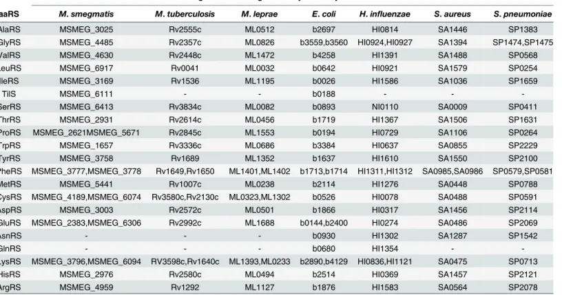
Among the 24 genes found to code for the various aaRS, PheRS is encoded by 2 genes, one cod-ing forαand the other for theβsubunit of this enzyme. Four out of the other five additional genes were found to code for an additional glutamyl, cysteinyl, prolyl and lysyl-tRNA synthe-tases, respectively, while the fifth additional genetilSis a tRNAIlelysidine synthetase (Table 2).
A similar search in KEGG for the pathogenic mycobacteria,M.tuberculosisandM.leprae
returned a list with 20 and 21 aaRS entries, respectively. The list indicated the presence of an additional CysRS and LysRS but not GluRS and ProRS (Table 2). Results from search per-formed against two Gram positive bacteria (S.aureusandS.pneumoniae) and two Gram nega-tive bacteria (E.coliandH.influenzae) showed the presence of additional genes for lysyl-tRNA synthetase in Gram negative bacteria only (Table 2).
Sequence analysis of tRNA
Ilelysidine synthetase and glutamyl-tRNA
synthetase
In the absence of genetic and biochemical characterization for these aaRSs fromM.smegmatis, pairwise sequence alignment was performed to understand the degree of homology with their orthologs fromM.tuberculosisandE.coli.
MSMEG_6111, annotated to code for tRNAIleLysidine synthetase was found to be 34% identical to theE.coliTilS. A search for homologs of MSMEG_6111 identified the presence of orthologs (annotated asmesJ) in all the other organisms analysed in this study. Although, experimental evidence is required to establish the physiological role and essentiality of MSMEG_6111 inM.smegmatis, it’s orthologs fromE.coliandM.tuberculosiswere Table 2. Genes encoding aminoacyl-tRNA synthetases*.
gene/s encoding aminoacyl-tRNA synthetases in
aaRS M.smegmatis M.tuberculosis M.leprae E.coli H.influenzae S.aureus S.pneumoniae
AlaRS MSMEG_3025 Rv2555c ML0512 b2697 HI0814 SA1446 SP1383
GlyRS MSMEG_4485 Rv2357c ML0826 b3559,b3560 HI0924,HI0927 SA1394 SP1474,SP1475
ValRS MSMEG_4630 Rv2448c ML1472 b4258 HI1391 SA1488 SP0568
LeuRS MSMEG_6917 Rv0041 ML0032 b0642 HI0921 SA1579 SP0254
IleRS MSMEG_3169 Rv1536 ML1195 b0026 HI1586 SA1036 SP1659
TilS MSMEG_6111 - - b0188 - -
-SerRS MSMEG_6413 Rv3834c ML0082 b0893 NI0110 SA0009 SP0411
ThrRS MSMEG_2931 Rv2614c ML0456 b1719 HI1367 SA1506 SP1631
ProRS MSMEG_2621MSMEG_5671 Rv2845c ML1553 b0194 HI0729 SA1106 SP0264
TrpRS MSMEG_1657 Rv3336c ML0686 b3384 HI0637 SA0855 SP2229
TyrRS MSMEG_3758 Rv1689 ML1352 b1637 HI1610 SA1550 SP2100
PheRS MSMEG_3777,MSMEG_3778 Rv1649,Rv1650 ML1401,ML1402 b1713,b1714 HI1311,HI1312 SA0985,SA0986 SP0579,SP0581
MetRS MSMEG_5441 Rv1007c ML0238 b2114 HI1276 SA0448 SP0788
CysRS MSMEG_4189,MSMEG_6074 Rv3580c,Rv2130c ML0323,ML1302 b0526 HI0078 SA0488 SP0591
AspRS MSMEG_3003 Rv2572c ML0501 b1866 HI0317 SA1456 SP2114
GluRS MSMEG_2383,MSMEG_6306 Rv2992c ML1688 b0144,b2400 HI0274 SA0486 SP2069
AsnRS - - - b0930 HI1302 SA1287 SP1542
GlnRS - - - b0680 HI1354 -
-LysRS MSMEG_3796,MSMEG_6094 RV3598c,Rv1640c ML1393,ML0233 b2890,b4129 HI0836,HI1121 SA0475 SP0713
HisRS MSMEG_2976 Rv2580c ML0494 b2514 HI0369 SA1457 SP2121
ArgRS MSMEG_4959 Rv1292 ML1127 b1876 HI1583 SA0564 SP2078
*KEGG genes database
demonstrated to be essential [31,32]. Biochemical characterization of theE.coliortholog has led to the understanding that Tils is needed to synthesize tRNAIleLysidine in a reaction where the CAU anticodon of tRNAIlegets converted to LAU when the enzyme transfers a lysine
moi-ety on to the cytidine residue present on the anticodon. This activity is required to maintain translation fidelity because it prevents mis-charging of tRNAIleby MetRS [33,34].
M.smegmatiswas found to possess twogltXgenes, one coding for glutamyl-tRNA synthe-tase (GluRS) and another one for glutamyl-Q-tRNAAspsynthetase (Glu-Q-RS), similar to
those inE.coli. Pairwise sequence alignment ofM.smegmatisGluRS and Glu-Q-RS indicated only about 39% identity between them in their N-terminal 250 amino acids, a result similar to the one observed betweenE.coliGluRS and Glu-Q-RS. However, Only GluRS orthologs could be found in other mycobacterial species and the gram positive and gram negative bacteria ana-lysed in this study. InE.coli, both GluRS and Glu-Q-RS were shown to activate glutamate. While the activated glutamate is transferred to tRNAGluby GluRS to synthesize Glu-tRNAGlu,
Glu-Q-RS transfers it to tRNAAspto synthesize Glu-tRNAAsp[35,36]. Glu-Q-RS was consid-ered non-essential as its product Glu-tRNAAspcould not bind EF-Tu to participate in the
translation process. This was substantiated later by the experimentally derived essential and dispensable nature of GluRS and Glu-Q-RS respectively inE.coli[37,38]. Although, the situa-tion could be very similar inM.smegmatis, biochemical and genetic evidence is required to establish the role and essentiality ofM.smegmatisGluRS and Glu-Q-RS. However, the evolu-tionary significance of the conservation of the non-essential Glu-Q-RS across different bacte-rial genera has remained unclear.
Sequence and structural analysis of prolyl-tRNA synthetase
Both MSMEG_2621 and MSMEG_5671 have been annotated as Prolyl-tRNA synthetase in the KEGG genes database. MSMEG_2621, coding for a 585 amino acid prolyl-tRNA synthetase has been indicated to possess motifs for a catalytic, an anti-codon binding and a tRNA editing activity, similar to the ones found in the canonical prolyl-tRNA synthetase ofE.coliandM.
tuberculosis. On the other hand, MSMEG_5671, which codes for a 159 amino acid protein has been suggested to contain a tRNA editing motif. BlastP analysis of MSMEG_5671 protein sequence indicated that it belongs to YbaK-like superfamily of proteins with homologs present in several bacterial genera. YbaK, anE.coliprotein annotated as Cys-tRNA (Pro)/Cys-tRNA (Cys) deacylase has been shown to deacylate the mischarged Cys-tRNAProto keep tRNAPro available for acylation with proline [39]. In a similar reaction, INS domain present within the canonical ProRS has been shown to deacylate mischarged Ala-tRNAProto make the tRNAPro available for acylation with proline [40,41]. Thus, the two editing motifs, one cis-acting INS domain and the other trans-acting YbaK like protein deacylate the wrongly charged tRNAPro, help the cells to maintain translation fidelity [42]. Surprisingly, no significant homology to any part of MSMEG_2621 was observed when MSMEG_5671 was aligned with MSMEG_2621 in spite of both having tRNA editing motifs. Even the sequence identity between MSMEG_5671 andE.coliYbaK was only 27% over a stretch of 85 amino acids. Therefore, we decided to gen-erate a 3-D model structure of MSMEG_5671 and compare it with theE.coliYbaK structure available in PDB.
MSMEG_5671 structure prediction analysis
one with highest Z-score value of 3.5 (PDB ID: 2CX5, chain A) was taken as the best template based on which five model structures were produced with a reported TM score of 0.85±0.08. A model with a confidence score of 1.03 was selected as the best model for MSMEG_5671. The Ramachandran plot obtained using PROCHECK program [47] showed that 86.6% and 13.4% of residues in the modelled structure of MSMEG_5671 were in favored and allowed regions, respectively. The ERRAT [48] score of 94.702 was found to fall within the range of a high qual-ity model suggesting that the backbone conformation and non-bonded interactions in the model were reasonable. VERIFY-3D [49] results showed that 91.82% of the residues in the modeled structure have an average score of>0.2 (Fig D inS1 File), thereby, confirming the
model to be of good quality. Thus, evaluation of the MSMEG_5671 modelled structure through PROCHECK, ERRAT and VERIFY-3D programs established that the model possessed high geometric quality and a good energy profile.
MSMEG_5671 and
E.
coli
YbaK have comparable 3-D structure
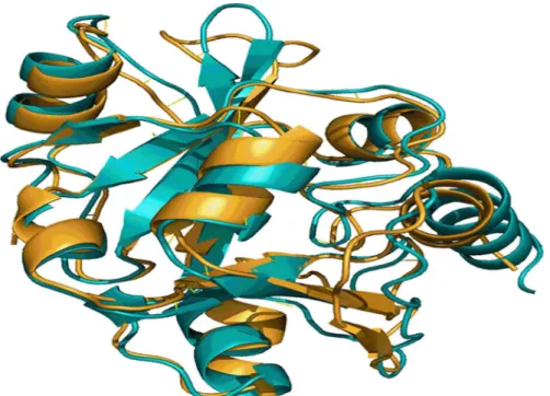
In the absence of biochemical evidence, meaningful alignments generated through 3-D structure comparison methods enable understanding proteins and their functions. A pairwise structure comparison performed with MSMEG_5671 andE.coliYbaK using DALI server showed that the two structures align well with a Dali-Z score of 21.7 [50]. Superimposition of the two structures as depicted inFig 1demonstrated that they possess similar folds indicating that they could be performing same function. Thus, the generation of a 3-D model structure for MSMEG_5671 and comparing it with the structure ofE.coliYbaK have clearly indicated that MSMEG_5671 is the most likely candidate performing Cys-tRNAProdeacylase activity inM.smegmatis.
The search in KEGG genes database for aminoacyl-tRNA synthetase genes did not yield additional prolyl-tRNA synthetase genes inM.tuberculosisandM.leprae. However, a careful examination of the list of orthologs of MSMEG_5671 in KEGG indicated presence of its Fig 1. A snapshot of structural alignment of MSMEG_5671 andE.coliYbaK.Structure ofE.coliYbaK (gold) and the modeled structure of MSMEG_5671 (cyan) were superimposed using DALI server.
homologs in these pathogenic organisms as well. Two proteins, RVBD_3224B (72 amino acids) ofM.tuberculosisH37Rv and ML0799 (135 amino acids) ofM.lepraehave been cur-rently annotated as hypothetical proteins and about 40% identical over a stretch of 50 residues and 30% identical over a stretch of 84 residues, respectively with MSMEG_5671. The genera-tion of experimental data is required to prove that these proteins perform Cys-tRNAPro
deacy-lation despite being shorter than YbaK and MSMEG_5671.
Sequence analysis of cysteinyl-tRNA synthetase
Among the several organisms analysed in this study for cysteinyl-tRNA synthetases, the two Gram positive and two Gram negative bacteria were found to have a single gene each encoding this enzyme. However, all the three mycobacterial species had two genes each (Table 2). Unlike GluRS and ProRS, the two CysRS ofM.smegmatiswere of similar size (MSMEG_4189–412 and MSMEG_6074–477 amino acids). A pairwise sequence alignment of these two proteins revealed only about 37% identity between them in their N-terminal 300 amino acids stretch indicating existence of significant sequence differences in their C-terminal portion. Chemical and transposon mutagenesis studies by Rawatet alhad enabled reannotation of MSMEG_4189 as MshC [51]. As a penultimate step in the mycothiol biosynthesis pathway, this enzyme is known to catalyse the activation of cysteine (like a canonical CysRS) which is then transferred to 1D-myo-inosityl-2-amino-2-deoxy-alpha-D-glucopyranoside (GlcN-Ins) in an ATP-depen-dent reaction unlike the canonical CysRS which transfers activated Cysteine to tRNACys[52,
53]. Although, these studies indicated non-essentiality of MshC to the survival ofM. smegma-tis, the inability to knockout its homolog inM.tuberculosisErdman strain [54] suggested dif-ferences in the essentiality of the same enzyme function in two related species. An independent mutagenesis study had suggested canonical CysRS ofM.tuberculosisto be essential [32], thereby, suggesting that MshC and CysRS could not complement each other’s function. We decided to investigate the essentiality of CysRS by employing a conditional expression strain to see if the situation is similar inM.smegmatis.
Sequence and transmembrane segment analysis of lysyl-tRNA
synthetase
The two Gram negative and the three mycobacterial species analysed in this study seem to code for two LysRS proteins each, while the two Gram positive bacteria code for a single LysRS gene each (Table 2). Among the two LysRS ofE.coli, LysS is constitutively expressed, while LysU expression was found to be inducible by heat and other stress factors [21]. A third puta-tive Lysyl-tRNA synthetase encoded byepmAwas shown to lysylate the elongation factor P (EF-P) and hence the annotation, elongation factor P Lys34-lysyltransferase [55]. The protein sequence alignment of all three LysRS ofE.coliindicated thatepmAgene product has about 30% identity with the other two LysRS which among themselves have about 90% identity. However, Yanagisawaet al., were able to demonstrate its structural features resembled those of class II aaRS [55]. At the primary structure level, thelysSandgenXproducts ofH.influenzae
were found to be about 70% and 62% identical to that oflysSandepmAofE.colirespectively. In addition to Lys-tRNALyssynthesis activity, in many bacteria LysRS was found to be the most efficient among the various aaRS to synthesize diadenosine tetraphosphate (Ap4A) and its
Genes encoding LysRS from three mycobacterial species,H.influenzaeandE.coliare listed inTable 3. The lengths of LysRS protein indicated the presence of two types of lysyl-tRNA syn-thetases in mycobacteria, one with about 500 amino acids (group A) and the other with about 1000 amino acids (group B).
A high degree of similarity with about 80% identity was observed within the group A and within the group B mycobacterial LysRS when their amino acid sequences were aligned (Table 3). However, alignment of group A LysRS (shorter) sequence with that of group B LysRS (longer) revealed that the C-terminal half of group B sequence (from 600thamino acid
till the end) had significant homology (about 47% identity) along the entire length of group A protein. Fig E inS1 Filerepresents one such analysis using theM.smegmatisLysRS
(MSMEG_3796 and MSMEG_6094) sequences. About 40% sequence identity was observed for group A LysRS and for C-terminal half of group B LysRS withE.coliLysS suggesting that in
mycobacteria, group A enzymes (MSMEG_6094, ML_0233, Rv3598c) are the cognate LysRS and the C-terminal domain of group B enzymes (MSMEG_3796, ML_1393, Rv1640c) also har-bour the lysyl-tRNA synthetase activity.
The LysX ofM.tuberculosis, belonging to group B LysRS has been described to possess an
additional function. Maloneyet al. have shown that the N-terminal half of this protein is involved in the transfer of lysine moiety to phosphatidyl glycerol (PG) to synthesize lysinylated phosphatidyl glycerol (L-PG) which confers resistance to cationic anti-microbial peptides (CAMPs). Maloneyet al. had also shown that the intact LysX protein with both domains are
required for the synthesis of L-PG, i.e., the N-terminus ofM.tuberculosisLysX codes for MprF (multiple peptide resistance factor) like protein which catalyses the transfer of lysyl moiety to PG from Lys-tRNALyssynthesized through the action of C-terminal half of LysX. The gene
dis-ruption studies had demonstrated that although LysX is not necessary forin vitrogrowth ofM.
tuberculosis, it is needed during the course of infection to efficiently counter the effect of host produced CAMPs [59]. By virtue of being very similar in its primary structure and possibly sec-ondary and tertiary structures toM.tuberculosisLysX, MSMEG_3796 probably performs a similar function inM.smegmatis. A primary structure alignment of N-terminal domain of MSMEG_3796 with MprF (encoded byfmtC) ofS.aureusrevealed an overall 23% identity sim-ilar to that seen with N-terminal domain ofM.tuberculosisLysX. Thus, the additional LysRS present in mycobacteria was found to be different from the additional CysRS in that it activates Table 3. List of lysyl tRNA synthetase genes*.
Description Length(amino acids) Genename Organism
lysS; lysine-tRNA synthetase,constitutive (EC:6.1.1.6); class II 505 b2890 E.coli
lysU; lysine-tRNA synthetase,inducible (EC:6.1.1.6); class II 505 b4129 E.coli
epmA; Elongation Factor P Lys34 lysyltransferase; lysyl-tRNA synthetase, class II 325 b4155 E.coli
lysS, lysyl-tRNA synthetase, class II 502 HI1211 H.Influenzae
genX; lysyl-tRNA synthetase, class II 323 HI0836 H.Influenzae
lysS; lysyl-tRNA synthetase (EC:6.1.1.6); class II 507 MSMEG_6094 M.smegmatis
lysS; lysyl-tRNA synthetase (EC:6.1.1.6); class II 1089 MSMEG_3796 M.smegmatis
lysS; lysyl-tRNA synthetase (EC:6.1.1.6); class II 507 ML_0233 M.leprae
lysS; lysyl-tRNA synthetase (EC:6.1.1.6); class II 1039 ML_1393 M.leprae
lysS; Lysyl-tRNA synthetase 1 LysS (EC:6.1.1.6); class II 505 Rv3598c M.tuberculosis
lysX; bifunctional lysine-tRNA ligase/phosphatidyl glycerol lysyltransferase (EC:6.1.1.6); lysyl-tRNA synthetase, class II
1172 Rv1640c M.tuberculosis
*KEGG genes database
the cognate amino acid, transfers the activated amino acid moiety to cognate tRNALys, and
sub-sequently uses this charged tRNA as a substrate to transfer the lysyl group to PG.
S.aureusMprF, a virulence factor was shown to confer resistance to cationic antimicrobial peptides (CAMPs) like defensins [60]. Homologs offmtCgene have been found in other patho-genic organisms likeP.aeruginosaandE.faecalisas well [61]. Unlike the mycobacterial LysX, the MprF protein lacks a lysyl-tRNA synthetase domain and is believed to use the Lys-tRNALys from the cellular pool to transfer the lysine moiety onto PG. The membrane bound MprF pro-tein was shown to contain two domains with the lysyl transferase activity at its C-terminal half and a flippase domain which is required to flip the newly synthesized L-PG across the mem-brane at its N-terminal half. Ernstet al., had demonstrated that these two domains can be expressed independently of each other and still bring about the synthesis and translocation of L-PG across the membrane [62]. This study also showed that N-terminal domain of MprF con-tains up to 14 transmembrane segments of which the first eight segments are sufficient to cata-lyse the flipping of L-PG. A re-examination of the pairwise alignment of MSMEG_3796 withS.
aureusMprF indicated that the homologous region between them lies from 234thto 530th
amino acid of MSMEG_3796. Alignment of MSMEG_3796 with MSMEG_6094 had indicated that the segment from 600thto 1080thamino acid of MSMEG_3796 possesses lysyl-tRNA
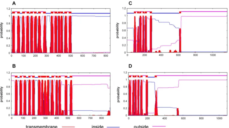
syn-thetase activity (Fig E inS1 File). This leaves its N-terminal 230 amino acids segment with no assigned function. BlastP analysis of the N-terminal 230 amino acids of MSMEG_3796 did not yield any sequences with significant homology. We performed a transmembrane segment (TMS) prediction analysis of MSMEG_3796 andM.tuberculosisLysX sequences using TMHMM tool to see if their extreme N-terminal regions had any transmembrane segments similar to the ones inS.aureusMprF.Fig 2shows the transmembrane segments as predicted by TMHMM for all the proteins analysed. This data indicated the presence of 5–6 transmem-brane segments in bothM.tuberculosisLysX (Fig 2C) and MSMEG_3796 (Fig 2D) at their extreme N-terminal region spanning about 200 amino acids. Although, this may be sufficient to keep them membrane bound, it remains to be experimentally proven whether these 5–6 TMs are sufficient for flipping the newly synthesized L-PG across the mycobacterial membrane as demonstrated by Ernstet al., forS.aureusMprF [62]. Interestingly, both the mycobacterial proteins were found to possess membrane spanning segments despite the absence of any homology at the primary structure level with the similar region fromS.aureusMprF.
Similar to the additional activities found with bacterial LysRS, the human homolog (KRS) was also shown to have additional function where it triggers the dissemination of cancer cells from the primary tumour when it associates itself with the plasma membrane. Namet al., had demonstrated that KRS is indeed involved in the intracellular signal transduction resulting in invasive dissemination of colon cancer spheroids [63] and suggested that it could serve as a suitable target for the development of anti-metastatic therapy.
Maloneyet al., had confirmed that LysX is not essential for thein vitrosurvival ofM. tuber-culosisand that LysS could not complement the absence of lysyl-tRNA synthetase function from LysX [59]. Conversely, the transposon mutagenesis study inM.tuberculosishad indicated LysS to bein vitroessential and that LysX could not complement its function [32]. The essenti-ality data could be very similar inM.smegmatis, however, the absence of direct experimental evidence triggered us to generate and evaluate conditional expression strains of both MSMEG_3796 and MSMEG_6094 to assess their essentiality.
LeuRS is essential for the growth of
M.
smegmatis
expression strain to grow in the absence of added inducer would indicate the essentiality of a gene under investigation. However, a tightly regulated inducible expression system is a basic necessity to derive unambiguous conclusion regarding the essentiality of a target gene under investigation. Acetamide and tetracycline regulated systems have been employed earlier for this purpose in mycobacteria [64–67]. We and others have reported the successful application of a pristinamycin-inducible system for conditional expression of genes in bothM.smegmatis
andM.tuberculosis[26,27,68]. Recently, we had reported the generation and validation of an IPTG-inducible conditional expression system for mycobacteria [25]. Using this system, we confirmed the essentiality of several clinically validated targets in mycobacteria. The assess-ment of essentiality ofM.smegmatisleucyl-tRNA synthetase was performed as a validation
step prior to evaluating the essentiality of CysRS and LysRS. There were multiple reasons for the selection of LeuRS as a validation tool: (1) it is a single gene inM.smegmatiswith no
known redundancy, (2) it belongs to the same family of proteins as others being investigated in this study, (3) it is a single gene in its transcription unit and hence its disruption is unlikely to cause any downstream polar effects, (4) it is highly homologous (about 80% identity) to its counterpart inM.tuberculosiswhich has been shown to be essential via genetic and chemical means [32,12].
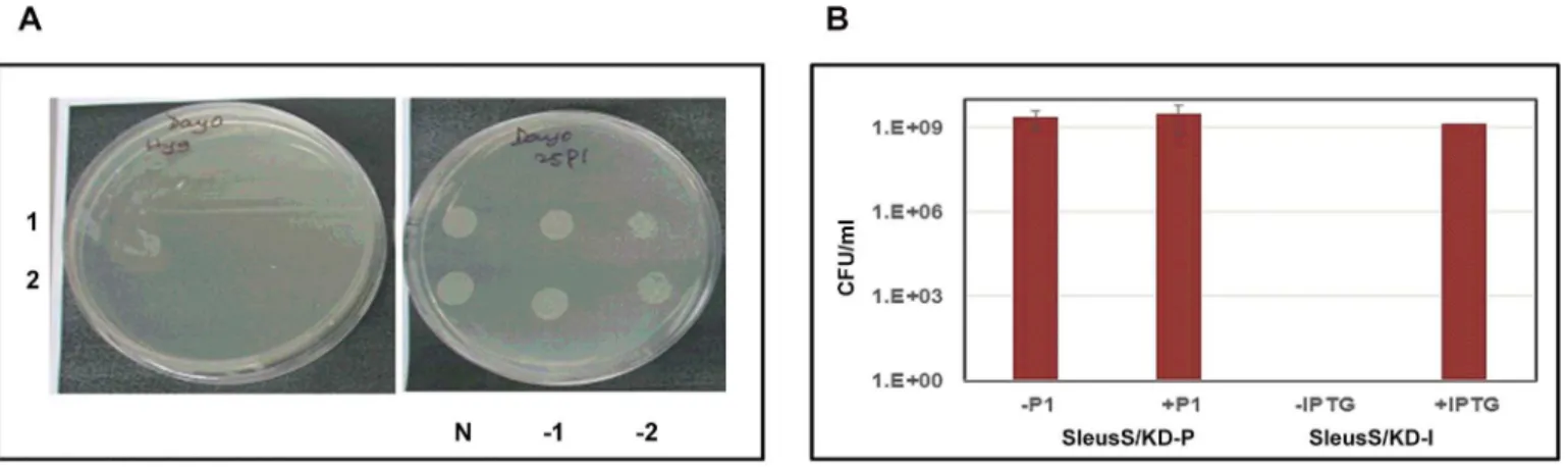
In order to compare the data across the two species, we generated conditional expression strains of LeuRS of bothM.tuberculosisandM.smegmatisusing the pristinamycin-inducible expression system. The recombinant strains, SleuS/KD-P and TleuS/KD-P were grown in the presence of 300 ng/ml of pristinamycin 1 (P1) till they reached mid-log phase. The cells were
washed 3 times with plain 7H9 broth and spotted on 7H11 plates without and with different Fig 2. Transmembrane segment prediction analysis.TMHMM plots forS.aureusMprF (A),P.aeruginosaPA0290 (B),M.tuberculosisLysX (C) and MSMEG_3796 (D).
concentrations of P1to assess their inducer dependency for growth. All the recombinant
TleuS/KD-P colonies tested, showed an absolute P1dependency for growth confirming its
essentiality for the growth ofM.tuberculosis in vitro.Fig 3Arepresents the results for two
inde-pendent colonies which showed no growth in the absence of P1but showed a good colony
mor-phology at 25 ng/ml P1. However, theM.smegmatisrecombinant colonies grew equally well
whether or not P1was present in the growth medium (data not shown). To confirm this result,
one of the SleuS/KD-P recombinant colonies was grown and processed as described earlier to prepare an inoculum for plating several dilutions on 7H11 plates with and without P1. We
hypothesized that the growth of a well isolated colony from a diluted culture would demon-strate cleaner inducer dependency than the culture spots from a broth culture. However, the strain showed no difference in the growth phenotype whether or not P1was present in the
plates (Fig 3B) suggesting either the pristinamycin system is leaky inM.smegmatisor LeuRS is not essential forM.smegmatis. On the other hand, SleuS/KD-I strain could not grow unless IPTG was provided in the growth medium, suggesting the essentiality of LeuRS (Fig 3B). The data from the two inducible systems were contradicting, however, we decided to consider the data from the IPTG-inducible system to be more reliable because of the tightness in regulation of expression it had demonstrated in the previous study [25] and for the reasons stated earlier. The results also suggested that the pristinamycin-inducible system is probably not as well-regu-lated inM.smegmatisas inM.tuberculosis. Therefore, we decided to employ the
IPTG-induc-ible system to generate and evaluate conditional expression strains of MSMEG_6074, MSMEG_3796 and MSMEG_6094 to investigate their essentiality.
Canonical LysRS and CysRS are essential for growth of
M.
smegmatis
Conditional expression strains ofM.smegmatisCysRS (ScysS/KD-I) and both LysRS (S3796/ KD-I and S6094/KD-I) were generated using an IPTG-inducible system as described in materi-als and methods. A minimum inducer concentration requirement test performed by plating the culture at different concentrations of the inducer (0, 5, 10, 50, 100 and 500μM IPTG)
indi-cated 100μM IPTG as the optimal concentration for the growth of these strains (data not
Fig 3. Inducer dependency for the growth ofM.tuberculosisandM.smegmatisLeuRS conditional expression strains.Several confirmed
recombinant colonies were grown in the presence of inducer till they reached mid-log phase, cells were washed, resuspended in fresh 7H9 broth and the cultures were either spotted or various dilutions plated for enumerating colony forming units (CFUs). Plates were incubated at 37°C for 28 days forM.
tuberculosisstrains and 48 hours forM.smegmatisstrains, respectively. (A). Recombinant colonies (1 and 2) of TleuS/KD-P analysed for growth in the absence (left) and the presence (right) of P1; N, -1 and -2 are the undiluted, 10−1and 10−2dilutions. (B). Culture dilutions of SleuS/KD-P and SleuS/KD-I
strains were plated with and without P1and IPTG, respectively. Bars in the graph represent CFU/ml calculated from the colony numbers that appeared on
plates under each of the growth condition specified.
shown). The strains were then grown in 7H9 broth supplemented with 50μg/ml hygromycin
and 100μM IPTG till they reached mid-log phase. Inducer free cell suspensions were prepared
by washing the cells with fresh broth and used as inoculum to plate on 7H11 plates supple-mented with 100μM IPTG or no IPTG. The plates were observed for growth after 48 hours of
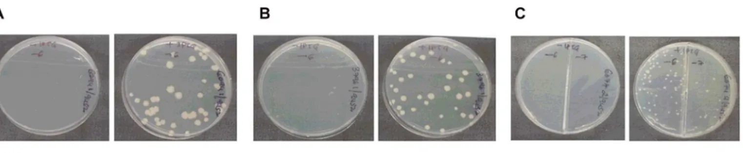
incubation at 37°C. Although, there were no colonies on plates without IPTG in the case of S6094/KD-I and ScysS/KD-I, tiny colonies could be seen in the case of S3796/KD-I (Fig 4). All the plates were incubated for an additional 48 hours to see if differences in the phenotype would be more pronounced following an extended period of incubation. While no colonies grew in S6094/KD-I and ScysS/KD-I plates without IPTG after 96 hours, in the case of S3796/ KD-I, the tiny colonies observed on plates without IPTG at 48 hours grew into well-formed colonies (data not shown).
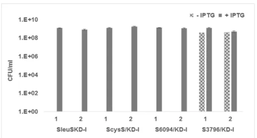
The delayed growth phenotype of S3796/KD-I conditional expression strain in the absence of inducer was different from that observed by Maloneyet al., [59] withM.tuberculosisLysX knockout strain which didn’t show any growth defectin vitro. In order to confirm the results, the experiment was repeated with two colonies for each of the conditional expression strain. Two colonies ofM.smegmatisLeuRS conditional expression strain were also included in the experiment. Dilutions of the culture suspensions prepared as described in materials and meth-ods were plated on 7H11 plates supplemented with 100μM IPTG or no IPTG. At the end of 48
hours of incubation, conditional expression strains showed the same phenotype as described earlier. After 96 hours of growth, the colonies appearing on each plate were counted, the CFU/ ml calculated and plotted against the growth condition for each of the strain. While the SleuS/ KD-I, S6094/KD-I, and ScysS/KD-I strains grew only in the presence of IPTG, S3796/KD-I strain showed growth even in the absence of IPTG at 96 hours indicating the reproducibility of the earlier observation (Fig 5). These results further confirm the essentiality ofM.smegmatis
LeuRS, CysRS, MSMEG_6094 and the non-essentiality of MSMEG_3796.
Summary and Conclusion
A search for genes encoding aminoacyl-tRNA synthetases ofM.smegmatisin KEGG genes database yielded 24 entries. Analysis of the primary structures of some of the additional pro-teins—TilS, Glu-Q-RS, Prolyl-tRNA editing protein (YbaK homolog), MshC and
MSMEG_3796 (LysX homolog) ofM.smegmatiswith their orthologs fromE.coliand / orM.
tuberculosisfollowed by literature survey enabled understanding their physiological roles. Although non-essential, it remains unclear and confusing why Glu-Q-RS is conserved across different bacterial genera when its activity could lead to the synthesis of several proteins with
Fig 4. Analysis of inducer dependence for growth.Identified recombinant colonies of S6094/KD-I, S3796/KD-I and ScysS/KD-I were grown with 100μM
IPTG until they reached mid-log phase, washed and the culture suspension in plain 7H9 broth was diluted and plated on plates with and without IPTG. The plates were incubated at 37°C for 48 hours and photographed. In all the cases, plate on left is without IPTG and plate on right is with IPTG. (A) S6094/KD-I; (B) S3796/KD-I and (C) ScysS/KD-I.
conserved changes–glutamate for aspartate. The essential nature of the activities catalysed by TilS and prolyl-tRNA editing protein substantiated their retention. The conservation of MshC and LysX homolog across different mycobacterial species indicated the importance of their function for mycobacterial survival.
The dispensability of MSMEG_3796 and essentiality of the canonical CysRS and LysRS for the survival ofM.smegmatiscould be established using their conditional expression strains. Thein vivoessentiality in the pathogenic mycobacteria and thein vitronon-essentiality of LysX homologs in the pathogenic and saprophytic mycobacteria could be readily explained based on the observation that LysX is required to counter the cationic anti-microbial peptides the bacteria would encounter within their host. However, reasons for the differential essential-ity of MshC in the pathogenic versus the saprophytic mycobacteria is not clear as it has been demonstrated to confer mycobacteria the ability to resist several alkylating agents and anti-mycobacterials [51].
The intriguing fact about MSMEG_3796 and MSMEG_6094 was that despite having the entire LysRS domain in both these proteins, they could not complement each other’s absence. The Lys-tRNALyssynthesized by each of these enzymes seems to get utilized towards different physiological functions, L-PG synthesis and protein synthesis, respectively. The difference in the localization of these proteins could be the reason for this non-redundant function. Bioinformatics analysis enabled us to predict the presence of transmembrane segments in MSMEG_3796 suggesting that it could be membrane bound while MSMEG_6094, a homolog of canonical LysRS required for translation process could be cytosolic. It is highly likely that the membrane bound MSEMG_3796 cannot access the Lys-tRNALyssynthesized by cytosolic MSMEG_6094 and vice versa. On the other hand it would be interesting to identify the factor which has enabled the membrane bound MprF inS.aureusto utilize the LystRNALys synthe-sized by the cytosolic LysRS
This study also enabled us to notice several interesting facts about aminoacyl-tRNA synthe-tases and their homologs. A number of them were found to be involved in non-translational functions such as synthesis of diadenosine tetraphosphate (Ap4A) and analogues in several
Fig 5. Inducer dependency for growth ofM.smegmatisLeuRS, LysRS and CysRS conditional expression strains.The conditional expression strains SleuS/KD-I, SCysS/KD-I, S6094/KD-I and S3796/ KD-I were grown in the presence of 100μM IPTG till they reached mid-log phase, the cells were harvested,
washed to remove traces of inducer and resuspended in fresh 7H9 broth to be used as inoculum. Several dilutions of these cultures were plated on 7H11 plates with and without IPTG. Plates were incubated at 37°C for 96 hours and the colonies were counted both at the end of 48 hours and 96 hours of incubation.
bacteria, synthesis of MshC and LP-G in mycobacteria, lysinylation of elongation factor by epmA/genX products in Gram negatives and involvement in cardiovascular development, immune response, signalling events as in triggering metastatic events in human cancer [69,70].
Thus, the literature and bioinformatics analysis along with 3-D structure modelling enabled us to understand the likely functions of the‘additional’aminoacyl-tRNA synthetases found in
M.smegmatis. The information gathered from this study also indicated the unique activity of these‘additional’aminoacyl-tRNA synthetases and very importantly the absence of redun-dancy in the function of canonical aaRS despite the presence of common functional domains. These studies have thus offered valuable insights into the role of various aminoacyl-tRNA syn-thetases in the growth and survival of mycobacteria which in turn could provide avenues for further research into the design of specific anti-mycobacterials agents.
Supporting Information
S1 File. Plasmid maps of pAZI9452 and pAZI947 (Fig A). List of primers used in the study (Table A). Genomic organization in the conditional expression strain (Fig B). Generic reac-tion scheme for aminoacyl-tRNA synthetases (Fig C). The VERIFY-3D Average score of modelled MSMEG_5671 structure (Fig D). Homology amongM.smegmatislysyl-tRNA synthetases (Fig E).
(DOCX)
Acknowledgments
We would like to acknowledge Ms. Naina Mudugal, Ms. Anubrita Das, Ms. Aditi A Mutgikar and Ms. Tanya Jain for the support provided towards generating conditional expression plas-mids and strains respectively. The authors would like to acknowledge the guidance and support received from Drs. Santanu Datta, V. Balasubramanian, Shridhar Narayanan through the course of this research.
Author Contributions
Conceived and designed the experiments: S Ravishankar A Anbarasu S Ramaiah VKS. Per-formed the experiments: S Ravishankar A Ambady RGS. Analyzed the data: S Ravishankar A Anbarasu S Ramaiah VKS. Contributed reagents/materials/analysis tools: S Ravishankar A Ambady RGS A Anbarasu S Ramaiah. Wrote the paper: S Ravishankar A Anbarasu S Ramaiah VKS.
References
1. Robinson A, van Oijen AM, Bacterial replication, transcription and translation: mechanistic insights from single molecule biochemical studies. Nature Reviews Microbiology 2013, 11:303–315 doi:10.
1038/nrmicro2994PMID:23549067
2. Wilson DN, The A–Z of bacterial translation inhibitors. Critical Reviews in Biochemistry and Molecular
Biology 2009, 44: 393–433. doi:10.3109/10409230903307311PMID:19929179
3. Blanchard SC, Cooperman BS, Wilson DN. Probing Translation with Small-Molecule Inhibitors. Chem-istry & Biology 2010, 17: 633–645.
4. Parenti MA, Hatfield SM, Leyden JJ. Mupirocin: a topical antibiotic with a unique structure and mecha-nism of action. Clin. Pharm. 1987, 6:761–770. PMID:3146455
5. Hoffmann M, Torchala M. Search for inhibitors of aminoacyl-tRNA synthases by virtual click chemistry. J. Mol. Model. 2009, 15:665–672 doi:10.1007/s00894-008-0421PMID:19048310
7. Yu XY, Finn J, Hill JM, Wang ZG, Keith D, Silverman J, et al. A series of heterocyclic inhibitors of pheny-lalanyl-tRNA synthetases with antibacterial activity. Bioorg. Med. Chem. Lett. 2004, 14:1343–1346.
PMID:14980695
8. Beyer D, Kroll HP, Endermann R, Schiffer G, Siegel S, Bauser M, et al. New class of bacterial phenyla-lanyl-tRNA synthetase inhibitors with high potency and broad-spectrum activity. Antimicrob. Agents Chemother. 2004, 48:525–532. PMID:14742205
9. Tandon M, Coffen DL, Gallant P, Keith D, Ashwell MA. Potent and selective inhibitors of bacterial methionyl-tRNA synthetase derived from an oxazolone-dipeptide scaffold. Bioorg. Med. Chem. Lett. 2004, 14:1909–1911. PMID:15050625
10. Jarvest RL, Berge JM, Berry V, Boyd HF, Brown MJ, Elder JS, et al. Nanomolar inhibitors of Staphylo-coccus aureusmethionyl-tRNA synthetase with potent antibacterial activity against Gram-positive path-ogens. J. Med. Chem. 2002, 45:1959–1962. PMID:11985462
11. Yu XY, Hill JM, Yu GX, Yang YF, Kluge AF, Keith D, et al. A series of quinoline analogues as potent inhibitors ofC.albicansprolyl-tRNA synthetase. Bioorg. Med. Chem. Lett. 2001, 11:541–544. PMID:
11229766
12. Stefanska AL, Coates NJ, Mensah LM, Pope AJ, Ready SJ, Warr SR. SB-219383, a novel tyrosyl-tRNA synthetase inhibitor from aMicromonosporasp. I. Fermentation, isolation and properties. J. Anti-biot. (Tokyo) 2000, 53:345–350.
13. Qing-Hua H, Ru-Juan L, Zhi-Peng F, Zhang J, Ying-Ying D, Tan M, et al. Discovery of a potent benzox-aborole based anti-pneumococcal agent, targeting leucyl-tRNA synthetase. Scientific Reports 2013, 3: 2475 doi:10.1038/srep02475PMID:23959225
14. Teng M, Hilgers MT, Cunningham ML, Borchardt A, Locke JB, Abraham S, et al. Identification of Bacte-ria-Selective Threonyl-tRNA Synthetase Substrate Inhibitors by Structure-Based Design. J. Med. Chem 2013, 56: 1748–1760. doi:10.1021/jm301756mPMID:23362938
15. Ochsner UA, Sun X, Jarvis T, Critchley I, Janjic N. Aminoacyl-tRNA synthetases: essential and still promising targets for new anti-infective agents. Expert Opin. Investig. Drug. 2007, 16: 1599–1609.
16. Gurcha S. S., Usha V, Cox JAG, Fu K, Abrahams KA, Bhatt A, et al. Biochemical and Structural Char-acterization of Mycobacterial Aspartyl-tRNA Synthetase AspS, a Promising TB Drug Target. PLoS One, 2014, 9: e113568. doi:10.1371/journal.pone.0113568PMID:25409504
17. Woese CR, Olsen GJ, Ibba M, Söll D. Aminoacyl-tRNA Synthetases, the Genetic Code, and the Evolu-tionary Process. Microbiol. Mol. Biol. Rev. 2000, 64: 202–236. PMID:10704480
18. Ibba MSo¨ ll D. Aminoacyl-tRNAs: setting the limits of the genetic code. Genes & Development 2004,
18: 731–738.
19. Eriani G, Delarue M, Poch O, Gangloff J, Moras D: Partition of tRNA synthetases into the two classes based on mutually exclusive sets of sequence motifs. Nature 1990, 347: 203–206. PMID:2203971
20. Burbaum JJ, Schimmel P. Structural Relationships and the Classification of Aminoacyl-tRNA Synthe-tases. The Journal of Biol. Chem. 1991, 266: 16965–16968.
21. Onesti S, Miller AD, Brick P. The crystal structure of the lysyl-tRNA synthetase (LysU) fromEscherichia coli. Structure 1995, 3:163–176 PMID:7735833
22. Breton R, Sanfaçon H, Papayannopoulos I, Biemann K, Lapointe J. Glutamyl-tRNA synthetase of
Escherichia coli. Isolation and primary structure of the gltX gene and homology with other aminoacyl-tRNA synthetases. J Biol. Chem. 1986, 261: 10610–10617. PMID:3015933
23. Blaise M, Becker HD, Lapointe J, Cambillau C, Giegà R, Kern D. Glu-Q-tRNA(Asp) synthetase coded
by the yadB gene, a new paralog of aminoacyl-tRNA synthetase that glutamylates tRNA(Asp) antico-don. Biochimie 2005, 87: 847–861. doi:10.1016/j.biochi.2005.03.007PMID:16164993
24. http://www.genome.jp/dbget-bin/www_bfind_sub?mode=bfind&max_hit=1000&dbkey=
msm&keywords=tRNA+synthetase&mode=bfind
25. Ravishankar S, Ambady A, Ramu H, Mudugal NV, Tunduguru R, Anbarasu A, et al. An IPTG Inducible Conditional Expression System for Mycobacteria. PLoS One 2015, 10(8): e0134562. doi:10.1371/ journal.pone.0134562PMID:26247874
26. Ravishankar S, Ambady A, Awasthy D, Mudugal NV, Menasinakai S, Jatheendranath S, et al. Genetic and Chemical Validation IdentifiesM.tuberculosisTopoisomerase I as an Attractive Anti-tubercular
Target. Tuberculosis 2015, doi:http://dx.doi.org/10.1016/j.tube.2015.05.004
27. Reddy BKK, Landge S, Ravishankar S, Patil V, Shinde V, Tantry S, et al. Assessment of Mycobacte-rium tuberculosisPantothenate Kinase vulnerability through Target Knockdown and Mechanistically
Diverse Inhibitors. Antimicrob. Agents. Chemother. 2014, 58: 3312–3326. doi:10.1128/AAC.00140-14
PMID:24687493
28. Stover CK, de la Cruz VF, Fuerst TR, Burlein JE, Benson LA, Bennett LT, et al. New use of BCG for
29. Wards BJ, Collins DM. Electroporation at elevated temperatures substantially improves transformation efficiency of slow-growing mycobacteria. FEMS Microbiol. Lett. 1996; 145:101–105. PMID:8931333
30. Wolf Y, Aravind L, Grishin N, Koonin E. Evolution of aminoacyl-tRNA synthetases-analysis of unique domain architectures and phylogenetic trees reveals a complex history of horizontal gene transfer events. Genome Res. 1999; 9:689–710. PMID:10447505
31. Soma A, Ikeuchi Y, Kanemasa S, Kobayashi K, Ogasawara N, Ote T, et al. An RNA-Modifying Enzyme that Governs Both the Codon and Amino Acid Specificities of Isoleucine tRNA. Mol. Cell, 12: 689–698.
PMID:14527414
32. Sassetti CM, Boyd DH, Rubin EJ. Genes required for mycobacterial growth defined by high density mutagenesis. Mol. Microbiol. 2003, 48: 77–84. PMID:12657046
33. Salowe SP, Wiltsie J, Hawkins JC, Sonatore LM. The catalytic flexibility of tRNA Ile-lysidine synthetase
can generate alternative tRNA substrates for isoleucyl-tRNA synthetase. J. Biol. Chem. 2009, 284: 9656–9662. doi:10.1074/jbc.M809013200PMID:19233850
34. Suzuki T, Miyauchi K. Discovery and characterization of tRNAIlelysidine synthetase (TilS). Febs
Let-ters. 2010, 584: 272–277. doi:10.1016/j.febslet.2009.11.085PMID:19944692
35. Blaise M, Becker HD, Keith G, Cambillau C, Lapointe J, Giege R, et al. A minimalist glutamyl-tRNA syn-thetase dedicated to aminoacylation of the tRNAAspQUC anticodon. Nucleic Acids Res. 2004, 32: 2768–2775. PMID:15150343
36. Blaise M, Olieric V, Sauter C, Lorber B, Roy B, Karmakar S, et al. Crystal Structure of Glutamyl-Queuo-sine tRNAAspSynthetase Complexed with L-Glutamate: Structural Elements Mediating tRNA-Indepen-dent Activation of Glutamate and Glutamylation of tRNAAspAnticodon. J. Mol. Biol. 2008, 381: 1224
–
1237. doi:10.1016/j.jmb.2008.06.053PMID:18602926
37. Dubois DY, Blaise MI, Becker HD, Campanacci V, Keith G, Giege R, et al. An aminoacyl-tRNA
synthe-tase-like protein encoded by theEscherichia coliyadB gene glutamylates specifically tRNAAsp. Proc. Natl. Acad. Sci. USA. 2004, 101: 7530–7535 PMID:15096594
38. Salazar JC, Ambrogelly A, Crain PF, McCloskey JA, Söll D. A truncated aminoacyl-tRNA synthetase
modifies RNA. Proc. Natl. Acad. Sci. USA. 2004, 101: 7536–7541. PMID:15096612
39. Ruan B, Soll D. The bacterial YbaK protein is a Cys-tRNAProand Cys-tRNACysdeacylase. The J. Biol. Chem. 2005, 280: 25887–25891.
40. Kumar S, Das M, Hadad CM, Musier-Forsyth K. Substrate Specificity of Bacterial Prolyl-tRNA Synthe-tase Editing Domain Is Controlled by a Tunable Hydrophobic Pocket. The J. Biol. Chem. 2012, 287: 3175–3184. doi:10.1074/jbc.M111.313619PMID:22128149
41. Bartholow TG, Sanfor BL, Cao B, Schmit HL, Johnson JM, Meitzner J, et al. Strictly conserved lysine of prolyl-tRNA synthetase editing domain facilitates binding and positioning of misacylated tRNAPro. Bio-chemistry. 2014, 53: 1059–1068. doi:10.1021/bi401279rPMID:24450765
42. Martinis SA, Boniecki MT. The balance between pre- and post-transfer editing in tRNA synthetases. Febs Lett. 2010, 584: 455–459. doi:10.1016/j.febslet.2009.11.071PMID:19941860
43. Zhang H, Huang K, Li Z, Banerjei L, Fisher KE, Grishin NV, Eisenstein E, et al. Crystal strucuture of YbaK protein fromHaemophilus influenza(HI1434) at 1.8Åresolution: functional implications. Proteins 2000, 40: 86–97. PMID:10813833
44. http://www.ebi.ac.uk/pdbe/entry/pdb/2DXA
45. Zhang Y. I-TASSER server for protein 3D structure prediction. BMC Bioinformatics 2008, 9: 40. doi: 10.1186/1471-2105-9-40PMID:18215316
46. Roy A, Kucukural A, Zhang Y. I-TASSER: a unified platform for automated protein structure and func-tion predicfunc-tion. Nat Protoc. 2010, 5: 725–738. doi:10.1038/nprot.2010.5PMID:20360767
47. Laskowski RA, Rullmannn JA, MacArthur MW, Kaptein R, Thornton JM. AQUA and PROCHECK-NMR: programs for checking the quality of protein structures solved by NMR. J Biomol. NMR 1996, 8: 477–486. PMID:9008363
48. Colovos C, Yeates TO. Verification of protein structures: patterns of nonbonded atomic interactions. Protein Sci. 1993, 2: 1511–1519. PMID:8401235
49. Eisenberg D, Luthy R, Bowie JU. VERIFY3D: assessment of protein models with three-dimensional
profiles. Methods Enzymol. 1997, 277: 396–404. PMID:9379925
50. Holm L, Park J. DaliLite workbench for protein structure comparison. Bioinformatics 2000, 16: 566–
567. PMID:10980157
52. Sareen D, Steffek M, Newton GL, Fahey RC. ATP-dependent L-cysteine:1D-myo-inosityl 2-amino-2-deoxy-alpha-D-glucopyranoside ligase, mycothiol biosynthesis enzyme MshC, is related to class I cysteinyl-tRNA synthetases. Biochemistry 2002, 41: 6885–6890. PMID:12033919
53. Tremblay LW, Fan F, Vetting MW, Blanchard JS. The 1.6ÅCrystal Structure ofMycobacterium smeg-matisMshC: The Penultimate Enzyme in the Mycothiol Biosynthetic Pathway. Biochemistry 2008, 47: 13326–13335. doi:10.1021/bi801708fPMID:19053270
54. Sareen D, Newton GL, Fahey RC, Buchmeier NA. Mycothiol is essential for growth ofMycobacterium tuberculosisErdman. J. Bacteriol. 2003, 185: 6736–6740. PMID:14594852
55. Yanagisawa T, Sumida T, Ishii R, Takemoto C, Yokoyama S. A paralog of lysyl-tRNA synthetase ami-noacylates a conserved lysine residue in translation elongation factor P. Nature Stru. Mol. Biol. 201, 17: 1136–1143.
56. Wright M, Azhar MA, Kamal A, Miller AD. Syntheses of stable, synthetic diadenosine polyphosphate analogues using recombinant histidine-tagged lysyl-tRNA synthetase (LysU). Bioorg. Med. Chem. Lett. 2014,http://dx.doi.org/10.1016/j.bmcl.2014.03.064.
57. Oka M, Takegawas K, Kimura Y. Enzymatic characterization of a class II lysyl-tRNA synthetase, LysS fromMyxococcus xanthus. Arch. Biochem. Biophys. 2015.http://dx.doi.org/10.1016/j.abb.2015.05. 014.
58. Tshori S, Razin E, Nechushtan H. Aminoacyl-tRNA synthetases generate dinucleotide polyphosphates as second messengers: Functional implications. A chapter in Aminoacyl-tRNA synthetases in Biology and Medicine. Kim S (editor) 2013, Springer-Verlag Berlin Heidelberg.
59. Maloney E, Stankowska D, Zhang J, Fol M, Cheng QJ, Lun S, et al. The Two-Domain LysX Protein of
M.tuberculosisis required for Production of Lysinylated Phosphatidylglycerol and Resistance to Cat-ionic Antimicrobial Peptides. 2009, 5: e1000534.
60. Peshel A, Jack RW, Otto M, Collins LV, Staubitz P, Nisholson G, et al.Staphylococcus aureus resis-tance to human defensins and evasion of neutrophil killing via the novel virulence factor MprF is based on modification of membrane lipids with L-Lysine. J. Exp. Med. 2001, 193: 1067–1076. PMID:
11342591
61. Oku Y, Kurukawa K, Ichihashi N, Sekimizu K. Characterization of theStaphylococcus aureusmprF gene, involved in lysinylation of phosphatidylglycerol. Microbiology 2004, 150: 45–51. PMID:
14702396
62. Ernst CM, Kuhn S, Slavetinsky CJ, Krismer B, Kraus D, Wagner S, et al. The lipid-modifying multiple peptide resistance factor is an oligomer consisting of distinct interacting synthase and flippase subunits. mBio. 2015, 6: e02340–14. doi:10.1128/mBio.02340-14PMID:25626904
63. Nam SH, Kim D, Lee MS, Lee D, Kwak TK, Kang M, et al. Noncanonical roles of membranous lysyl-tRNA synthetase in transducing cell-substrate signalling for invasive dissemination of colon cancer spheroids in 3D collagen I gels. Oncotarget 2015. 6: 21655–21674. PMID:26091349
64. Raghunandan TR, Bishai WR, Chen P. Towards establishing a method to screen for inhibitors of essential genes in mycobacteria: evaluation of the acetamidase promoter. J. Antimicrobial Agents 2006, 8: 36–41.
65. Ehrt S, Guo XV, Hickey CM, Ryou M, Monteleone M, Riley LW, et al. Controlling gene expression in mycobacteria with anhydrotetracycline and tet repressor. Nucleic Acids Res. 2005, doi:10.1093/nar/ gni013
66. Carroll P, Muttucumaru DGN, Parish T. Use of a Tetracycline-Inducible System for Conditional Expres-sion inMycobacterium tuberculosisandMycobacterium smegmatis. Applied and Environmental Microbiol. 2005, 71: 3077–3084.
67. Boldrin F, Casonato S, Dainese E, Sala C, Dhar N, Palù G, et al. Development of a repressible myco-bacterial promoter system based on two transcriptional repressors. Nucleic Acids Res. 2010, 38: e134. doi:10.1093/nar/gkq235PMID:20406773
68. Forti F, Crosta A, Ghisotti D. Pristinamycin-inducible gene regulation in mycobacteria. J. Biotechnology 2009, 140: 270–277.
69. Guo M, Schimmel P. Essential Non-Translational Functions of tRNA Synthetases, Nat Chem Biol. 2013; 9:145–153. doi:10.1038/nchembio.1158PMID:23416400
70. Guo M, Yang X, Schimmel P. New functions of aminoacyl-tRNA synthetases beyond translation.