Introduction
Citrus tristeza virus (CTV, genus Closterovirus, family Closteroviridae) is the causal agent of one of the most economically important diseases of
cit-Differentiation of Citrus tristeza virus (CTV) isolates by
cleavase fragment length polymorphism (CFLP) analysis of the
major coat protein gene
NATÁLIA T. MARQUES1, ANA M. BAILEY2, CHARLES L. NIBLETT3 and GUSTAVO NOLASCO1
1Universidade do Algarve - Centro de Desenvolvimento de Ciências e Técnicas de Produção Vegetal, Campus de Gambelas, 8005-139 Faro, Portugal
2Centro de Investigación y de Estudios Avanzados del Instituto Politécnico Nacional, Unidad Irapuato, Apdo. Postal 629, 6500, Irapuato, Mexico
3Horticultural Sciences Department, University of Florida, P.O. Box 110780, Gainesville, FL 32611-0780 USA
Summary. A panel of Citrus tristeza virus (CTV, genus Closterovirus, family Closteroviridae) isolates of different
origins and with different biological properties were compared for polymorphisms in the major coat protein (CP) gene by cleavase fragment length polymorphism (CFLP) and single stranded conformation polymorphism (SSCP) analy-sis. The similarity between the CFLP patterns, which consisted of 15 to 20 bands, was estimated by the Pearson coefficient. The clustering patterns from the CFLP data were very similar to those from sequence data in an experi-ment with 16 cloned standards of the CP gene. By SSCP analysis on the other hand, most of the clones were not clustered in the same way. To assess the ability of CFLP to analyse biological samples, which may consist of a mixture of genomic variants, the CP gene of 12 CTV isolates was obtained directly from infected plants by immunocapture/ RT-PCR and analysed. With few exceptions, the isolates were correctly clustered according to the sequences of the variants composing the isolates. In artificial mixed infections of mild and severe isolates the patterns obtained were more closely related to the severe isolate. Thus the CFLP method was an accurate method for the identification, typing and clustering of CTV isolates. The usefulness of this technique as an alternative to SSCP analysis is sug-gested and discussed.
Key words: CFLP, genomic variability, typing.
Corrresponding author: G. Nolasco Fax: +351 289818419
E-mail: [email protected]
rus. The virus has a single-stranded positive-sense RNA genome varying from 19,226 (Mawassi et al., 1996) to 19296 (Karasev et al., 1995) nucleotides, containing 12 ORFs, potentially encoding at least 17 protein products (Karasev et al., 1995). The major capsid protein (here referred to as coat pro-tein [CP]) has 25 KDa and covers about 95% of the virion (Febres et al., 1996). CTV isolates differ in the type and severity of the symptoms they induce in the various citrus hosts (Grant and Higgins,
1956). It is generally accepted that CTV infection has caused the decline and death of millions of cit-rus trees grafted on sour orange rootstocks. De-cline is rapid in certain cases, but slow deDe-cline over a period of months or years is most frequent. Strains causing quick decline or stem pitting on sweet orange or grapefruit are usually called se-vere strains. Those that do not cause these symp-toms are mild strains. Attempts have been made to relate the biological properties of the virus to its biochemical characteristics (Niblett et al., 2000). The rapid identification of severe isolates is im-portant to control the disease. CTV isolates have been characterized in a number of ways: by sero-logical tests (Manjunath et al., 1993; Pappu et al., 1993), double-stranded RNA analysis (Moreno et al., 1990; Guerri et al., 1991; Moreno et al., 1993; Moreno et al., 1996), restriction fragment length polymorphism (Gillings et al., 1993; Nolasco and Sequeira, 1997), single stranded conformation pol-ymorphism (SSCP) (Rubio et al., 1996; Nolasco and Sequeira, 1997; Ayllón et al., 1999; Gago-Zachert et al., 1999; Kong et al., 2000), hybridisation with complementary long DNA probes (Rosner et al., 1986; Narváez et al., 2000) or short DNA probes (Niblett et al., 2000; Zemzami et al., 2002), and by analysis of peptide maps (Guerri et al., 1990; Albi-ach-Martí et al., 2000).
Cleavase fragment length polymorphism (CFLP) analysis is based on the activity of the endonucle-ase cleavendonucle-ase, which recognizes the secondary struc-ture of hairpins and cuts the DNA strand at the 5’ end of the stem part. The DNA molecules to be ana-lysed are thermally denatured and allowed to cool to a certain temperature so that on each DNA strand a certain amount of the secondary structure builds up by self-base pairing. Cleavase activity produces a collection of fragments that are resolved by dena-turing gel electrophoresis. CFLP analysis has al-ready been used to study variability in diverse ge-netic systems, such as human somatic genes (O’Connell et al., 1999), bacteria (Brow et al., 1996), plants (Fofana et al., 1998) and animal viruses (Mar-shall et al., 1997), but not plant viruses.
In this work we describe the use of CFLP as an alternative to sequencing or SSCP for typing pur-poses.
Materials and methods
Virus isolates and clones of the CP gene
Two sources of the CP gene were used in the assays: the cloned CP gene and infected plant ma-terial. Sixteen CP gene clones used as standards were obtained from different sources as minipreps. The origin and the biological characterisation by
Table 1. Characterisation of the 12 Citrus tristeza virus isolates (Bonacalza, 1998).
Biological characterisation Isolate Origin SP-SwO c SP-ML d SP-GF e 002 Madeira Island + + + 008 Madeira Island + + ? 013 Madeira Island + + -014 Madeira Island + + -015 Madeira Island + + -019 a Spain - + -025 a Spain - + -028 Portugal - + +
144 Reunion Island - + n/a
196 b =25+15 Artificial mixed infection + n/a n/a
200 b =25+8 Artificial mixed infection + n/a n/a
206 b =25+212 Artificial mixed infection + n/a n/a
212 Reunion Island + +
-a Plants 19 and 25 were also tested for quick decline symptoms, with negative results. b Plants double inoculated with different isolates.
c SP-SwO, stem pitting on sweet orange; d SP-ML, stem pitting on Mexican lime; e SP-GF, stem pitting on grapefruit. n/a, not assayed.
means of the biological indexing of the isolates from which these clones were obtained are shown in Tables 1 and 2. A further twelve CTV isolates were taken from a collection maintained in insect-proof greenhouse-grown seedlings of sweet orange (Ci-trus sinensis [L.] Osb.) cv. Madam vinous that had been biologically characterised in an earlier study (Bonacalza, 1998). In some cases mixed infections were obtained by double inoculation in which the second inoculation was carried out after ELISA confirmed the establishment of the first infection.
Isolating and cloning the CP gene
The CP gene of the 12 CTV isolates was ampli-fied by Immunocapture reverse transcriptional (RT) PCR, as described in Nolasco et al. (1993). Each RT-PCR reaction was done in 50 µl of a mix-ture containing: 4 mM MgCl2, 200 µM of each dNTP, 200 nM of each primer, 3 units of RNase inhibitor (GE Healthcare, Chalfont St. Giles, UK, ref. 27-0815-01), 7.5 units of MuLV Rtase (Applied Bio-systems, Foster City, USA, ref. N808-0018) and 1.25 units of Taq DNA polymerase (Fermentas Inc., Hanover, USA, ref. EPO402). The reaction tubes were incubated in a termocycler with the follow-ing cyclfollow-ing conditions: 38°C for 45 min, 94°C for 2
min, 35 cycles at 92°C for 30 s, 52°C for 30 s, and 72°C for 1 min, with a final step of 72°C for 7 min. The primers used were: CTV 1 [5´–ATGGACGAC-GAAACAAAGAA–3´] (forward primer) and CTV 10 [5´–ATCAACGTGTGTTGAATTTCC–3´] (reverse primer), generating a 673 bp fragment correspond-ing to the whole CP gene, the stop codon and an extra nucleotide at the 3´ end. PCR amplifications using the cloned CP gene as template were done using 1 µl of a miniprep in the same conditions as above, except for the RT reagents and the incuba-tion step, which were omitted. Usually, one-tenth of the amplified product was analysed in 1% agar-ose gel. When necessary, the PCR product was TA cloned in a pCRII vector (Invitrogen, Carlsbad, CA, USA) or a pGEM T-Easy vector (Promega, Madi-son, USA), according to manufacturer’s instruc-tions. Competent INVαF´ Escherichia coli cells were transformed and colonies containing the in-sert were selected in the presence of X-gal and con-firmed by PCR. Minipreps were done according to standard procedures.
CFLP analysis
CFLP analysis was done using the CFLP“ Scanning System (Takara Shuzo Co. Ltd., Otsu
Table 2. Description of 16 cloned coat protein gene standards.
Clone Geographicalorigin Biological characterisation of the isolatefrom which the CP clone was obtainedb bibliographic reference of sequence dataGenBank accession number or
T36 Florida, USA QD M76485
B53 Spain / Japana QD, SP-GFc, SP-SwO Pappu et al., 1993; Akbulut et al., 1996
19.121 Spain Isolate 19, Table 1 AF1841144
13C Madeira Island Isolate 13, Table 1 AF184113
B185 Japan SP-GF, SP-SwO Pappu et al., 1993
T3 Florida, USA QD, SP-SwO Pappu et al., 1993
15.118 Madeira Island Isolate 15, Table 1 AY660009
T30 Florida, USA SP-ML AF260651
B274 Colombia SP-ML Acosta et al., 1994
B249 Venezuela QD; SP-SwO Peñaranda et al., 1996
2.93 Madeira Island Isolate 2, Table 1 AF184116
2.98 Madeira Island Isolate 2, Table 1 AF184117
25.120 Spain Isolate 25, Table 1 AF184115
TR11a Reunion Island SP-GF AY660010
28.121 Portugal Isolate 28, Table 1 AF184118
B128 Colombia QD, SP-GF, SP-SwO Pappu et al., 1993
a The origin of this isolate is reported by several authors as Spain, by others as Japan.
b QD, quick decline; SP-GF, stem pitting on grapefruit; SP-SwO, stem pitting on sweet orange; SP-ML, stem pitting on Mexican lime.
Shiga, Japan, ref. 6627). PCR products were pre-cipitated with ethanol and re-suspended in 15 µl of water. Depending on previous gel electrophore-sis quantification, 1 or 2 µl of the PCR products was diluted to a final volume of 15 µl in 5 mM MOPS, pH 7.5, denatured at 95°C for 30 s, then cooled and maintained at the optimised reaction temperature (45°C). The pre-warmed cleavase enzyme solution (5 µl) was then added. Final com-position of the reaction mix was 10 mM MOPS (pH 7.5), 0.2 mM of MnCl2, and contained 0.05% (w:v) Tween 20, 0.05% (w:v) Nonidet P-40 and 1.25 U of cleavase. Cleavase digestion proceeded for 12 minutes until stopped by the addition of 16 µl of stop solution (95% formamide, 10 mM EDTA [pH 8], 0.05% xylene cyanol, 0.05% bromophenol blue). The DNA fragments were resolved by elec-trophoresis in 0.75 mm mini polyacrylamide gels (8% with 19:1 cross-linking) containing 7 M urea in 0.5⫻TBE buffer (Tris-borate 44.5 mM; EDTA 1 mM; pH 8.3). Before loading the gel the samples were denatured at 85°C for 1–2 min and the gel was pre-run for 30 min at 300 V. Electrophoretic separation was done for 45 min at 300 V. Silver staining and drying of the gels was done accord-ing to standard procedures. Digested DNA from CTV clone 15.118 was included in each assay as a reference sample. Analysis of band patterns was done with the help of the Bionumerics software package (Applied Maths, Kortrijk, Belgium) us-ing the Pearson similarity coefficient and Un-weighted Pair Group Average Method (UPGMA) clustering. Cophenetic correlation values were determined to assess the consistence of the clus-ters. To avoid interference caused by small varia-tions in the length of the electrophoresis run, or problems caused by the software having difficul-ty in recognising faint bands at the lower end of the gel, only bands corresponding to more than 200 bases were compared. Sequence alignments and nucleotide distances were determined using the BioEdit sequence alignment editor software package (Hall, 1999).
SSCP analysis
The SSCP analysis was done on PCR products obtained from the 16 cloned standards (Table 2), as described in Rubio et al. (1996). The CP gene previously amplified by primers CTV1/CTV10 was reamplified with primers CTV 43
[5´–ATGTTGTT-GCNGCNGAGTC–3´] (forward primer) and CTV 42 [5´–CTCAAATTGCGRTTCTGTCT–3´] (reverse primer), generating a fragment with 415 bp. Elec-trophoretic separation was done on 8% polyacryla-mide gels run at 200 V for 100 min. Silver staining and drying of the gels was done according to stand-ard procedures.
Results
Optimal temperature of cleavase digestion
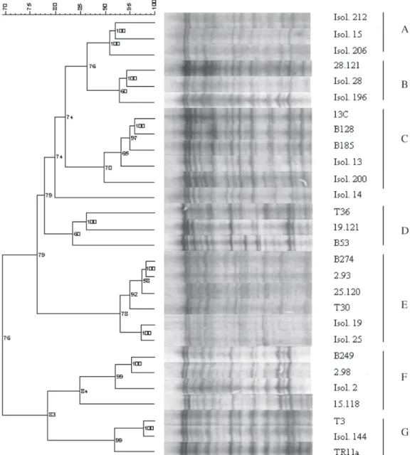
Electrophoretic separation of the fragments produced by cleavase digestion of the CP gene PCR product caused patterns with several bands differ-ing in signal intensity (Fig. 1). Reaction tempera-ture may affect the number of DNA fragments pro-duced. The optimal temperature for the cleavase reaction was found to be 45°C. At this tempera-ture a greater number of bands (15–20) was ob-tained with the uppermost band present, signify-ing absence of over-digestion.
CFLP analysis of cloned CP gene standards
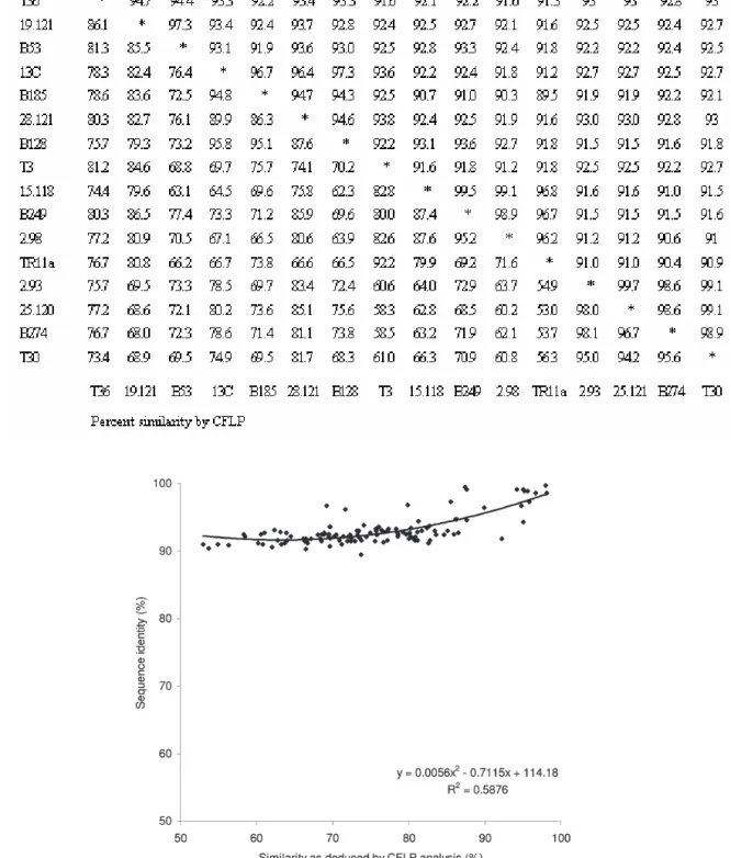
The 16 standards whose nucleotide sequence was known (sharing from 89.5% to 99.7% nucleotide identity) were assayed (Fig. 1), and the CFLP pat-tern similarities computed by the Pearson coefficient (Table 3). The ability of CFLP analysis to estimate the sequence similarity was inferred by comparing both sets of data (Fig. 2), showing a quadratic (sec-ond-degree polynomial) correlation with a statisti-cally significant correlation coefficient (P<0.01). As can be seen in Fig. 2, CFLP was highly sensitive in detecting nucleotide differences above 90%: for in-stance, it was able to discriminate two clones, 2.93 and 25.120, which differed in only 2 positions. The two closest clones deduced by CFLP (2.93 and B274) differed in 10 nucleotides, and their patterns had a Pearson similarity coefficient of 98.1%.
The similarity data from CFLP (Table 3) were used to construct a dendrogram (Fig. 1) in which several clusters could be recognised. The 16 cloned standards analysed are represented on clusters B to G. The most striking aspect here was that in general the standards were grouped according to the biological properties of the isolates from which they were obtained. Standards obtained from se-vere sweet orange stem-pitting isolates (B128, 13C, B185) clustered in group C or in group F (15.118, 2.98, B249), while clones from mild isolates (T30,
Fig. 1. Dendrogram and respective CFLP patterns of the 12 isolates and 16 cloned standards. Major clusters are indicated on the right (A, B, C, D, E, F and G). The rule-bar at the top-left represents the similarity (%) between patterns. Numbers in the dendrogram refer to the cophenetic correlation values. Isolates 19 and 25 were the only ones that did not induce stem pitting in sweet orange (SP-SwO) or grapefruit, nor quick decline, thus being consid-ered mild ones.
2.93, 25.120, B274) clustered in group E. Clones from severe quick-decline isolates were clustered in a less distinct mode in groups G and D. A very similar dendrogram was obtained when the stand-ards were clustered according to sequence similar-ity (Fig. 3), the only exception being standards T3 and Tr11a, which now appeared separated. To test reproducibility of CFLP, the set of 16 clones of the
CP gene was re-analysed at two dates about 6 months apart and the dendrogram from the CFLP patterns was mostly identical at both dates.
CFLP analysis targeted to the CTV CP gene of infected plant material
Infected plant material from biologically char-acterised sources (Table 1) was analysed by
immu-Table 3. Percent nucleotide identity obtained from sequence data (above diagonal) and percent similarities as esti-mated by CFLP (below diagonal) of the 16 cloned standards of the coat protein gene.
nocapture RT-PCR followed by CFLP analysis tar-geted to the CP gene. The similarities between band patterns were computed and used to cluster the isolates in the same dendrogram as for the stand-ards (Fig. 1). With the exception of isolate 14, which was not clearly assigned to any group, the remain-ing 11 isolates appeared in different clusters, fre-quently grouped with isolates or standards with which they shared some kind of geographic or bio-logical relationship. For instance, the mild isolates 19 and 25 (from Spain) were grouped with clones of cluster E, which came from plants described as having mild symptoms. Isolate 144 from Reunion Island was grouped in cluster G, together with a
clone from Reunion Island. Isolate 28 and one clone originated from it, clone 28.121, were grouped clos-er in the same clustclos-er B. Isolate 13 was grouped in cluster C along with standards obtained from plants which caused stem pitting on sweet orange (SP-SwO), and with a standard (13C) that was ob-tained from itself. Isolate 2, which is a SP-SwO isolate from Madeira Island, produced two differ-ent clones, clone 2.98 and clone 2.93. Clone 2.98 was grouped together with isolate 2 and other clones that were produced by SP-SwO isolates. Clone 2.93 was grouped in cluster E with mild iso-lates and mild-origin clones (in accordance with the sequence clustering in Fig. 3).
Fig. 3. Dendrogram showing UPGMA clustering of the coat protein (CP) gene sequences of the standards used. CP clones 15a and 8–1 were not analysed by CFLP. Letters on the right refer to the cluster as determined by CFLP.
As it was clear that some isolates harboured a mixture of sequence variants, we used CFLP to screen isolates that had been subjected to inocu-lations of known origin. Plants 196, 200 and 206 were inoculated with a mixture of mild and stem pitting isolates and showed SP-SwO symptoms. Both mild and SP-SwO components were later retrieved by cloning and sequencing the genomic variants, thus confirming the mixed character of the infection (results not shown). A new cluster (A) appeared, which did not contain any of the previous cloned standards. In this cluster some isolates were grouped known to harbour a mix-ture of haplotypes: isolate 206 which is an artifi-cial mixture of isolates 25 and 212; isolate 15 (from which were obtained the haplotypes 15.118, group F, and 15a, group C - see Fig. 1). Isolate 212 was also in this group. All the isolates from this clus-ter caused SP-SwO. Isolate 200, which was an artificial mixture of isolates 25 and 8, was clus-tered in group C. Although isolate 8 was not ana-lysed by CFLP, the sequence of a haplotype ob-tained from this isolate clustered in group C (Fig. 3). The only isolate which clustered in an unre-lated group was isolate 196.
SSCP analysis of cloned CP gene standards
The SSCP analysis of the 16 cloned standards is shown in Fig. 4. Only conspicuous bands were considered to group the patterns. Six standards had unique patterns that could not be grouped. The remaining ten standards fell into three different
patterns, named 1 to 3. Only group 2 had a close relationship to group E as defined by CFLP, the other two did not.
Discussion
Optimising the CFLP technique and cleavase digestion
CFLP analysis usually relies on the radioac-tive labelling of one DNA strand. However, O’Connell et al. (1999) obtained discriminating patterns by labelling both strands. Using the band patterns generated by both strands makes it pos-sible to silver stain the gels, thus avoiding radio-active labelling, and this was done in this work. In this way the protocol is much simpler and more user-friendly. Silver staining was tried in an ear-lier study (Rossetti et al., 1997) but without much success because of the limited number of bands produced with the CFLP protocol used. In the present study the CFLP patterns for DNA frag-ments with 673 bp consisted of 15 to 20 bands. Different patterns were formed by sets of bands differing in signal intensity. When establishing the optimum conditions for the CFLP reaction it is desirable to obtain patterns with a maximum number of bands. According to the manufacturer, a major parameter that determines the number of bands is the temperature of the cleavase diges-tion. Temperatures that are too low do not pro-duce enough bands, while temperatures that are too high lead to over-digestion and the disappear-ance of the slower migrating bands. Avoiding
digestion is important since it ensures that the whole set of fragments will be present. In this work results obtained at 45°C were deemed to be adequate.
Comparative ability of CFLP and SSCP to distinguish and cluster genomic variants
The similarity of cloned standards obtained by CFLP analysis is in close relationship with simi-larities computed at the nucleotide level. This re-sulted in very similar dendrograms obtained with both methods. Moreover, as can be seen in Fig. 2, the CFLP technique was sensitive enough to de-tect minor changes at the nucleotide level even between very similar sequences (e.g. those with a nucleotide similarity greater than 90%). As a re-sult, all the 16 clones showed different patterns with CFLP and in the pool of sequences tested, the smallest difference detected was of only 2 nucle-otides. This sensitivity makes CFLP a good meth-od to compare viral genes, as for instance the coat protein gene, in which variants of the same spe-cies usually have a greater than 85% nucleotide identity.
SSCP is also suitable for the detection of poly-morphisms in CTV (Rubio et al., 1996; Nolasco et al., 1997) and other RNA viruses and is now very commonly used. A short report on the SSCP analy-sis of the 16 standards used in this study present-ed at an earlier date (Marques et al., 2002) showpresent-ed that this technique had only a limited ability to group genomic variants. Of the three groups that could be formed, only one was in agreement with the clusters formed by CFLP and nucleotide se-quence analysis. With SSCP the emphasis is usu-ally on its capacity to distinguish between sequence variants. For instance, Rubio et al. (1996) used this method to discriminate between certain CTV var-iants that differed in only 1 nucleotide. However, CTV has a high capacity for genomic variation, with each isolate being associated with genetically re-lated variants (Rubio et al., 2001). Thus the main aim when using SSCP to compare two CTV iso-lates should be to ascertain their degree of simi-larity, rather than to determine whether the iso-lates are different or not (which however they usu-ally are). SSCP is therefore a very limited tech-nique since it does not relate the migration of mo-lecular conformations to the nucleotide sequence (Orita et al., 1989). Thus the information obtained
with SSCP is less useful than that obtained with CFLP when the objective is to determine the rela-tionships between sequences. Another advantage of the CFLP technique, reported also by O’Connell et al. (1999) and Brow et al. (1996), is that it yields reproducible results, which SSCP does not always do (O’Connell et al., 1999).
CFLP and the typing of CTV isolates
Cost-effective molecular methods to character-ise CTV variants and establish their relation to already characterised strains are vital for early CTV control. An important aspect of CFLP analy-sis is that it clusters the majority of CTV isolates together with cloned standards of the same ori-gin and also with isolates having similar biologi-cal properties. This characteristic seems to be the direct result of the ability of CFLP to cluster iso-lates according to the nucleotide sequence rela-tionship, which makes this technique very prom-ising for CTV typing. Under natural conditions trees are frequently infected with mixtures of CTV strains, which may be distributed irregularly (Grant and Higgins, 1956). When isolates are com-posed of a mixture of sequence variants, the re-sults need to be interpreted carefully. For in-stance, isolate 2 is composed of a mixture of se-quence variants 2.98 and 2.93 which cluster in different groups; in this case the CFLP pattern of isolate 2 clustered close to the pattern obtained from clone 2.98 in a group of severe isolates. On the contrary, the CFLP pattern from isolate 19 clustered according to biological properties, far from the only clone from this isolate (19.121) that was analysed by CFLP. However, further work (not presented) showed that clone 19.121 is a mi-nor variant present in isolate 19. In the other cas-es, when mixtures of mild and SP-SwO compo-nents were studied, the CFLP patterns clustered close to the stem-pitting component. Since se-quencing showed both variants were present, probably the SP-SwO component multiplied at a higher rate, being easily detected. Further stud-ies using artificial mixtures of cloned standards should be carried out to estimate the proportion of components that will cause the pattern to shift from one type of cluster to another. From a prac-tical point of view, being able to detect the severe components so that they can be eradicated is very important.
CFLP thus is an alternative to SSCP analysis, providing useful information that enables the re-lationships between isolates to be established ac-cording to their degree of sequence similarity, and, indirectly, according to their biological activity.
Acknowledgements
Parts of this work were supported by the grant POCI/AGR-2004/59090 from the Fundação para a Ciência e Tecnologia, Portugal.
Literature cited
Acosta O., A. Alegria, M. Guzman, R. Lee, C. Niblett and J. Peñaranda, 1994. El Virus de la Tristeza de los
Citri-cos: Una Grave Amenaza para la Citricultura Colombi-ana. Evidencias Epidemiologicas y Moleculares. 1st
edi-tion, Editorial Científica, Santafé de Bogotá, Colombia, 58 pp.
Akbulut M., S. Baloglu, E.A. Momol, H.R. Pappu, S.S. Pap-pu, V.J. Febres, M.A. Yilmaz, R.F. Lee and C.L. Niblett, 1996. Comparison of the capsid protein gene sequences of five Turkish isolates of Citrus tristeza virus. In:
Pro-ceedings of the 13th Conference of the International Or-ganization of Citrus Virologists, International
Organi-zation of Citrus Virologists (J.V. da Graça, P. Moreno, R.K. Yokomi, ed.), Riverside, CA, USA, 318–322. Albiach-Martí M.R., J. Guerri, M. Cambra, S.M. Garnsey
and P. Moreno, 2000. Differentiation of Citrus tristeza
virus isolates by serological analysis of p25 coat
pro-tein peptide maps. Journal of Virological Methods 88, 25–34.
Ayllón M.A., L. Rubio, A. Moya, J. Guerri and P. Moreno, 1999. The haplotype distribution of two genes of Citrus
tristeza virus is altered after host change or aphid
trans-mission. Virology 255, 32–39.
Bonacalza B., 1998. Tipificação Biomolecular de
Isolamen-tos do Virus da Tristeza dos Citrinos em Portugal. MSc
Thesis, Instituto Superior de Agronomia, Universidade Técnica de Lisboa, Lisboa, Portugal, 112 pp.
Brow M.A.D., M.C. Oldenburg, V. Lyamichev, L.M. Heisler, N. Lyamicheva, J.G. Hall, N.J. Eagan, D.M. Olive, L.M. Smith, L. Fors and J.E. Dahlberg, 1996. Differentia-tion of bacterial 16S rRNA genes and intergenic regions and Mycobacterium tuberculosis katG genes by struc-ture-specific endonuclease cleavage. Journal of
Clini-cal Microbiology 34(12), 3129–3137.
Febres V.J., L. Ashoulin, M. Mawassi, A. Frank, M. Bar-Joseph, K.L. Manjunath, R.F. Lee and C.L. Niblett, 1996. The p27 protein is present at one end of Citrus
tristeza virus particles. Phytopathology 86, 1331–1335.
Fofana B., J.C. Martiat, J.P. Baudoin and P. du Jardin, 1998. Cleavase fragment length polymorphism (CFLP): a methodology to further exploit polymorphisms from PCR products of plastid DNA (ptDNA) in Phaseolus.
Plant Molecular Biology Reporter 16, 271–282.
Gago-Zachert S.P., N.B. Costa, L. Semorile and O. Grau, 1999. Sequence variability in p27 gene of Citrus
triste-za virus (CTV) revealed by SSCP analysis. Electronic Journal of Biotechnology 2(1), 16–25.
Gillings M., P. Broadbent, J. Indsto and R. Lee, 1993. Char-acterisation of isolates and strains of citrus tristeza clos-terovirus using restriction analysis of the coat protein gene amplified by the polymerase chain reaction.
Jour-nal of Virological Methods 44, 305–317.
Grant T.J. and R.P. Higgins, 1956. Occurrence of mixtures of tristeza virus strains in Citrus. Phytopathology 47, 273–276.
Guerri J., P. Moreno and R.F. Lee, 1990. Identification of
Citrus tristeza virus strains by peptide maps of virion
coat protein. Phytopathology 80(8), 692–698.
Guerri J., P. Moreno, N. Muñoz and M.E. Martinez, 1991. Variability among Spanish Citrus tristeza virus isolates revealed by double-stranded RNA analysis. Plant
Pa-thology 40, 38–44.
Hall T.A., 1999. BioEdit: a user-friendly biological sequence alignment editor and analysis program for Windows 95/ 98/NT. Nucleic Acids Symposium Series 41, 95–98. Karasev A.V., V.P. Boyko, S. Gowda, O.V. Nikolaeva, M.E.
Hilf, E.V. Koonin, C.L. Niblett, K. Cline, D.J. Gumpf, R.F Lee, S.M. Garnsey, D.J. Lewandowski and W.O. Dawson, 1995. Complete sequence of the Citrus
triste-za virus RNA genome. Virology 208, 511–520.
Kong P., L. Rubio, M. Polek and B.W. Falk, 2000. Popula-tion structure and genetic diversity within California
Citrus tristeza virus (CTV) isolates. Virus Genes 21(3),
139–145.
Manjunath K.L., H.R. Pappu, R.F. Lee, C.L. Niblett and E.L. Civerolo, 1993. Studies on the coat protein genes of four isolates of citrus tristeza closterovirus from In-dia: cloning, sequencing and expression. In:
Proceed-ings of the 12th Conference of the International Organ-ization of Citrus Virologists, International OrganOrgan-ization
of Citrus Virologists (P. Moreno, J.V. da GraÁa, L.W. Timmer, ed.), Riverside, CA, USA, 20–26.
Marques N., C.L. Niblett and G. Nolasco, 2002. Typing of citrus tristeza variants by Cleavase Fragment Length Polymorphism analysis. In: Proceedings of the 15th
Conference of the International Organization of Citrus Virologists, International Organization of Citrus
Virol-ogists (N. Duran-Vila, R.G. Milne, J.V. da GraÁa, ed.), Riverside, CA, USA, 438 (abstract).
Marshall D.J., L.M. Heisler, V. Lyamichev, C. Murvine, D.M. Olive, G.D. Ehrlich, B.P. Neri and M. de Arruda, 1997. Determination of Hepatitis C Virus genotypes in the United States by Cleavase Fragment Length Polymor-phism analysis. Journal of Clinical Microbiology 35, 3156–3162.
Mawassi M., E. Mietkiewska, R. Gofman, G. Yang and M. Bar-Joseph, 1996. Unusual sequence relationships be-tween two isolates of Citrus tristeza virus. Journal of
General Virology 77, 2359–2364.
Moreno P., J. Guerri, M.R. Albiach, J.F. Ballester-Olmos and M.E. Martínez, 1996. Interference between Citrus
tristeza virus (CTV) isolates detected by analysis of
dou-ble stranded RNA (dsRNA). In: Proceedings of the 13th
Conference of the International Organization of Citrus Virologists, International Organization of Citrus
Virol-ogists (J.V. da GraÁa, P. Moreno, R.K. Yokomi, ed.), Riv-erside, CA, USA, 54–63.
Moreno P., J. Guerri, J.F. Ballester-Olmos, R. Albiach and M.E. Martínez, 1993. Separation and interference of strains from a Citrus tristeza virus isolate evidenced by biological activity and double-stranded RNA (dsRNA) analysis. Plant Pathology 42, 35–41.
Moreno P., J. Guerri and N. Muñoz, 1990. Identification of Spanish strains of Citrus tristeza virus by analysis of double-stranded RNA. Phytopathology 80(5), 477–482. Narváez G., B.S. Skander, M.A. Ayllón, L. Rubio, J. Guerri and P. Moreno, 2000. A new procedure to differentiate
Citrus tristeza virus isolates by hybridisation with
dig-oxigenin-labelled cDNA probes. Journal of Virological
Methods 85, 83–92.
Niblett C.L., H. Genc, B. Cevik, S. Halbert, L. Brown, G. Nolasco, B. Bonacalza, K.L. Manjunath, V.J. Febres, H.R. Pappu and R.F. Lee, 2000. Progress on strain dif-ferentiation and its application to the epidemiology of
Citrus tristeza virus. Virus Research 71, 97–106.
Nolasco G., C. de Blas, V. Torres and F. Ponz, 1993. A meth-od combining immunocapture and PCR amplification in a microtiter plate for the detection of plant viruses and subviral pathogens. Journal of Virological
Meth-ods 45, 201–218.
Nolasco G. and O.A. de Sequeira, 1997. New approaches in analysing genomic variability of Citrus tristeza virus. In: Filamentous Viruses of Woody Plants (P.L. Monette, ed.), Research Signpost, Trivandrum, India, 109–120. O’Connell C.D, D.H. Atha, M.C. Oldenburg, J. Tian, M.
Sie-bert, R. Handrow, K. Grooms, L. Heisler and M. de Ar-ruda, 1999. Detection of p53 gene mutation: Analysis by single-strand conformation polymorphism and Cleav-ase fragment length polymorphism. Electrophoresis 20, 1211–1223.
Orita M., Y. Suzuki, T. Sekiya and K. Hayashi, 1989. Rapid and sensitive detection of point mutations and DNA
polymorphisms using the polymerase chain reaction.
Genomics 5, 874–879.
Pappu S., C. Niblett, R. Lee and E. Civerolo, 1993. Com-parative sequence analysis of the coat protein of biolog-ically distinct citrus tristeza closterovirus isolates.
Vi-rus Genes 7(3), 255–264.
Peñaranda J., O. Acosta, M. Guzmán-Barney, A. Alegria, H.R. Pappu, S.S. Pappu, K.L. Manjunath, V.J. Febres, R.F. Lee and C.L. Niblett, 1996. Incidence and charac-terisation of mild and severe isolates of Citrus tristeza
virus from Colombia. In: Proceedings of the 13th Con-ference of the International Organization of Citrus Vi-rologists, International Organization of Citrus
Virolo-gists (J.V. da Graça, P. Moreno, R.K. Yokomi, ed.), Riv-erside, CA, USA, 71–77.
Rosner A., R.F. Lee and M. Bar-Joseph, 1986. Differential hybridisation with cloned cDNA sequences for detect-ing a specific isolate of Citrus tristeza virus.
Phytopa-thology 76, 820–824.
Rossetti S., S. Englisch, E. Bresin, P.F. Pignatti and A.E. Turco, 1997. Detection of mutations in human genes by a new rapid method: cleavage fragment length polymor-phism analysis (CFLPA). Molecular and Cellular Probes 11, 155–160.
Rubio L., M.A. Ayllón, J. Guerri, H. Pappu, C. Niblett and P. Moreno, 1996. Differentiation of citrus tristeza clos-terovirus (CTV) isolates by single-strand conformation polymorphism analysis of the coat protein gene. Annals
of Applied Biology 129, 479–489.
Rubio L., M.A. Ayllón, P. Kong, A. Fernández, M. Polek, J. Guerri, P. Moreno and B.W. Falk, 2001. Genetic varia-tion of Citrus tristeza virus isolates from California and Spain: evidence for mixed infections and recombination.
Journal of Virology 75, 8054–8062.
Zemzami M., C.M. Soares, A.M. Bailey, C.L. Niblett and G. Nolasco, 2002. Molecular characterization and classifi-cation of Morrocan isolates of citrus tristeza closterovi-rus. In: Proceedings of the 15th Conference of the
Inter-national Organization of Citrus Virologists, InterInter-national
Organization of Citrus Virologists (N. Duran-Vila, R.G. Milne, J.V. da Graça, ed.), Riverside, CA, USA, 8–12.