https://doi.org/10.1177/1179548420956365
Creative Commons Non Commercial CC BY-NC: This article is distributed under the terms of the Creative Commons Attribution-NonCommercial 4.0 License (https://creativecommons.org/licenses/by-nc/4.0/) which permits non-commercial use, reproduction and distribution of the work without further permission provided the original work is attributed as specified on the SAGE and Open Access pages (https://us.sagepub.com/en-us/nam/open-access-at-sage).
Clinical Medicine Insights: Circulatory, Respiratory and Pulmonary Medicine Volume 14: 1–6
© The Author(s) 2020 Article reuse guidelines: sagepub.com/journals-permissions DOI: 10.1177/1179548420956365
Introduction
Pulmonary embolism (PE) is the third most fatal cardiovascu-lar disorder. The main contributing factor is a persistently high and increasing incidence of PE cases in the general population,
with around 0.4 symptomatic cases per 1.000 person-years.1
Fatality rates of PE are still unacceptably high and are
responsible for a short-term case fatality rate of 3.9% to 12%1-4
among clinically recognizable cases, not to mention the sudden death clinical presentation cases, that may represent 20% to
25% of all PE cases.5,6 As these events can be silent and
misdi-agnosed, an early and accurate diagnosis of acute PE is vital for initiating emergent directed therapy and preventing deaths. The delay in the diagnosis of acute PE is a common feature of the disease as almost one third of patients are diagnosed only
after 5 days from the onset of symptoms,7 equally divided
between patient delays in getting medical care and long time from medical assistance to diagnosis. The heterogeneity and lack of specific symptoms and signs of PE clinical presentation may contribute to the physicians’ attributable delay to diagnosis alongside limited access to the definite diagnostic test, only
assured by the pulmonary CT scanning.8 The current
diagnos-tic approach to acute PE, entailing a laborious and non-specific integration of clinical judgment, laboratory D-dimers results
and diagnostic imaging, could be revolutionized by the discov-ery of new sensitive and readily available biomarkers.
In recent years, platelet indices have been studied on cardio-vascular diseases, and an elevated mean platelet volume (MPV), in particular, has been associated with coronary artery disease and with cardiovascular risk factors, of which diabetes and
metabolic syndromes gathered the most evidence.9-11 Although
MPV value is automatically available to the prescribing physi-cian from any ordinary electronic blood cell count, this param-eter is only exceptionally used in the clinical practice. Nevertheless, recent attentions have focused on MPV as a potential diagnostic and prognostic biomarker in several
con-ditions.12 A recent meta-analysis reported a higher value of
MPV in deep vein thrombosis patients, compared to control groups, but it included studies with mixed deep vein thrombo-sis and pulmonary embolism populations, and heterogenous clinical presentations such as acute and non-acute phases of the
diseases.13 Contradictory results and shortness of studies justify
why the role of MPV in diagnosing acute PE is still uncertain and, therefore, undervalued in clinical practice.
This study aims to systematically review publications on MPV determinations in acute PE patients and to conduct a meta-analysis on differences of MPV between acute PE
Diagnostic Role of Mean-Platelet Volume in
Acute Pulmonary Embolism: A Meta-analysis
and Systematic Review
Cláudia Febra
1and Ana Macedo
2,31Faculdade de Medicina, Universidade do Porto, Porto, Portugal. 2Keypoint, Consultoria Científica, Lda. 3Departamento de Ciências Biomédicas e Medicina, Universidade do Algarve, Faro, Portugal.
ABSTRACT
BACkgRounD: Acute pulmonary embolism (PE) is the third most fatal cardiovascular disease. PE is frequently misdiagnosed due to its
clinical presentation’s heterogeneity and the inexistence of biomarkers for its immediate diagnosis. Mean platelet volume (MPV) has shown a potential role as a biomarker in acute PE. In this analysis, we aimed to systematically compare the MPV in patients with and without defi-nite diagnosis of PE, in emergency departments.
METhoDS: Embase, PubMed and Medline were searched for relevant publications, in English. The main inclusion criteria were studies
which compared MPV in patients with acute PEA versus a control group.
RESulTS: Thirteen studies consisting of a total number of 2428 participants were included. Of the participants included, 1316 were patients
with confirmed acute PE, and 1112 were assigned to the control group. MPV was significantly higher in patients with acute PE than in con-trols (RR: 0.84, 95% CI: 0.76 – 0.92; P < .00001). There was a significant heterogeneity in the data.
ConCluSionS: This analysis showed higher MPV to be associated with acute PE immediate diagnosis. These data show promise for the
use of MPV as a readily available biomarker for the diagnosis of acute PE at the emergency department.
kEywoRDS: MPV, mean platelet volume, thrombosis/embolism, biomarker, coagulation, pulmonary embolism
RECEiVED: May 13, 2020. ACCEPTED: August 13, 2020. TyPE: Meta-Analysis
FunDing: The author(s) received no financial support for the research, authorship, and/or
publication of this article.
DEClARATion oF ConFliCTing inTERESTS: The author(s) declared no potential
conflicts of interest with respect to the research, authorship, and/or publication of this article.
CoRRESPonDing AuThoR: Cláudia Febra, Faculdade de Medicina da Universidade do
Porto, Rua Comandante José Simões Bento 67, 1495-696 Cruz Quebrada, Portugal. Email: [email protected]
Material).
Clinical question
The PICO model was used to define our clinical question:
Population – Clinical studies of patients with acute PE; Intervention – MPV detected by any laboratory method; Comparison – Control patients who are (1) Emergency
department controls (ruled out for acute PE diagnosis after imaging tests), or (2) Non-emergency department controls (inpatient or outpatient clinic patients without acute PE suspi-cion, or healthy); Outcome measure – Primary outcome: effect size (MPV in acute PE patients compared to controls). Secondary outcomes: assessment of quality and heterogeneity between studies.
Study design
All study types in which MPV was measured and compared to a control.
Search strategy and data extraction
A highly sensitive search strategy was conducted in EMBASE, PubMed and Medline without date restrictions. The keywords used for systematic searches were deep vein thrombosis, pul-monary embolism (PE), and MPV. Search terms were (Pulmonary Embolism [MeSH] OR Venous Thrombosis [MeSH] OR Venous Thromboembolism [MeSH]) AND (Mean Platelet Volume [MeSH] OR Blood Platelets [MeSH]). The last search was performed on 02/03/2020 and we re-ran the searches at final analyses’ procedures.
Study selection and inclusion criteria
We looked for studies assessing the measurement of MPV for acute PE in adult patients. Two independent reviewers (the authors) conducted title and abstract screening procedures of the initial search results and independently evaluated eligibility of the retrieved articles. Only those in which MPV was meas-ured at the exact moment of the acute PE provisional diagnosis and whose PE diagnosis was confirmed by contrast thoracic angiographic tomography were finally included for analysis. This review was limited to studies comparing acute PE patients with control individuals, and only included studies where the association of MPV and acute PE was explicitly investigated.
moderation.
Data items
Data extracted from each study included: authors, year of pub-lication, type of population (emergency department patients, or other clinical settings), type of study, number of patients and controls, characterization of controls (recruitment and inclu-sion criteria), age of patients and controls, MPV measurement method, and values of MPV for patients and controls.
Quality assessment
Quality assessment of the observational retrospective and pro-spective studies was carried out by the Newcastle-Ottawa scale
(NOS)15 where scores were attributed in number of stars
quan-titatively related to positive parameters.
Risk of bias and applicability were evaluated in Review Manager (RevMan 5.3, The Cochrane Collaboration, Oxford, UK). Domains were graded as low risk, high risk, or uncertain risk respecting biases. The studies were stratified in terms of risk of bias: low risk, when a maximum of 1 domain was tain” or “high”; moderate risk when 2 or 3domains were “uncer-tain” or “high”; and high risk when at least 4 domains were
“uncertain” or “high.”16
Statistical analysis
Standardized mean differences (SMDs) were considered for MPV within each study. SMD > 0 demonstrates an increased level of MPV.
Heterogeneity across studies was quantified using the I2
sta-tistics and an I2 ⩾ 50% pointed to significant heterogeneity.17
Publication bias was analyzed trough Egger’s Regression test and bias visualization was assessed using funnel plot.
Statistical analysis was carried out by the RevMan (software version 5.3) and R (software version 3.6.1, package “metaphor” and “PRROC”). The data are expressed as a value (95% confi-dence interval [CI]). P < .05 was considered statistically significant.
Results
Study selection
From a total of 42 studies, we isolated 13 studies which fulfilled all the inclusion criteria and had complete data on MPV values
at the time of acute PE diagnostic hypothesis for a patients’ group. The complete searching process is shown in Figure 1.
Study characteristics
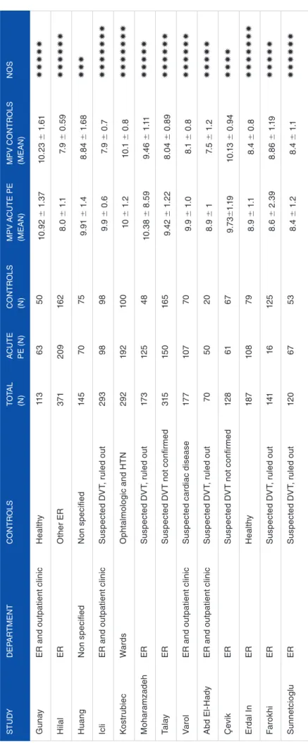
A total of 13 studies were included in the meta-analysis18,19,20-30.
There were 4 prospective cohort studies and 9 retrospective case-control studies for a total of 2428 participants, including 1316 patients and 1112 controls. In 7 studies, controls were specified as patients presenting suspected pulmonary embo-lism, although not confirmed after imaging tests results, which represented 576 individuals of the control group. The other control sub-groups were represented by healthy adults (229 individuals) or other emergency department patients (307 patients). 11 studies were performed in an emergency depart-ment and 1 study was conducted in the hospital ward. Only 1 study did not specify the clinical department.
Risk of bias within studies
No publication bias was identified through Egger’s test (P = .696), and low evidence of publication bias was observed through a funnel plot, as shown in Figure 2.
Quality assessment
The overall assessment of the 13 studies revealed a classifica-tion of 5 or more stars in 11 studies (Table 1), but only 2 studies included healthy and community controls (the same studies) and none was blinded to cases.
Results for MPV
Meta-analysis of acute PE versus controls using standard mean difference found an effect estimate of 0.84 (95% CI: 0.76–0.92) and a Z-score of 20.25 (P < .00001) demonstrat-ing a significant increase in MPV of patients with acute PE
compared to controls (Figure 3). However, Chi2 was 439.06
and I2 was 97%, showing significant heterogeneity in this
dataset.
ROC curve analysis
The ROC curve analysis of MPV when predicting acute PE were constructed on an exploratory basis (Supplementary Material). The optimal cut-off values for MPV when predict-ing acute PE was 8.5 fl (sensitivity 80%; specificity 60%). Figure 1. Search process.
Tab le 1 . Gener al pr oper ties and MPV f eatur es of the studies . S TU d y dEP AR TME n T Con TR ol S To TA l (n) A CUTE PE (n) Con TR ol S (n) MPV A CUTE PE (MEA n) MPV C on TR (MEA n) Gun ay E R a nd o ut pa tie nt c lin ic H ea lthy 11 3 63 50 10 .9 2 ± 1. 37 10 .2 3 ± 1. H ila l ER o th er ER 371 20 9 16 2 8.0 ± 1 .1 7. 9 ± 0. Hu ang n on s pe cifie d n on s pe cifie d 14 5 70 75 9. 91 ± 1. 4 8. 8 4 ± 1. Ic li E R a nd o ut pa tie nt c lin ic S us pe ct ed d V T, r ul ed o ut 29 3 98 98 9. 9 ± 0 .6 7. 9 ± 0 K os tr ub ie c Wa rd s o ph talm ol og ic an d HT n 29 2 19 2 10 0 10 ± 1. 2 10 .1 ± 0. M oh ar am zad eh ER S us pe ct ed d V T, r ul ed o ut 17 3 12 5 48 10 .3 8 ± 8 .59 9. 46 ± 1 Ta la y ER S us pe ct ed d V T n ot c on fir me d 315 15 0 16 5 9. 42 ± 1. 22 8.0 4 ± 0. Va ro l E R a nd o ut pa tie nt c lin ic S us pe ct ed c ar dia c d ise ase 17 7 10 7 70 9. 9 ± 1. 0 8.1 ± 0. A bd El -H ad y E R a nd o ut pa tie nt c lin ic S us pe ct ed d V T, r ul ed o ut 70 50 20 8. 9 ± 1 7. 5 ± 1. Ç ev ik ER S us pe ct ed d V T n ot c on fir me d 12 8 61 67 9.7 3± 1.1 9 10 .1 3 ± 0. Er da l I n ER H ea lthy 18 7 10 8 79 8. 9 ± 1 .1 8.4 ± 0. F aro kh i ER S us pe ct ed d V T, r ul ed o ut 141 16 12 5 8. 6 ± 2. 39 8. 86 ± 1 S un ne tc io gl u ER S us pe ct ed d V T, r ul ed o ut 12 0 67 53 8.4 ± 1. 2 8.4 ± 1 Ab br eviations: d VT , deep v ein thr ombosis; ER, emer genc y r oom; HT n , h yper tension; MPV , mean plat elet v alue; no S , n ew castlle-o tta w a scale; PE, pulmonar y embolism.
Discussion
To our knowledge, this is the first meta-analysis on the MPV values of acute PE at emergency department presentation. Our analysis showed significantly higher MPV in acute PE patients. However, likely due to diverse laboratory methods in MPV measurement, clinical manifestations’ heterogeneity in acute PE, and diverse controls, we found substantial heterogeneity and risk of bias across the studies. Furthermore, different time-frames from sampling to storage – which were not documented – may have influenced MPV values. Nonetheless, diagnostic criteria for patient selection did not vary between studies.
Data on the role of MPV in acute PE is misleading. We know that variation of platelet size occurs in different arterial and venous thrombotic conditions, but it is not clear whether the sizing changes are a cause of a pro-thrombotic status or the consequence of the presence of an intra-vascular thrombus, which may lead to an increase of younger and bigger platelets on blood samples. We found the best cut-off value for the pre-dictive value of MPV for acute PE to be 8.5 fl and this can be interpreted as a consequence of increased platelet aggregation.
Along with evidence on the influence of genetic
polymor-phisms31 and lifestyle factors32 on MPV (although these being
assumed to contribute to pathogenic mechanisms of cardiovas-cular diseases known to develop under their influence), studies on acute phases of venous thrombosis rise additional associa-tive hypotheses. MPV has been shown to increase in acute thrombotic conditions after platelet consumption and
acceler-ated platelet turnover,33 making it a potentially good
hyper-acute diagnostic marker for PE. Additionally, inflammatory mediators seem to give feedback to the bone marrow that results in changes towards a higher MPV and prothrombotic phenotype. This reinforces that higher MPV is a consequence of acute thrombosis and may be detected in the beginning of
acute PE pathologic processes.34,35
The pragmatic question raised with this meta-analysis results is: how can we gather MPV to other variables in the early diagnosis of acute PE?
Some particular aspects deserve special considerations when it comes to biomarkers, and to MPV as an acute PE biomarker specifically. First, technical conditions for MPV determination represent a great amount of results variability. MPV values may be affected by venipuncture and sample stor-age conditions, indicated by significant different results in
samples stored at room temperature for more than a 3-hour
period,36 which corresponds to an easily accepted time-frame
of delayed refrigeration for real clinical conditions. Similarly, platelet exposure to EDTA, the uniformly used anticoagulant in clinical laboratories, results in an increase of up to 50% on
MPV determinations.37 In the analytical phase, several other
factors contribute to differences in the MPV measurements by different automated analyzers, either impedance or optical
counters,38 and, in our opinion, the daily use of MPV as a
diagnostic marker for acute PE shall not precede the optimi-zation of guidelines for its quantification.
Second, we share Norris et al12 concerns about the small
difference on MPV absolute results between both arterial and venous thrombosis patients, and controls. The narrow diag-nostic range of MPV for acute PE complicates the definition of unquestionable cut-off values for its use in clinical practice, although this being already the case for d-dimers, the only blood biomarker used to determine PE diagnostic probability before imaging exams. In our opinion, this points out that investigation on the diagnostic efficacy of well-defined MPV measurements for acute PE in an early stage of the disease is justifiable by the present evidence. In summary, we believe that MPV values should not be used as a single diagnostic tool of acute PE. However, this parameter deserves further investiga-tion as a potential biomarker of this potential suddenly fatal event, in the light of clinical probability scores’ validation efforts.
Our meta-analysis has certain limitations. First, the majority of studies included represent small and somewhat heterogene-ous populations. Although we were very strict to select only absolutely clear acute PE events in the patient’s group, we had to accept some heterogeneity of controls. In fact, while some controls were healthy individuals, others were acute patients who presented to an emergency department and were suspected of having acute PE at admission. Even though these varied con-trols may have had some impact on the studies’ results, their exclusion would have implied a significant loss of patients included in the meta-analysis. Second, most of such studies were retrospective, which may have limited their conclusions. Moreover, missing information on counters and technical pro-tocols, on some demographic variables, on risk factors to venous thrombosis, or on time from symptoms to MPV determination, limited our evaluation on clinical heterogeneity. Third, there Figure 3. Forest plot for mean platelet volume: acute PE patients versus controls.
lives every year, worldwide. Conclusion
Our systematic review and meta-analysis reveal that MPV may be a promising biomarker for the immediate diagnosis of acute PE. Given the current gap of diagnostic biomarkers of acute PE, further research regarding MPV’s utility in this context shall be pursued.
Acknowledgements
The authors wish to thank Dr. Ricardo Racha-Pacheco for his writing assistance and Nilza Gonçalves for her support in exploratory statistical analyses.
Supplemental material
Supplemental material for this article is available online. RefeRenCeS
1. Alotaibi GS, Wu C, Senthilselvan A, McMurtry MS. Secular trends in inci-dence and mortality of acute venous thromboembolism: The AB-VTE popula-tion-based study. Am J Med. 2016;129:879.e19-879.e25. doi:10.1016/j. amjmed.2016.01.041.
2. Næss IA, Christiansen SC, Romundstad P, Cannegieter SC, Rosendaal FR, Hammerstrøm J. Incidence and mortality of venous thrombosis: A population-based study. J Thromb Haemost. 2007;5:692-699. doi:10.1111/j.1538-7836.2007. 02450.x.
3. Furlan A, Aghayev A, Chang CCH, et al. Short-term mortality in acute nary embolism: Clot burden and signs of right heart dysfunction at CT pulmo-nary angiography. Radiology. 2012;265:283-293. doi:10.1148/radiol.12110802. 4. White RH. The epidemiology of venous thromboembolism. Circulation.
2003;107(23 suppl 1):I4-I8. doi:10.1161/01.CIR.0000078468.11849.66. 5. Heit JA, Silverstein MD, Mohr DN, et al. The epidemiology of venous
thrombo-embolism in the community. Thromb Haemost. 2001;86:452-463.
6. Beckman MG, Hooper WC, Critchley SE, Ortel TL. Venous thromboembo-lism: a public health concern. Am J Prev Med. 2010;38(4 suppl):S495-S501. doi:10.1016/j.amepre.2009.12.017
7. Ageno W, Agnelli G, Imberti D, et al. Factors associated with the timing of diagnosis of venous thromboembolism: results from the MASTER registry.
Thromb Res. 2008;121:751-756. doi:10.1016/j.thromres.2007.08.009.
8. Elliott CG, Goldhaber SZ, Jensen RL. Delays in diagnosis of deep vein throm-bosis and pulmonary embolism. Chest. 2005;128:3372-3376. doi:10.1378/ chest.128.5.3372
9. Sansanayudh N, Anothaisintawee T, Muntham D, McEvoy M, Attia J, Thak-kinstian A. Mean platelet volume and coronary artery disease: a systematic review and meta-analysis. Int J Cardiol. 2014;175:433-440. doi:10.1016/j. ijcard.2014.06.028.
10. Maluf CB, Barreto SM, dos Reis RCP, Vidigal PG. Platelet volume is associated with the Framingham risk score for cardiovascular disease in the Brazilian Lon-gitudinal Study of Adult Health (ELSA-Brasil). Clin Chem Lab Med. 2016;54:879-887.
11. Sansanayudh N, Muntham D, Yamwong S, Sritara P, Akrawichien T, Thakkin-stian A. The association between mean platelet volume and cardiovascular risk factors. Eur J Intern Med. 2016;30:37-42. doi:10.1016/j.ejim.2015.11.028. 12. Noris P, Melazzini F, Balduini CL. New roles for mean platelet volume
measure-ment in the clinical practice? Platelets. 2016;27:607-612. doi:10.1080/09537104. 2016.1224828.
13. Kovács S, Csiki Z, Zsóri KS, Bereczky Z, Shemirani AH. Characteristics of platelet count and size and diagnostic accuracy of mean platelet volume in
17. Huedo-Medina TB, Sánchez-Meca J, Marín-Martínez F, Botella J. Assessing heterogeneity in meta-analysis: Q statistic or I 2 Index? Psychol Methods. 2006;11:193-206. doi:10.1037/1082-989X.11.2.193.
18. Günay E, Sarinc Ulasli S, Kacar E, et al. Can platelet indices predict obstruction level of pulmonary vascular bed in patients with acute pulmonary embolism? Clin
Respir J. 2014;8(1):33-40. doi:10.1111/crj.12031.
19. Hilal E, Neslihan Y, Gazi G, Sinan T, Zeynep Ayfer A. Does the mean platelet volume have any importance in patients with acute pulmonary embolism? Wien
Klin Wochenschr. 2013;125:381-385. doi:10.1007/s00508-013-0380-9.
20. Huang J, Chen Y, Cai Z, Chen P. Diagnostic value of platelet indexes for pulmo-nary embolism. Am J Emerg Med. 2015;33:760-763. doi:10.1016/j.ajem.2015. 02.043.
21. Icli A, Aksoy F, Turker Y, et al. Relationship between mean platelet volume and pulmonary embolism in patients with deep vein thrombosis. Hear Lung Circ. 2015;24(11):1081-1086. doi:10.1016/j.hlc.2015.04.170.
22. Kostrubiec M, Łabyk A, Pedowska-Włoszek J, et al. Mean platelet volume pre-dicts early death in acute pulmonary embolism. Heart. 2010;96:460-465. doi:10.1136/hrt.2009.180489.
23. Moharamzadeh P, Rahmani F, Foroughifar S, Shahsavarinia K. Reliability of platelet indices for diagnosing pulmonary embolism; a Brief report. Adv J Emerg
Med. 2019;3:e27. doi:10.22114/ajem.v0i0.137.
24. Talay F, Ocak T, Alcelik A, et al. The new diagnostic marker for acute pulmo-nary embolism in emergency department; mean platelet volume. Afr Health Sci. 2014;14:94-99. doi:10.4314/ahs.v14i1.15
25. Varol E, Icli A, Uysal BA, Ozaydin M. Platelet indices in patients with acute pulmonary embolism. Scand J Clin Lab Invest. 2011;71:163-167. doi:10.3109/003 65513.2010.547596.
26. Abd El-Hady Abd El-Ghany E, Abdelaziz A, Abd El-Raof Abd El-Fatah R, et al. Role of hemogram parameters in diagnosis and assessing severity of pulmo-nary embolism. Egypt J Chest Dis Tuberc. 2019;68:194-202. doi:10.4103/ejcdt. ejcdt_36_18.
27. Çevik İ, Narcı H, Dündar GA, Ayrık C, Babuş SB. Is there a diagnostic value for the platelet indices patients in pulmonary embolism? Hong Kong J Emerg Med. 2018;25:91-94. doi:10.1177/1024907917743489.
28. In E, Deveci F, Kaman D, et al. The importance of mean platelet volume and red cell distribution width in acute pulmonary embolism. Acta Medica Mediterr. 2015;31:1209-1216.
29. Farokhi M, Seashore J, Epelbaum O. Complete blood count parameters in hos-pitalized patients with suspected pulmonary embolism. Am J Resp Crit Care Med. 2015;191:A4890.
30. Sunnetcioglu A, Karadas S. Mean platelet volume as an indicator of disease in patients with acute pulmonary embolisms. East J Med. 2014;19:107-111. 31. Gieger C, Radhakrishnan A, Cvejic A, et al. New gene functions in
megakaryo-poiesis and platelet formation. Nature. 2011;480:201-208. doi:10.1038/ nature10659.
32. Panova-Noeva M, Schulz A, Hermanns MI, et al. Sex-specific differences in genetic and nongenetic determinants of mean platelet volume: results from the Gutenberg health study. Blood. 2016;127:251-259. doi:10.1182/blood-2015-07-660308. 33. Montoro-García S, Schindewolf M, Stanford S, Larsen OH, Thiele T. The role
of platelets in venous thromboembolism. Semin Thromb Hemost. 2016;42:242-251. doi:10.1055/s-0035-1570079.
34. Burstein SA, Peng J, Friese P, et al. Cytokine-induced alteration of platelet and hemostatic function. Stem Cells. 1996;14(suppl. 1):154-162. doi:10.1002/ stem.5530140720.
35. Harrison P, Goodall AH. Studies on Mean Platelet Volume (MPV) – New Edi-torial Policy. Platelets. 2016;27:605-606. doi:10.1080/09537104.2016.1225467 36. Daves M, Zagler EM, Cemin R, et al. Sample stability for complete blood cell
count using the Sysmex XN haematological analyser. Blood Transfus. 2015;13(4):576-582. doi:10.2450/2015.0007-15.
37. Diaz-Ricart M, Brunso L, Pino M, et al. Preanalytical treatment of EDTA-anticoagulated blood to ensure stabilization of the mean platelet volume and component measured with the ADVIA counters. Thromb Res. 2010;126(1):e30-e35. doi:10.1016/j.thromres.2010.04.002.
38. Lippi G, Pavesi F, Pipitone S. Evaluation of mean platelet volume with four hematological analyzers: Harmonization is still an unresolved issue. Blood Coagul