Contents lists available atScienceDirect
Environment International
journal homepage:www.elsevier.com/locate/envintImpacts of in vivo and in vitro exposures to tamoxifen: Comparative e
ffects on
human cells and marine organisms
T.G. Fonseca
a,b, T. Carriço
a, E. Fernandes
a, D.M.S. Abessa
b, A. Tavares
c, M.J. Bebianno
a,⁎ aCIMA, Centro de Investigação Marinha e Ambiental, Universidade do Algarve, Campus Gambelas, 8005-135 Faro, PortugalbNEPEA, Núcleo de Estudos em Poluição e Ecotoxicologia, Aquática, Universidade Estadual Paulista (UNESP), Campus do Litoral Paulista, São Vicente, SP 11330-900, Brazil
cDepartamento de Ciências Biomédicas e Medicina, Universidade do Algarve, Campus Gambelas, 8005-135 Faro, Portugal
A R T I C L E I N F O Handling Editor: Martí Nadal Keywords: Tamoxifen Marine organisms Human cells Biomarkers Cytotoxicity A B S T R A C T
Tamoxifen (TAM) is afirst generation-SERM administered for hormone receptor-positive (HER+) breast cancer
in both pre- and post-menopausal patients and may undergo metabolic activation in organisms that share similar receptors and thus face comparable mechanisms of response. The present study aimed to assess whether
en-vironmental trace concentrations of TAM are bioavailable to thefilter feeder M. galloprovincialis (100 ng L−1)
and to the deposit feeder N. diversicolor (0.5, 10, 25 and 100 ng L−1) after 14 days of exposure. Behavioural
impairment (burrowing kinetic), neurotoxicity (AChE activity), endocrine disruption by alkali-labile phosphate (ALP) content, oxidative stress (SOD, CAT, GPXs activities), biotransformation (GST activity), oxidative damage (LPO) and genotoxicity (DNA damage) were assessed. Moreover, this study also pertained to compare TAM cytotoxicity effects to mussels and targeted human (i.e. immortalized retinal pigment epithelium – RPE; and
human transformed endothelial cells – HeLa) cell lines, in a range of concentrations from 0.5 ng L−1 to
50μg L−1. In polychaetes N. diversicolor, TAM exerted remarkable oxidative stress and damage at the lowest
concentration (0.5 ng L−1), whereas significant genotoxicity was reported at the highest exposure level
(100 ng L−1). In mussels M. galloprovincialis, 100 ng L−1TAM caused endocrine disruption in males,
neuro-toxicity, and an induction in GST activity and LPO byproducts in gills, corroborating in genotoxicity over the exposure days. Although cytotoxicity assays conducted with mussel haemocytes following in vivo exposure was
not effective, in vitro exposure showed to be a feasible alternative, with comparable sensitivity to human cell line
(HeLa).
1. Introduction
In recent years, the occurrence of pharmaceuticals in the aquatic environment is recognized as one of the worldwide emerging issues regarding environmental quality and human health (Heberer, 2002). Increasing consumption of medicines leads to the continuous load of pharmaceuticals´ parent compounds and metabolites into wastewater effluents and treatment plants (WWTPs), where low removal effi-ciencies have been reported (Daughton, 2016;Furuhagen et al., 2014; Hernando et al., 2006; Pereira et al., 2017). Therefore, it is well es-tablished that several hundreds of pharmaceutical compounds are ubiquitous in WWTPs effluents, ground and surface waters, ultimately reaching estuarine and marine ecosystems (Frédéric and Yves, 2014; Verlicchi and Zambello, 2014).
Among the variety of pharmaceuticals classes released into the aquatic environment, anticancer drugs are one of the critical groups
that raise concern regarding the ecotoxicological potential risk of long-term exposure to non-target organisms (Besse et al., 2012;Booker et al., 2014;Rowney et al., 2009). Overall, the mode of action (MoA) of these molecules rely on the interaction with DNA, cellular growth factors, proteins and signalling pathways that lead to apoptosis, thus promoting the tumor cell death (Gačić et al., 2014). Nevertheless, detrimental effects of anticancer drugs are not restricted to the highly proliferative cells or onto cancer drivers, thus presenting cytotoxic, mutagenic, carcinogenic and teratogenic potential to all normal growing cells (Mater et al., 2014;Parrella et al., 2014).
Endocrine therapy is an alternative anticancer treatment that uses selective mechanisms of interaction with cancer targets, applied in-dividually or in combination with cytotoxic molecules (Hoskins et al., 2009). It consists of the administration of a structurally diverse group of compounds that binds to the estrogen receptorsα (ERα) and β (ERβ), and may produce estrogen agonist or antagonist effects, depending of
https://doi.org/10.1016/j.envint.2019.05.014 Received 22 January 2019; Accepted 6 May 2019
⁎Corresponding author.
E-mail address:[email protected](M.J. Bebianno).
0160-4120/ © 2019 The Authors. Published by Elsevier Ltd. This is an open access article under the CC BY-NC-ND license (http://creativecommons.org/licenses/BY-NC-ND/4.0/).
the targeted tissue (Goldstein et al., 2000;Paterni et al., 2014). These molecules are referred as selective estrogen receptor modulators (SERM) (An, 2016;Goldstein et al., 2000;Rodenas et al., 2015). The antagonist effect following the formation of estrogen receptor-SERM complexes is crucial for the management of hormone-dependent breast cancer and tumor cell survival (Goldstein et al., 2000;Zheng et al., 2007;Criscitiello et al., 2011).
Tamoxifen (TAM) is afirst generation-SERM administered for hor-mone receptor-positive (HER+) breast cancer in both pre- and post-menopausal patients (Goetz et al., 2007). Its antitumor ability derives from the antagonism towards the proliferative action of estrogen through competitive binding to ERs (Goldstein et al., 2000). As a pro-drug, TAM requires a complex metabolic activation to elicit its designed pharmacological activity (Kisanga et al., 2005). The 4-hydro-xytamoxifen and 4-hydroxy-N-desmethyltamoxifen (endoxifen) are the main products of hepatic oxidation, elicited by CYP2D6 and CY3A4, with high anti-estrogen potential in humans (Murdter et al., 2012; Johnson et al., 2008) (Fig. 1). Since pharmaceuticals active compounds are designed to have a specific MoA, targeting a specific metabolic and molecular pathway in humans, their biological activity may occur in organisms that share similar receptors and face comparable mechan-isms of responses (Fent et al., 2006). Conservation of biomolecules during evolution may trigger similar responses of chemicals in different non-targeted biological systems (Franzellitti et al., 2013;Zhang et al., 2017). Although organisms may lack homologues of estrogen receptors, they may contain genes for estrogen-related receptors (ERRs) that, ac-cording to (Borgatta et al., 2015) are structurally close to human ERα and ERβ.
Assumptions based on physico-chemical properties indicate that TAM has a high hydrophobic potential (Kow= 6.3), thus prone to ad-sorb to organic particles (Orias et al., 2015) and settle onto bottom sediments. However, once in coastal areas, TAM as other pharmaceu-ticals molecules, can be subject to physico-chemical and biological conditions different from those found in freshwater environments (e.g.
salinity, pH, temperature, turbidity, organic compounds, microbial population) and thus TAM may present different chemical behaviour and unexpected partition over water, suspended particles and sedi-ments, therefore a distinct persistence (Lara-Martín et al., 2014;Schmitt et al., 2008;Weigel et al., 2002).
Ecotoxicity assays conducted with TAM, in freshwater and marine species, showed inhibition in the larval development of the sea urchin Spherechinus granularis (Pagano et al., 2001), a decrease in egg pro-duction of thefish Tautogolabrus adspersus (Mills et al., 2015), and an alteration of vitellogenin gene expression in thefish Sparus aurata L. (García-Hernández et al., 2016) (see details inTable 1). These effects were detected at a concentration range from ng L−1 toμg L−1, en-compassing TAM concentrations detected in aquatic compartments (Table 1). Despite the increasing efforts devoted to comprehend the risks of TAM occurrence in aquatic compartments, there is still a lack of environmental concentrations screening in coastal and marine ecosys-tems, as well as information around its bioavailability and potential routes of exposures that may derive toxicological effects to non-target species (Lara-Martín et al., 2014;Roberts and Thomas, 2006;Thomas and Hilton, 2004).
Bivalves have been extensively used in in vivo studies to assess biological effects in relation to pharmaceuticals exposures (Cortez et al., 2012;Gonzalez-Rey and Bebianno, 2012, 2013, 2014;Oliveira et al., 2017a, 2017b). Regardless the MoA of pharmaceuticals, the marine mussel Mytilus galloprovincialis has been used to assess ecotoxicological effects including impacts at cellular level, oxidative stress, embry-otoxicity, immunotoxicity, genotoxicity and metabolomics/tran-scriptomic outcomes (Gonzalez-Rey and Bebianno, 2012, 2013, 2014; Oliveira et al., 2017a, 2017b; Schmidt et al., 2011; Teixeira et al., 2017). The cytotoxic effects of cisplatin in M. galloprovincialis after in vivo exposure arise insights over the subtle impairments that anticancer drugs may induce at very low concentrations, over a long-term ex-posure time (Trombini et al., 2016). On the other hand, in vitro toxicity assays conducted with bivalves primary cell cultures, showed to be a Fig. 1. Metabolic pathways of tamoxifen in humans, following activation by cytochrome P450.
simple and sensitive approach compared to whole animal models (Binelli et al., 2009;Katsumiti et al., 2018;Katsumiti et al., 2014) and provide basic information on the MoA of tested agents towards mole-cular and cellular mechanisms (Zhang et al., 2017) and underpin how different cellular models respond to complex environmental stressors (Balbi et al., 2017). To date, there is a lack of information related to the effects of marine organisms' cells in in vitro exposures to anticancer drugs. Haemocytes play a key role in the immune defence system, by means of phagocytosis of foreign particles or microorganisms, and in-tracellular production of ROS following exposure to xenobiotics (Cajaraville and Gómez-Mendikute, 2003; Lacaze et al., 2015). They may be applied as a sensitive marker to assess human-derived impacts on the aquatic health status (Toufexi et al., 2013).
In light of the trend of increasing concentrations of anticancer drugs in the aquatic environment, even though below human therapeutic doses, it is important to understand whether pharmaceuticals are likely to pose a risk to the biota via similar metabolic pathways detected in humans (Furuhagen et al., 2014;Mater et al., 2014). Estrogen receptor expression varies across species and can be differentially expressed according to tissues (McGinnis and Crivello, 2011). Human cell lines such as the retinal pigment epithelium (RPE) cell line have shown to express ER subtypes (Koulisis et al., 2016;Wang et al., 2015), in con-trast to cervical carcinoma HeLa cells, in which ER does not mediate estrogen responsive reporter expression (Klinge, 2015;Maminta et al., 1991;Shanle and Xu, 2010).
Polychaetes are sediment dwelling organisms from estuarine and marine benthic zones which possess high ecological relevance, besides being an important component of an ecotoxicological toolbox to assess the impact of contaminants in sediments due to their life habits that provide a constant contact with chemicals adsorbed onto particles and interstitial water (Freitas et al., 2015). Putative detrimental effects of anticancer drugs in sediments have been depicted through biochemical impairments in the polychaete N. diversicolor, exposed to the cytotoxic drug cisplatin (0.1 to 100 ng Pt L−1) (Fonseca et al., 2017) and cyclo-phosphamide (10 to 1000 ng L−1) (Fonseca et al., 2018), and the am-phipod Corophium volutator exposed to the drug methotrexate (1 to 1000 ng L−1) (Moreira et al., 2016). Due the paucity of research to effects of TAM to marine organisms, particularly considering their routes of uptake, one of the aims of the present study was to investigate whether environmental trace concentrations of TAM are bioavailable to thefilter feeder M. galloprovincialis and the deposit feeder N. diversi-color. The in vivo approach involves the assessment of behavioural im-pairment (burrowing kinetic imim-pairment), neurotoxicity (AChE ac-tivity), endocrine disruption by alkali-labile phosphate (ALP) content, oxidative stress (SOD, CAT, GPXs activities), biotransformation (GST activities), oxidative damage (LPO) and genotoxicity (DNA damage).
Moreover, this study also pertained to compare TAM cytotoxicity ef-fects to mussels and targeted human (i.e. immortalized retinal pigment epithelium– RPE; and human transformed endothelial cells – HeLa) cell lines, in a range of concentrations from 0.5 ng L−1to 50μg L−1, that encompass environmental relevant levels and those considered a worst-case scenario.
2. Materials and methods 2.1. Chemicals
Tamoxifen analytical standard (CAS 10540-29-01) was purchased from Sigma-Aldrich (Portugal). For safety handling of the drug, the experimental work was performed using class II biological safety ca-binet, with appropriate clothing (open-back, impervious chemotherapy protection gown, double powder-free latex gloves and safety goggles). Since TAM is not readily soluble in water, the stock solution was pre-pared in the carrier solvent dimethyl sulfoxide (DMSO). Experimental solutions were prepared from sequential dilutions in ultrapure Milli-Q water, with afinal concentration of 0.001% (v/v) DMSO in order to avoid a solvent toxic effect.
2.2. In vivo bioassay with polychaetes Nereis diversicolor
Polychaetes N. diversicolor and sediment samples, for sediment characterization purposes, were collected in an intertidal mudflat in Mira River Estuary (SW, Portugal), during low tide, and transported alive to the laboratory. Sediment grain size distribution and organic matter content were determined according to methods described by Royse (1970)andGross (1971)(seeFonseca et al., 2018).
Animals were maintained in glass aquariafilled with aerated nat-ural seawater (salinity 35) and sediments from the site of origin, during 5 days before testing. A total of 75 polychaetes was randomly selected and added to each treatment, in a triplicate design (25 animals per aquaria), in 10 L glass aquaria containing sediments and seawater (1:4 ratio, respectively). Specimens were unexposed: controls (CT0 (day 0) and CT14 (day 14) and exposed to DMSO (0.001%), and to TAM (0.5, 10, 25 and 100 ng L−1) for 14 days. Experimental set-up was kept under constant aeration and light period (12:12 h), and physico-chemical parameters registered (pH 7.92 ± 0.2; temperature 19 ± 1 °C; salinity 35 ± 1). During the experiment, water was carefully renewed every 48 h avoiding sediment resuspension, with addition of DMSO (0.001%) and TAM concentrations into water column aftermath. Animals were not fed during the bioassay.
For burrowing and comet assays, specimens were immediately handled and managed for respective analysis, while those regarding Table 1
TAM Concentrations in water (ng L−1), suspended solids and sediments (ng g−1) from ground, riverine and marine waters.
Study area Groundwater River Marine Reference
Surface water Suspended solids Sediment Seawater Suspended solids Sediment
France – < 5.8–25 – – – – – Coetsier et al. (2009)
Japan – – 658 0.042 – – – Azuma et al. (2017)
Germany 6–16.5 – – – – – – Reh et al. (2013)
England – 27–212 – – – – – Roberts and Thomas (2006)
England – < 10 – – – – – Ashton et al. (2004)
England – 13–71 – – – – – Thomas and Hilton (2004)
England – < 10–23 – – – – – Hilton et al. (2003)
Spain – 12.4–26.8 – – – – – López-Serna et al. (2012)
Spain 11.2–223 – – – – – – López-Serna et al. (2013)
Spain – 12–38 – – – – – Fernando-Climent et al. (2014)
Spain – 0.5–25 – – – – – Franquet-Griell et al. (2017)
USA – – – – – 8–44 7 Lara-Martín et al., 2014
USA – n.d. - 11.2 – – – – – Lara-Martín et al. (2015)
USA – – – – 93 – – Nödler et al. (2014)
biochemical end-points (AChE, SOD, CAT, GST, GPx, LPO) were rinsed with clean seawater and stored at−80 °C until further use. For the DNA damage analysis coelomic cells of N. diversicolor (5 specimens per aquaria, in triplicate; total of 15 organisms per treatment) were ex-tracted from the posterior region of the polychaete body into 20μL of PBS buffer with a 0.5 mL-syringe fitted with hypodermic needle (adapted fromLewis and Galloway, 2008).
2.2.1. Burrowing assay
The behavioural endpoint of the burrowing assay was verified only over the exposure of N. diversicolor. Polychaetes from control condi-tions, DMSO and TAM-exposed were submitted to a burrowing test after the 14-days bioassay, according toFonseca et al. (2017). Polychaetes (5 specimens per aquaria, in triplicate; total of 15 organisms per treat-ment) were carefully and individually displaced from their test con-tainers and transferred to 150 mL-plastic flasks, filled with natural seawater and 5 cm of sediments. The vertical position of polychaetes in sediment column was recorded every 2 min, to assess the time for fully burrowing in the 30 min-assay. The results are expressed as the per-centage (%) of un-burrowed specimens, over time (min).
2.3. M. galloprovincialis in vivo bioassays
Mussels (5.2 ± 0.4 cm shell length) were collected in the Ria Formosa coastal lagoon (Portugal; N 37° 0′ 29.4546″, W 7° 59′ 41.265″) and acclimated over 5 days in tanksfilled in natural seawater (salinity 35) under constant aeration and controlled photoperiod (12 h: 12 h light/dark). Animals were fed with Tetraselmis chuii only during the acclimation time, and water was renewed every other day in this period. Afterwards, 150 mussels were randomly distributed in 20 L glass tanks, in a duplicate design (75 mussels per treatment). They were exposed over 14 days to DMSO (0.001%) and 100 ng L−1 of TAM, jointly with unexposed mussels (CT0 and CT14).
Physico-chemical parameters were daily registered (pH 7.86 ± 0.19; temperature 19 ± 1 °C; salinity 37.4 ± 0.5) and water renewed every 48 h, following re-dosing of the drug or DMSO to the water column. Mussels were fed only with the plankton present in the natural seawater. By the end of the bioassay, no mortality was re-gistered. Mussels were collected at the beginning of the experiment (T0) and after 3, 7 and 14 days of exposure. Gills, digestive glands and go-nads were dissected and immediately frozen in liquid nitrogen and stored at −80 °C until further analysis of biochemical biomarkers (AChE, ALP, SOD, CAT, GST, GPx, LPO).
2.3.1. M. galloprovincialis in vitro bioassays
Sixty mussels M. galloprovincialis (5.0 ± 0.5 cm shell length) were handpicked in the Ria Formosa coastal lagoon and transported alive to the laboratory, where they were placed in a 20 L-tank (1 animal per 0.33 L−1)filled with natural seawater (salinity 35 ± 1) and kept under constant aeration. Animals were maintained over 48 h until haemo-lymph extraction. Cells collection was performed according to the procedure described inSection 2.3.2.
2.3.2. Sampling of mussel haemocytes
Cell extraction and incubation methods used were based on mod-ifications of protocols developed byGómez-Mendikute and Cajaraville (2003)andKatsumiti et al. (2014). Haemolymph was extracted from the posterior adductor muscle of the mussels with a sterile hypodermic syringe (1 mL) (25 G needle). About 300μL of haemolymph was in-dividually collected for cytotoxicity (3 specimens per aquaria, in du-plicate; total of 6 organisms per treatment) and genotoxicity (3 speci-mens per aquaria, in duplicate; total of 6 organisms per treatment), then transferred into a 1.5 mL microtube on ice (Gómez-Mendikute and Cajaraville, 2003). The handling procedure was performed under aseptic conditions in a vertical laminar airflow cabinet.
2.3.3. Cytotoxicity assessment
2.3.3.1. Cell counting and Trypan Blue assay. Firstly, 10μL of haemolymph was individually diluted in anti-aggregation solution (171 mM NaCl; 0.2 M Tris; 0.15% v/v HCl 1 N; 24 mM EDTA) in order to avoid cell clumping and agglomeration (Katsumiti et al., 2014). Cell staining was carried out through addition of trypan blue dye (0.4% in physiological solution; v/v) in a proportion of 1:2 (cell suspension: Trypan Blue 0.4%). Total cell counting was performed in a Neubauer chamber (200 cells per specimen) with the use of a haemocytometer and light microscopy (Olympus CH30). Cell viability was determined by the following equation:
= × ×
−
Concentration (cell mL ) Number of cells 10,000
Number of squares dilution factor 1
2.3.3.2. Neutral Red (NR) Assay. The NR assay was adapted from the protocol described in Olabarrieta et al. (2001)and Katsumiti et al. (2014). After total cell counting, cell suspensions were diluted at a density of 2 × 10⁵cells mL−1(in anti-aggregation solution), and 100μL seeded into 96-well microplates (6 replicates per animal). Microplates were centrifuged at 1200 rpm (10 min, 4 °C), in order to assist cells to settle and adhere. Supernatant was discarded, and cells of in vivo-exposed animals were re-suspended in 100μL of cell culture media Dulbecco's Modified Eagle Medium (DMEM, pH 7.4).
Further, cells were incubated with 50μL of neutral red working solution (0.4%, pH 7) over 1 h, at 18 °C, in order to allow uptake of the dye. Afterwards, cells were centrifuged at 1200 rpm for 10 min, at 4 °C, and carefully washed with PBS. The dye was then extracted from the viable cells in acetic acid/ethanol solution (1:100), following samples transference to U-bottom 96-well microplates and centrifuged at 1200 rpm, for 10 min, at 4 °C. Supernatants were placed inflat bottom microplates and absorbance measured at 550 nm (Infinite M200 Pro, TECAN®).
2.3.4. Mussels haemocytes
Cell suspensions were diluted at a density of 2 × 10⁵cells mL−1(in anti-aggregation solution), and 100μL seeded into 96-well microplates (6 replicates per animal; 3 animals per treatment). The in vitro cyto-toxicity approach consisted in the incubation of haemocytes, extracted from drug-unexposed mussels, exposed to DMSO and to increasing concentrations of TAM (0; 0.0005, 0.025, 0.05, 0.1, 0.5, 1, 10 and 25μg TAM L−1, prepared in DMEM at 0.001% DMSO). Microplates were incubated for 24 h at 18 °C. Afterwards, culture media was discarded, following NR procedure described in detail inSection 2.3.3.1. 2.3.5. Human cells
The RPE and HeLa cell lines were cultured in Dulbecco's Modified Eagle Media (DMEM). Immortalized RPE cell clones consist of a transfected cell line with the catalytic component of human telomerase (hTERT), which transient expression conserves telomers length without converting to a neoplastic transformed phenotype (Bodnar et al., 1998). HeLa cell line immortalized cells derived from cervical cancer tissues, with a remarkable growth potential (Lucey et al., 2009).
Cells were allowed to grow until confluence and then detached by trypsinization, then added to DMEM media, and maintained at 36 °C under. Cell viability was determined by Trypan Blue exclusion before cytotoxicity assessment. Further, the media was removed by aspiration and the cells washed twice with PBS. A 100μl aliquot of cell suspension, containing 2 × 10⁴cells, was seeded per well in 96-well microplates following incubation for 24 h at 36 °C for cell adherence. Afterwards, supernatant was removed and TAM solution, prepared in DMEM culture medium (at 0.001% DMSO), was seeded at an increasing range of TAM concentrations (0, 0.5, 25, 50, 100, 500, 1000, 10,000, 25,000, 50,000 ng L−1). Cells were incubated over 24 h, at 36 °C, following NR procedure (seeSection 2.3.3.1).
2.4. Biochemical analysis 2.4.1. Total protein
Total proteins concentrations (mg protein g−1 tissue) were de-termined according to the method described by Bradford (1976) adapted for microplate reader, using bovine serum albumin (BSA) as a standard.
2.4.2. ALP levels
The organic alkali-labile phosphate technique (Gagné and Blaise, 2000) was used to determine the vitellogenin content in the cytosolic fraction of gonadal tissues of M. galloprovincialis. Sex-differentiated gonads (3 specimens per aquaria, in duplicate; total of 6 organisms per treatment) were homogenized in 25 mM HEPES–NaOH buffer (pH 7.4) and centrifuged (12,000 ×g, at 2 °C for 30 min). Aliquots of super-natants were used for total protein content determination according to Bradford (1976)and ALP analysis, according to adaptations described bySchmidt et al. (2011).
2.4.3. Neurotoxicity
AChE activity was determined in whole tissues of N. diversicolor (2 specimens per aquaria, in triplicate; total of 6 organisms per treatment) and in gills of M. galloprovincialis (3 specimens per aquaria, in duplicate; total of 6 organisms per treatment). Samples were individually homo-genized in 100 mM Tris-HCl buffer (pH 8.0) with addition of 0.1% Triton, following centrifugation at 12,000g, for 30 min, at 4 °C. Supernatants were split into aliquots for total protein determination (Bradford, 1976) and AChE activity was analysed using the method of Ellman et al. (1961)adapted to microplate reader.
2.4.4. Antioxidant enzymes
Antioxidant enzymes activities were determined in whole tissues of N. diversicolor (2 specimens per aquaria, in triplicate; total of 6 organ-isms per treatment) and in both gills and digestive glands of M. gallo-provincialis (3 specimens per aquaria, in duplicate; total of 6 organisms per treatment). Samples were individually homogenized in 20 mM Tris-HCl buffer (0.5 M sucrose; 0.075 M KCl; 1 mM DTT; 1 mM EDTA; ad-justed to pH 7.6 with HCl), according to the protocol described by (Géret et al., 2002). Homogenates were centrifuged at 500 g, for 15 min, at 4 °C, and the supernatants obtained centrifuged again (12,000 g, 45 min, 4 °C). Enzymatic activities were analysed in the cytosolic frac-tion. SOD activity was assessed by measuring the decrease of absor-bance of the substrate cytochrome-c by xanthine oxidase/hypoxanthine system, at 550 nm (McCord and Fridovich, 1969), and the results are expressed as U mg−1protein. CAT activity was evaluated through the decrease of absorbance originated by hydrogen peroxide (H2O2) con-sumption at 240 nm, according toGreenwald (1985). GPX activity was measured at 340 nm by using cumene hydroperoxide and H2O2 as substrates for T-GPx and Se-GPx, respectively (adapted fromLawrence and Burk, 1976). The activity of CAT, Se- and T-GPx are expressed in nmol min−1mg−1protein.
2.4.5. Biotransformation enzyme activity
The cytosolic fraction for the determination of GST activity was obtained according the protocol described inSection 2.3.2. Measure-ment of this biotransformation enzyme activity was conducted in whole specimens of N. diversicolor (6 organisms per treatment) and tissues (gills and digestive glands) of M. galloprovincialis (6 organisms per treatment). Reaction mixture undergoes by the conjugation of 0.2 mM reduced glutathione (GSH) with 0.2 mM CDNB (molar coefficient of extinction = 0.6 mM−1cm−1) in a reaction mixture of 0.2 M KH2PO4/ K2HPO4buffer (pH 7.9), at 340 nm (adapted fromHabig et al., 1974). GST activity is determined in nmol min−1mg−1.
2.4.6. Oxidative damage
Polychaetes N. diversicolor (6 organisms per treatment) and tissues
(gills and digestive glands) of M. galloprovincialis (6 organisms per treatment) were individually homogenized in a Tris–HCl buffer (20 mM, pH 8.6) and butylated hydroxy toluene (BHT) (100:1μL, re-spectively). The resulting homogenates were centrifuged, for 45 min at 30,000g at 4 °C, in order to obtain the cytosolic fraction. Aliquots of supernatants were used for the quantification of total proteins (Bradford, 1976) and determination of oxidative damage (Erdelmeier et al., 1998), adapted for microplate reading. The absorbance of LPO by-products, the malondialdehyde (MDA) and (2E)-4-hydroxy-2-nonenal (4-HNE), was measured at 586 nm and expressed as nmol MDA + 4-HNE mg−1protein.
2.5. Genotoxicity assay
Genotoxicity was estimated by the alkaline Comet assay, as de-scribed inFonseca et al. (2018), following the determination of the amount of DNA in tail through the software Komet 5.5 (Kinetic Imaging Ltd). Results are expressed as mean ± standard deviation (S.D.). 2.6. Statistical analysis
Statistical analysis was carried out using the Statistica 8.0 software (Statsoft Inc., 2007, USA). The results were compared using parametric tests (ANOVA, followed by the Tukey's test), or non-parametric equivalent test (Kruskal-Wallis), according to data distribution and variances homogeneity (Shapiro-Wilk and Levene's tests, respectively). Results were significant when p < 0.05.
3. Results
3.1. Polychaete N. diversicolor 3.1.1. Burrowing behaviour
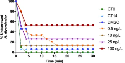
The percentage of polychaetes from CT0, CT14 and exposed to DMSO buried were 100%, 90% and 90%, respectively, within 6 min of bioassay. In contrast, organisms exposed to the different TAM con-centrations showed a general increase in burrowing time with the in-crease of TAM concentrations exposure. Polychaetes treated with 25 ng L−1 and 100 ng L−1failed to burrow after 4 min and 26% and 53% of polychaetes, respectively, remained unburied (Fig. 2). 3.1.2. AChE activity
No differences in AChE activity were detected among controls (i.e. CT0, CT14) and DMSO (p > 0.05) (Fig. 3). Polychaetes exposed to TAM showed an induction of AChE activity, except for those exposed to 10 ng L−1.
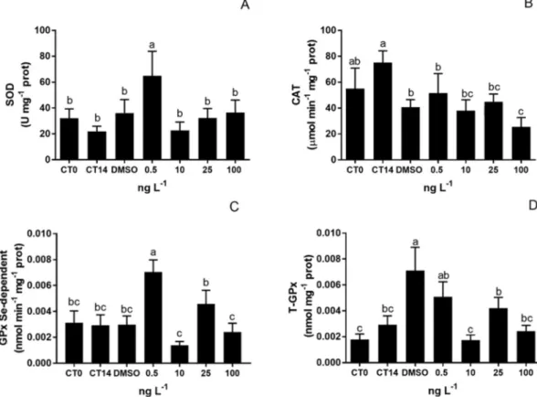
3.1.3. Antioxidant enzyme responses
Overall, enzymatic activities of unexposed specimens (i.e. CT0, CT14) and exposed to DMSO were similar (p > 0.05), with the ex-ception of T-GPx that indicated an increase in DMSO exposed group. In polychaetes exposed to the lowest TAM concentration (i.e. 0.5 ng L−1), SOD activity increased compared to all other treatments (p < 0.05) (Fig. 4-A). Although not significant (p > 0.05), there was an increasing trend of SOD activity in the other TAM concentrations. Regarding CAT activity, a significant inhibition occurred at the highest TAM con-centration compared to control conditions (p < 0.05) (Fig. 4-B). The activity of GPx-Se and T-GPx, like for SOD, also increased in poly-chaetes exposed to 0.5 ng L−1of TAM (p < 0.05) (Fig. 4-C and D) (2-fold compared to controls).
3.1.4. Biotransformation enzyme activity
Relatively to GST activity, no significant alteration was observed among treatments (p > 0.05) (Fig. 5).
3.1.5. Oxidative damage
LPO levels in polychaetes controls and exposed to DMSO were si-milar (p > 0.05). In contrast, the exposure of polychaetes to TAM duced the generation of LPO by-products since there was a similar in-crease of MDA + 4HNE concentrations in all exposed animals compared to controls (p < 0.05;Fig. 6).
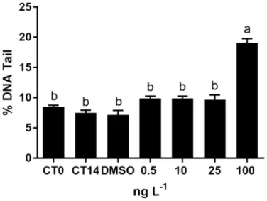
3.1.6. Genotoxicity
Coelomocytes from control conditions (CT0, CT14 and DMSO) in-dicated no significant changes in % of DNA tail (p > 0.05;Fig. 6). In TAM-treated polychaetes, DNA damage was only addressed in speci-mens exposed to the highest drug concentration (100 ng L−1) (p < 0.05;Fig. 6).
3.2. M. galloprovincialis bioassay 3.2.1. ALP levels
In female gonads, with exception of those exposed to DMSO, a de-creasing trend of ALP levels was observed over time, resulting in 1.4-fold decrease of CT and TAM-exposed mussels compared to the initial condition (p < 0.05). In males, although no significant differences over time were detected (p > 0.05), ALP levels of TAM-exposed mussels increased about 2-fold during the assay, with significant differences
compared to the respective controls, on days 7 and 14 (p < 0.05) while those exposed to DMSO decreased.
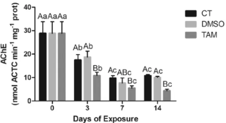
3.2.2. Neurotoxicity
A sharp decrease in AChE activity was observed in mussels from all treatments with significant differences over time (p < 0.05) (Fig. 9). In TAM-treated gills, AChE activity was inhibited, compared to controls, particularly at days 3 and 14 (p < 0.05).
3.2.3. Antioxidant enzymes
Enzymatic activity did not change over time in tissues of unexposed mussels (p > 0.05), with exception of a SOD increase in gills from animals exposed to DMSO in the last day of the experiment (p < 0.05) (Fig. 9-A). SOD activity in TAM treated groups did not change in gills during the 14 days of exposure (Fig. 10-A). In contrast, in digestive glands, SOD activity increased after 3 days of TAM-exposure, followed by its inhibition until the last day of experiment (p < 0.05) (Fig. 10-B). Regarding CAT activity, no changes were detected in gills of un-exposed mussels over the 14 days of experiment, although DMSO pre-sented a slight increase only at the 3rd day of experiment (p < 0.05). TAM-treated mussels showed significant fluctuations of CAT activity in gills, presenting a 2-fold increase at the 3rd day following a 4.7-fold inhibition at the 7th day (p < 0.05), until ultimately reaching levels similar to controls (Fig. 10-C). CAT activity in digestive glands of un-exposed mussels showed to decline over time, although not significant (p > 0.05). Regarding drug-exposed mussels, CAT activity increased 5-fold at the 3rd day, followed by a 4.7-5-fold decrease, at the 7th day, to levels similar to the control group (Fig. 10-D).
3.2.4. Biotransformation enzyme activity
GST activity in gills from unexposed mussels remained unchanged (p > 0.05). After 3 days of exposure, gills of TAM-contaminated mus-sels showed a significant increase in enzymatic activity compared to controls, following an inhibition trend until the end of the experiment, although not significantly different from controls (p > 0.05) (Fig. 11). A significant induction of GST activity was observed in digestive glands of mussels from control, and those exposed to DMSO and to TAM, re-garding the beginning of the experiment (p < 0.05).
3.2.5. Oxidative damage
Levels of LPO by-products were similar (p > 0.05) between mussels from control conditions, in both gills and digestive glands (Fig. 12). Regarding oxidative damage in gills of TAM-exposed mussels, LPO by-products were significantly higher than controls over the entire ex-periment (p < 0.05). In digestive glands of controls, levels of MDA and
Fig. 2. Percentage of unburied N. diversicolor over time (minutes) in controls (CT0; CT14) and exposed to DMSO and to TAM concentrations (0.5, 10, 25, 100 ng L−1).
Lines with symbols indicate the behaviour during the 30-min experiment.
Fig. 3. N. diversicolor AChE activity (mean ± S.D.) (ATC.min−1mg−1protein)
in unexposed (CT0; CT14) and exposed to DMSO and to TAM concentrations
(0.5, 10, 25 and 100 ng L−1). Different letters indicate significant differences
4-HNE did not change significantly, whereas in TAM-exposed mussels LPO levels increased over time with a 1.5-fold increment at the end of the experiment (p < 0.05).
3.2.6. Cell viability
The results regarding the mussel assay using trypan blue dye are depicted in Fig. 13. No significant differences in mussel cell viability were detected during the 14 days of experiment, except for TAM-treated mussels at the 7th and 14th days when compared with the beginning of the assay (p < 0.05).
Regarding the results of mussels´ cell viability through NR dye
(Fig. 14),fluctuations in absorbance were registered in all conditions with significant differences among times of exposure. Overall, Hae-mocytes indicated an increase in viability, except at the 7th day, with ultimate levels 3.3- and 4-fold higher than the initial condition, for controls and TAM-exposed mussels, respectively (p < 0.05).
3.2.7. Genotoxicity
The DNA damage expressed as % of tail was selected to assess genotoxicity caused by TAM exposure (Fig. 15). During the experiment, no significant changes were detected in DNA damage in haemocytes from unexposed mussels, although at the 14th day a slight increase was Fig. 4. N. diversicolor antioxidant enzymes activities (mean ± S.D.) expressed in unexposed polychaetes (CT0; CT14) and exposed to DMSO and to a range of TAM
concentrations (0.5, 10, 25 and 100 ng L−1): (A) SOD, (B) CAT, (C) GPx Se-dependent, (D) T-GPx. Different letters indicate significant differences among treatments
(One-way ANOVA; p < 0.05).
Fig. 5. N. diversicolor GST activity (mean ± S.D.) in unexposed (CT0; CT14)
and exposed to DMSO and to TAM concentrations (0.5, 10, 25 and 100 ng L−1)
Absence of letters indicate no differences among treatments (One-way ANOVA,
Kruskal-Wallis; p > 0.05).
Fig. 6. N. diversicolor LPO levels (mean ± STD) (MDA+ 4-HNE nmol·mg−1
protein) in unexposed (CT0; CT14) and exposed to DMSO and to different TAM
concentrations (0.5, 10, 25 and 100 ng L−1). Different letters indicate
registered in those exposed to the solvent (p > 0.05). Despite no sig-nificant differences detected between DMSO and TAM-exposed mussels, genotoxicity was always higher in haemocytes of mussels from the drug-treated group (p > 0.05).
3.3. In vitro cytotoxicity assessments 3.3.1. M. galloprovincialis
In haemocytes, TAM produced a significant reduction in cell via-bility at 0.5, 100, 500 ng L−1and 10μg L−1(p < 0.05). Although the wide range of increasing drug concentration, a dose-dependent cyto-toxicity was not observed (Fig. 16).
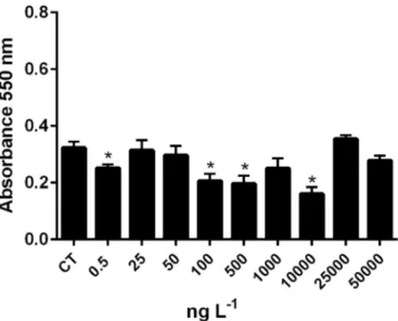
3.3.2. Human cell viability
RPE cells were not affected by any of the TAM concentrations (p > 0.05) (Fig. 17-A). In contrast, HeLa showed an increased viability after 24 h when exposed to 100 ng L−1of TAM. However, cytotoxicity occurred at 500 ng L−1, 10 and 50μg L−1of TAM (p < 0.05), with the absence of a dose-response profile (Fig. 17-B).
4. Discussion
Specific physicochemical properties of pharmaceuticals are known to provide predictable behaviour and fate in aquatic compartments. The high log Kowand low water solubility of TAM suggest a potential re-moval from wastewater through drug adsorption onto sewage sludge and sediments. However, detection of TAM in WWTPs effluents (con-centrations up to 369 ng L−1) (Roberts and Thomas, 2006) confirm its low removal efficiency (i.e. 17 to 43%) and drug recalcitrance over conventional technologies that use sorption processes ( Fernando-Climent et al., 2014). Once released in the aquatic environment, besides its residence in water column, pharmaceuticals compounds also sorb onto suspended organic matter, clay, sediments and microorganisms, that eventually settle in the bottom areas (Silva et al., 2011; Zenker et al., 2014;Rocha et al., 2015). Therefore, differential uptake, trophic transfer and bioaccumulation are experienced by aquatic species sub-jected to different ways of exposure, via water or sediment contact, although there is still a lack of information around TAM's bioavail-ability, that may derive toxicological effects to non-target species (Table 2) (Chapman and Wang, 2001;Du et al., 2014;Ribeiro et al., 2015;Torres et al., 2009;Yamamoto et al., 2009).
4.1. Effects of tamoxifen in N. diversicolor
The presentfindings indicated that SOD and Se-GPx activities sig-nificantly increase in polychaetes exposed to the lowest TAM con-centration (0.5 ng L−1) (p < 0.05) (Fig. 2). SOD upregulation illus-trates the prompt of the antioxidant system to overcome those toxic by-products, by means of converting superoxide radical into less toxic hydrogen peroxide (H2O2), following detoxification into water and oxygen by GPx's (Gonzalez-Rey and Bebianno, 2014;van der Oost et al., 2003). At the lowest TAM concentration, GPx may have exerted a cri-tical compensatory mechanism over the unchanged CAT activity (Cozzari et al., 2015;Marchi et al., 2017). The non-linear dose response characterized by the lower dose stimulation (0.5 ng L−1) observed in SOD and Se-GPx activities (Fig. 4), is referred as hormesis and may include mechanisms to overcompensate alterations in homeostasis and adaptive response based on inducible repair processes (Chapman, 2002; Hoffmann, 2009). In other words, a small exposure to a stressful sti-mulus increases the resistance of the cell or organism to a moderate or severe level of stress (Arumugam et al., 2006;Hoffmann, 2009). Besides repairing any damage, organisms also reduce background damage more effectively (Chapman, 2002). The hormetic curve is described as non-monotonic and biphasic shape, in which effects at low doses are op-posite to those at high doses (Hoffmann, 2009). This kind of relation-ship have been described from bacteria to vertebrates, as an outcome at Table
2 Ecotoxicological eff ects of TAM reported in coastal and marine species. Taxon Species Concentration range (ng L − 1) a Time (d) b Parameter Eff ect Reference Microalgae Isochrysis galbana 1– 5× 1 0 8 4 Growth inhibition EC50 = 3.5 × 10 7 Aguirre-Martínez et al. (2016a) Bivalve Mollusc Ruditapes phillipinarum 100 –50,000 14 Oxidative stress, oxidative damage, genotoxicity, oxidative damage 100 : *↑ EROD, LPO; *↓ GPx, DBF; 1000 : *↑ EROD, GPx, GR, LPO, DNA; *↓ DBF, GST; 10,000 : *↑ EROD, GPx, LPO ; *↓ DBF 50000 : *↑ EROD, GPx, GR, LPO ; *↓ AChE, GST, DBF Aguirre-Martínez et al. (2016a) Echinorderm Paracentrotus lividus 10 –10 9 1 b Fertilization EC 50 = 1.5 × 10 7 Aguirre-Martínez et al. (2016a) 10 –10 9 2 Larval Development EC 50 =1 ×1 0 9 10 − 8 -1 0 − 6M 0.5 b Fertilization 1 × 10 − 7M: *↓ Pagano et al. (2001) 10 − 8 -1 0 − 6M 0.5 b O ff spring Quality 1 × 10 − 6M: *↓ Spherechinus granularis 10 − 8 -1 0 − 6M 3 Larval Development 1 × 10 − 6M: Embryotoxicity *↑ Strongylocentrotus purpuratus – 4 Larval Development EC 50 =5 0 Roepke et al. (2005) Fish Tautogolabrus adspersus 5×1 0 5–5× 1 0 6a 17 Egg production 5 × 10 5:* ↓ Mills et al. (2015) Sparus aurata L. 100000 a 25 Gene expression Vitellogenin *↑ García-Hernández et al. (2016) 100000 a 25 Gene expression. Immune response (ilb1, tnfa, tgfb1,mhc1a, tlr9) *↑ 100000 a 25 Sperm concentration *↑ 100000 a 25 Sperm motility *↑
1000 different environmental stressors (Chapman, 2002;Mater et al., 2014), and is also attributed to the response of N. diversicolor exposed to the lowest CP concentration (10 ng L−1) (Fonseca et al., 2018).
The increase of AChE activity in polychaetes N. diversicolor, fol-lowing a concentration–response relationship (Fig. 3) was also observed on M. galloprovincialis exposed to 75 ng L−1 of fluoxetine over two weeks (Gonzalez-Rey and Bebianno, 2013), and on amphipods Ampe-lisca brevicornis exposed to sediments spiked with 0.5 ng L−1 of ibu-profen (Maranho et al., 2015). Likewise, Ruditapes phillipinarum showed an AChE induction at single exposures to 0.1μg L−1 of caffeine and 5μg L−1 of ibuprofen, following its inhibition at the highest con-centrations of pharmaceuticals (Aguirre-Martínez et al., 2016b). Ex-planation for such increased neurosynaptic effect was hypothesized according to particular drugs´ MoA, and also elucidated as a transitory mechanism to overcome an AChE inhibition (Milan et al., 2013).Deng et al. (2006)found that both anticancer drugs etoposide and excisanin A, administered in rats, upregulated AChE expression during apoptosis, in a mechanism driven by oxidative stress and activation of kinase signalling cascade, proposing that AChE activity is raised in the pre-sence of ROS. Ferreira et al. (2012)showed that an excess in AChE levels in rats may display a reduction of cholinergic neurotransmission efficiency and neurological dysfunctions, since the essential acet-ylcholine is rapidly hydrolysed in the synaptic cleft. In this sense, im-pairments in burrowing ability of polychaetes observed with the in-crease of drug concentration (Fig. 2) may be linked to such neurotoxic response. However, the burrowing impairment of N. diversicolor ad-dressed afterwards exposure to cisPt at 100 ng Pt L−1indicated a sig-nificant dose-response relationship and correlation to the inhibition of AChE activity (Fonseca et al., 2017), in contrast tofindings of poly-chaetes submitted to the alkylating agent cyclophosphamide, which behavioural changes occurred as a result of the oxidative stress dis-played by the drug rather than due to interferences of AChE levels (Fonseca et al., 2018). Therefore, even though anticancer drugs are generally classified as one therapeutic class, their specific MoAs may ultimately modulate environmental stimuli that, in the case of bur-rowing, has critical ecological consequences (Díaz-Jaramillo et al., 2013;Sovová et al., 2014).
In the case of TAM administration to human patients, an increase in enzymatic activity was registered for choline acetyltransferase, ac-countable for the synthesis of the acetylcholine neurotransmitter cri-tical for synapses (Silva et al., 2000). In mammalian brain, TAM acts as an ER agonist and exerts estrogen-like effects associated to modulation of neuroprotection and increase of human cognition, attention and verbal skills (Dicko et al., 1999;McEwen and Alves, 1999;Newhouse et al., 2013;Simpkins et al., 2009). Besides the beneficial effects of TAM in human neuroactivity, there are still confounding factors in the mammalian group itself related to features of hormonal status, age, tissue-specific activation of ERs, and whether ERs possess ability to bind estrogens or anti-estrogens at low concentrations (Lv et al., 2017; Newhouse et al., 2013). At a concentration range from 0.1 to 50μg L−1, TAM was unable to alter AChE activity in the freshwater clam Corbicula fluminea and disagree with the present observations, reinforcing that its role as a receptor-estrogen modulator in invertebrates is far from clear (Aguirre-Martínez et al., 2018).
The biotransformation enzyme, GST, conjugates reactions of active electrophilic metabolites or their parental compounds with reduced glutathione (GSH), enabling its transformation to more extractable hydrophilic metabolites (Luis et al., 2016;van der Oost et al., 2003). In addition, GST provides protection against ROS, playing a critical role in the defence against oxidative damage d peroxidative products of DNA and lipids (Nuwaysir et al., 1996). However, in human liver, TAM is extensively metabolized by phase I cytochrome P450 and phase II sulfotransferases (SULTs) and UDP-glucoronosyltransferases (UGTs) (Kiyotani et al., 2012), whereas the mechanism by which TAM affects GST expression is poorly studied. According toNuwaysir et al. (1996), TAM administration at relevant clinically therapeutic doses produce
significant suppression in GST mRNA expression in rat liver, resembling the outcomes associated to cisplatin MoA (Fuertes et al., 2003). Herein, the activity of GST did not change in polychaetes exposed to any TAM-concentration, in contrast to the significant inhibition in N. diversicolor specimens exposed to cisplatin at 100 ng Pt L−1(Fonseca et al., 2017). In the present case, either TAM metabolism may not be involved in GST phase II enzyme, or the concentration range applied in the bioassay was not enough to trigger this detoxification pathway.
In this study, TAM induced LPO in polychaetes exposed to all con-centrations (Fig. 6). TAM embeds itself in the lipid membranes and generates superoxide, which causes LPO and subsequent 4-HNE for-mation, following activation of caspase-3 cascade and cell death (Bekele et al., 2016). Such mechanism supports the contribution of oxidative damage on killing cancer cells during TAM therapy. Inter-ferences in the oxidative damage may be attributed either to the me-tabolic activation of TAM in N. diversicolor, following production of ROS, and/or by oxidative stress generated by TAM molecule intrusion in the lipid bilayer, which does not depend on the prodrug metabolism neither ERα expression and its activation (Bekele et al., 2016; Lushchak, 2011;Trachootham et al., 2009).
Accordingly, the noteworthy DNA damage herein revealed in spe-cimens exposed to the highest TAM concentration may be particularly linked to activation of prodrug at such great drug levels (Fig. 7), since the conversion of TAM into its intermediary putative active metabolite α-hydroxytamoxifen is a pre-requisite for DNA covalent binding and adducts formation (Boocock et al., 2002), as reported in human per-ipheral blood lymphocytes and MCF-7 cells using alkaline Comet Assay (Wozniak et al., 2007). Such explanation is plausible considering that the antioxidant disruption shown in coelomocytes from 100 ng L−1 treatment only occurred through inhibition of CAT activity in addition to levels of LPO by-products, that corroborate to those generated in all tested TAM concentrations, hence comparable grades of oxidative da-mage.
4.2. Effects of tamoxifen in M. galloprovincialis
Changes in ALP levels are positively related with those from vi-tellogenin-like proteins, which are naturally synthesized in females and, although present, are normally not expressed in males (Gonzalez-Rey and Bebianno, 2012, 2013;Matozzo et al., 2008;Sun et al., 2009). This biomarker has been used for endocrine disturbance assessment, since
Fig. 7. DNA damage (mean ± standard deviation) in coelomocytes of poly-chaetes N. diversicolor unexposed (CT0; CT14) and exposed to DMSO and to
different TAM concentrations (0.5, 10, 25 and 100 ng L−1). Different letters
indicate significant differences among treatments (One-way ANOVA;
the exposure of aquatic organisms to endocrine disruptors like xe-noestrogens and estrogens present in the water trigger the increase of vitellogenin levels in plasma (Sumpter and Jobling, 1995), indicating a sex-specific response (Sun et al., 2009). Males of M. galloprovincialis experienced a 2-fold increase of ALP after 14-days of exposure to ibu-profen at 250 ng L−1(Gonzalez-Rey and Bebianno, 2012, 2013). Simi-larly, despite exposure at an unrealistic environmental concentration, levels of vitellogenin-like proteins in males were induced after a 24-h exposure to gemfibrozil, at 1 mg L−1(Schmidt et al., 2011). Perturba-tions in endocrine homeostasis were also addressed in M. gallopro-vincialis collected in areas influenced by sewage effluents containing chemicals with estrogenic properties and vitellogenin-like proteins were induced both in male and female gonads (Pampanin et al., 2005). In mussels exposed to TAM (concentrations of 0.1 to 50μg L−1) an anti-estrogenic potential was detected in the freshwater clam Corbicula flu-minea, by reducing the levels of vitellogenin, although specimens were not sex-differentiated (Aguirre-Martínez et al., 2018). Moreover, the exposure of the adult male medaka Oryzias latipes to synthetic estrogen, EE2 (200 ng L−1), induce 4 × 10³-fold of vitellogenin to levels com-pared to unexposed males, whereas the co-exposure to TAM indicate an anti-estrogenic potential by reducing the presence of this egg protein in plasma (Sun et al., 2009). On the other hand, in the present study, ALP levels significantly increase in males exposed to TAM (at the 7th and 14th days) (Fig. 8). In accordance,Rodenas et al. (2015)detected an upregulation of hepatic vitellogenin mRNA in gilthead seabream after fifty days under a dietary administration containing TAM (100 μg g−1). These contrasting findings are in accordance to paradoxical agonist
interference of TAM towards ERs, able to trigger estrogenic disruptive mechanisms besides the anti-estrogenic designed MoA. The variable responses depend on particular interactions specific to cell types (García-Hernández et al., 2016), and it is also accountable for causing side effects in bones and uterus of patients under breast cancer treat-ment (Love et al., 1992;O'Regar and Gradishar, 2001). In this sense, TAM acts as a xenoestrogen in certain species and lead to the impair-ment of reproductive fitness or ultimately to feminization of male aquatic organisms (Gonzalez-Rey and Bebianno, 2012, 2013).
In contrast to N. diversicolor, M. galloprovincilis revealed an inhibi-tion of AChE activity during TAM exposure (Fig. 9). AChE inhibitors impair the cholinesterase enzyme from breaking down acetylcholine, critical for synapses and maintenance of memory processes, increasing both the level and duration of the neurotransmitter action (Colovic et al., 2013). Data available in the literature indicate that pharmaceu-ticals are relevant neurotoxic agents to aquatic organisms (Luis et al., 2016;Maranho et al., 2015;Milan et al., 2013;Munari et al., 2014). However, it is important to bear in mind that even though pharma-ceuticals are grouped as one type of emerging contaminants able to trigger neuronal disturbance by altering AChE activity, the vast array of therapeutic classes and their designed MoA in combination to species-specific properties (e.g. presence/absence of critical receptors, genes encoding enzymes for drug metabolism) lead to differential molecular docking, activation of biochemical pathways, detoxification metabo-lism and,finally, toxicological responses.
Similar to polychaetes, mussels exposed to TAM showed an induc-tion of SOD in digestive glands (Fig. 10-B). In the present study, after 7 days of exposure, CAT levels were significantly reduced in gills of TAM-treated specimens, whereas no SOD alteration occurred, as in polychaetes N. diversicolor submitted to 100 ng L−1 (Fig. 4-B). SOD activity increased in digestive glands at the 3rd day of exposure (Fig. 9 -B), corroborating the results obtained byTrombini et al. (2016), for M. galloprovincialis exposed CisPt (100 ng L−1). In addition, digestive glands of mussels exposed to TAM also showed a 3-fold increase in CAT activity (Fig. 9), and indicates an early activation of antioxidant me-chanisms, to counteract prooxidant forces and consequent damage. Since SOD represents a source of hydrogen peroxide, catalase is an extremely active and critical catalyst for the reduction of H2O2to H2O (Halliwell and Gutteridge, 2007), thus preventing these ROS to be converted to hydroxyl radicals through Fenton reaction, which are re-active initiators of membrane lipid peroxidation (Regoli and Giuliani, 2014).
Herein, gills of mussels exposed to TAM showed a significant GST induction at the 3rd day of exposure (Fig. 10-A), jointly with the CAT activity increase. In ROS-generating conditions, CAT and GST activities are pertinent to restore the cellular homeostasis before irreversible
Fig. 8. M. galloprovincialis ALP levels (mean ± STD) in sex-differentiated
go-nads (A: females; B: males), unexposed (CT) and exposed to DMSO and to TAM
(100 ng L−1) for 14 days. Capital and lower letters indicate significant
differ-ences among treatments of the same sampling day, and the same treatment over
different days (One-way ANOVA; p < 0.05).
Fig. 9. M. galloprovincialis AChE activity (mean ± STD) (ATC min−1mg−1
protein) in gills unexposed (CT) and exposed to DMSO and to TAM
(100 ng L−1), over 14 days. Capital and lower letters indicate significant
dif-ferences among treatments of the same sampling day, and the same treatment
damage occurs (Capolupo et al., 2016;Regoli and Giuliani, 2014). The basal GST activity in gills of M. galloprovincialis is three-fold higher than in digestive glands. Biotransformation response in digestive glands of Perna perna did not exhibit significant alterations at the diclofenac concentrations tested (20, 200 and 2000 ng L−1) (Fontes et al., 2018). In bivalves, digestive glands play a key role in absorption, metabolism and accumulation of xenobiotics, especially pharmaceuticals, and are thus considered an ideal tissue for estimating detoxification pathways, adverse effects of drugs to comprehend their MoA in this biological model (Canesi et al., 2007a, 2007b;Capolupo et al., 2016;Franzellitti et al., 2014). Despite proclaimed that induction of GST activity in gills could result from an attempt of ROS scavenging in this tissue rather than a biotransformation mechanism, acting in conjunction with CAT induction at the 3rd day (Regoli and Giuliani, 2014). In addition, the absence of upregulation of GST in digestive glands is also in agreement with the responses observed with N. diversicolor. Moreover, since GST and GPx use reduced glutathione (GSH) as electron donor for reducing H2O2to H2O, the absence of alterations in GST in digestive glands could be attributed to the competition for the substrate by both enzymes (Trombini et al., 2016). Therefore, further studies are needed to clarify comparisons regarding TAM pharmacodynamic in marine organisms and humans and comprehend if metabolic activation undergo via the same pathways.
Generation of LPO by-products occurred in gills, 4-fold higher than the oxidative damage elicited in digestive glands (Fig. 11). According to Bekele et al. (2016), TAM has a killing-cell potential that may be in-dependent of ERα expression, since the drug binds to lipid membranes following ROS formation and oxidative damage. Although the lower range of antioxidant and biotransformation activity in TAM-exposed digestive glands, compared to gills, mechanisms of defence were en-ough to counteract the action of reactive metabolites (Teles et al., 2016).
Regarding cytotoxicity obtained through Trypan Blue exclusion assay, no significant alterations in cell viability of TAM-exposed mussel were detected (Fig. 13). In contrast, results obtained by the NR method showed strongfluctuations and significant temporal differences in dye
retention (Fig. 14). In a cytotoxicity assessment conducted with gran-ulocytes of the European lobster Homarus gammarus exposed to com-mercial immunoestimulants, the NR assay exhibited a higher sensitivity relative to trypan blue exclusion, respectively indicating 0 and 56% of Fig. 10. M. galloprovincialis antioxidant enzymes activity (mean ± STD) of (A-B) SOD and (C-D) CAT in gills and digestive glands of unexposed (CT) and exposed to
DMSO and to TAM (100 ng L−1) for 14 days. Capital and lower letters indicate significant differences among treatments of the same sampling day, and the same
treatment over different days (One-way ANOVA; p < 0.05).
Fig. 11. M. galloprovincialis GST activity (mean ± STD) (nmol CDNB
min−1mg−1protein) in gills (A) and digestive glands (B) of unexposed (CT)
and exposed to DMSO and to TAM (100 ng L−1), over 14 days. Capital and
lower letters indicate significant differences among treatments of the same
sampling day, and the same treatment over different days (One-way ANOVA;
cell viability (Hauton and Smith, 2004). Such difference may be at-tributed to the fact that these methods measure different aspects of cell physiology. A reduction in NR uptake reflects a decrease in lipid membrane stability, which would usually precede cell death, whereas trypan blue only enters cells when the membranes become disrupted, a process which occurs at the time of, or shortly after, cell death. How-ever, in the present study, results indicated a continuous increase in cell viability in all treatments with time, ultimately culminating in a higher dye retention in TAM-treated mussels in comparison to controls.Miller et al. (2015)detected comparable results, in NR absorbance following in vivo exposure of Mytillus sp. to single (250 and 500μg L−1) and multi-walled nanocarbon (500μg L−1) were higher than unexposed ones.
Fig. 12. M. galloprovincialis LPO by-products (mean ± STD) (nmol
MDA + 4HNE mg−1protein) in gills (A) and digestive glands (B) of unexposed
(CT) and exposed to DMSO and to TAM (100 ng L−1) for 14 days. Capital and
lower letters indicate significant differences among treatments of the same
sampling day, and the same treatment over different days (One-way ANOVA; p < 0.05).
Fig. 13. M. galloprovincialis Cell viability (% of viable cells) using Trypan Blue
dye in haemocytes, unexposed (CT), DMSO and exposed to TAM (100 ng L−1),
at different times for 14 days. Capital and lower letters indicate significant
differences among treatments of the same sampling day, and the same
treat-ment over different days (One-way ANOVA; p < 0.05).
Fig. 14. M. galloprovincialis Cell viability (% of viable cells) using Neutral Red dye in haemocytes unexposed (CT), 0.001% DMSO and exposed to TAM
(100 ng L−1), for14 days. Capital and lower letters indicate significant
differ-ences among treatments of the same sampling day, and the same treatment over
different days (One-way ANOVA; p < 0.05).
Fig. 15. M. galloprovincialis DNA (mean ± standard deviation) in tail measured in mussel haemocytes unexposed (CT), DMSO (0.001%) and exposed to 100 ng
TAM L−1, for 14 days. Capital and lower letters indicate significant differences
among treatments of the same sampling day, and the same treatment over different days (One-way ANOVA; p < 0.05).
Fig. 16. M. galloprovincialis cell viability (Neutral Red Assay) of unexposed (CT) and exposed to TAM. Asterisks indicate significant differences between control and TAM-exposed cells (t-test; p < 0.05).
They suggested that this cytotoxic method performed in microplate was unable to detect a concentration-dependent toxic impairment to mussel haemocytes following in vivo exposure, contrarily to the successful re-sults reported in in vitro incubations. According to Weyermann et al. (2005)andFotakis and Timbrell (2006), different cytotoxicity assays can provide different results depending on the test agent. In addition, divergent of such profile of cytotoxicity is the genotoxicity responses, which demonstrated a higher % of DNA in tail of haemocytes exposed to TAM which were in accordance with damage effects. Partitioning of TAM into cell membrane generates a transmembrane signal transduc-tion cascade mediated by protein kinases, involved both in cell pro-liferation and apoptosis. Previous clinical trials revealed that TAM triggers apoptotic signalling cascade in human cell lines that either express and do not express ERs, and once activation of ERK, JNK and p38 kinases is suppressed, reducing cell viability blocked jointly with apoptosis (Mabuchi et al., 2004). Therefore, it is of relevant interest to speculate if mechanisms of kinase in mussels are activated by TAM, as an alternative mechanism to interfere in cell viability and apoptosis, disregarding from ERs sensitivity and consequent TAM metabolites generation.
4.3. Cytotoxic effects of tamoxifen on cells in vitro
In vitro techniques could provide valuable tools to rapidly screen the toxicity caused by pharmaceuticals and allow the detection of re-lationships between experimental animals and humans (IARC). Besides, it is of relevant importance to determine whether in vitro assays de-veloped with non-targeted species can predict drug metabolism from their in vivo exposure. However, to date, no studies pertaining exposure of primary cells of marine organisms to anticancer drugs were con-ducted. In the present study, the cytotoxicity observed in ER-negative HeLa cells, against the absence of cytotoxic effect in the ER-positive RPE cells, suggests that the mechanism accountable for the drug MoA was related to a pathway other than binding to membrane receptor. According toCabot et al. (1997), the interaction of TAM with mem-brane enzymes, like protein kinase C, or with the lipid phase of cell membranes, affects both its physical properties and chemical compo-sition, besides able to trigger membrane responses that ultimately in-fluence nuclear events (Cabot et al., 1997;Engelke et al., 2002).
Ma et al. (2015)addressed that TAM effects may be independent of estrogen signalling. The decrease in RPE cell viability obtained from WST-1 and Trypan Blue exclusion assays did not reveal a reliable clue of necrosis, demonstrating that TAM induced cytotoxicity via cell cycle arrest and pyknosis (i.e. fragmentation of nucleus, although cytoplasmic membrane is intact). Besides, it is important to bear in mind that the TAM concentration range of exposure, 106to 108lower than the ther-apeutic doses usually tested in in vitro studies conducted with human cell lines (i.e.1 to 100μM), may be the reason to the experienced lack of responses (Engelke et al., 2002).
In this sense, cytotoxic responses elicited by TAM to both ER-ne-gative cells, mussels´ haemocytes and HeLa, could be attributed to membrane-related mechanisms other than ER signalling, although further studies are needed in order to test the pathways involved. Despite indicating similar responses of cytotoxicity at the intermediary TAM concentrations (500 and 10,000 ng L−1) in vitro mussels exhibited a higher sensitivity to the drug by means of a non-monotonic response at the lowest concentration (0.5 ng L−1), which did not trigger any ef-fect in the human counterpart. Such low observed effect concentration (LOEC) comprises an environmental realistic level of TAM concentra-tion that highlights the real risk for non-target organisms. As far as we know, this is thefirst report on the cytotoxic effects of anticancer drugs on marine organisms in vitro, particularly in mussels´ cells.
In addition, research on biological effects of pharmaceuticals con-firmed non-monotonic dose responses at lower exposure doses, in a variety of endpoints (Mezzelani et al., 2018). According toLagarde et al. (2015), endocrine disrupting chemicals are regularly associated to this profile of dose-response, representing a challenge to fundamental concepts in toxicology and risk assessment that characterizes a hazard according to an increasing toxicant dose. Similarly, the non-monotonic profile of cytotoxicity detected in M. galloprovincialis was also detected in in vitro hepatic human cells (HEPG2) exposed to TAM (0.1 to 10μg L−1) (Mater et al., 2014), which reinforces the use of haemocytes in in vitro approaches screening the cytotoxicity of emergent con-taminants like anticancer drugs.
5. Conclusions
Our presentfindings confirmed that the prodrug TAM elicits detri-mental impacts at environdetri-mental realistic concentrations in both polychaetes and mussels, through in vivo and water-borne exposure, disregarding the aquatic compartment and form of uptake. TAM causes hormetic responses in N. diversicolor and M. galloprovincialis in vivo-exposed, as well as in haemocytes of M. galloprovincialis incubated in vitro, at 0.5 ng L−1. These results highlight the risks to which coastal species are chronically subjected. Although both biological models are ERs-positive, enabling drug metabolic activation and apoptotic out-comes, the generation of oxidative stress, membrane damage, and Fig. 17. Human Cell viability (Neutral Red Assay) of RPE (A) and HeLa (B) cells
exposed to TAM. Asterisks indicate significant differences between control (CT) and TAM-exposed cells (t-test; p < 0.05). (For interpretation of the references
to colour in thisfigure legend, the reader is referred to the web version of this