Universidade de Lisboa
Faculdade de Farmácia
Active targeted therapy to cancer diseases
mediated by lectins
Domingos João Baptista Pires
Mestrado Integrado em Ciências Farmacêuticas
2
Universidade de Lisboa
Faculdade de Farmácia
Active targeted therapy to cancer diseases
mediated by lectins
Domingos João Baptista Pires
Monografia de Mestrado Integrado em Ciências Farmacêuticas apresentada à Universidade de Lisboa através da Faculdade de Farmácia
Orientador: Prof. Dra. Ana Cristina Ferreira da Conceição Ribeiro
3
Resumo
As lectinas são proteínas com diversas funções biológicas existindo em todos os organismos vivos. Para cada organismo as lectinas endógenas exibem importantes funções na manutenção da homeostasia do organismo e na defesa frente a ameaças externas. As lectinas purificadas de outras fontes podem ser utilizadas de modo a exibirem as sua bioactividades em outros organismos.
Uma das mais marcantes propriedades das lectinas é a sua afinidade para hidratos de carbono aberrantes presentes em proteínas e lípidos na superfície das células. As alterações sofridas por estes hidratos de carbono é uma característica associada ao processo de mutação celular presente no cancro que afecta o processo de diferenciação celular, um dos processos de modificação pós-tradução mais importantes a nível das proteínas da superfície celular.
Esta especificidade associada à citotoxicidade descrita de diversas lectinas vegetais, permite que as lectinas vegetais induzam morte celular programa, levando ao desenvolvimento de estudos na área da terapêutica tumoral no sentido de utilizar as lectinas vegetais como agentes anticancerígenos com capacidade de actuar apenas em células malignas sem afectar células saudáveis.
Assim, esta monografia foca-se essencialmente nas aplicações terapêuticas das lectinas de leguminosas no cancro, tentando antes contextualizar o que são lectinas, as suas aplicações e mecanismos de acção, assim como a sua relevância actual e potencial futuro.
4
Abstract
Lectins are proteins that exist in all living organisms and possess a multitude of biological functions. In each organism endogenous lectins play a major role both in maintaining homeostasis and protection from external threats. Lectins purified from other sources can be utilized as exhibit they biological functions in different organisms from the one from which they were extracted.
One of the most defining properties of lectins is their affinity towards aberrant carbohydrates expressed by cell surface proteins and lipids. This carbohydrate aberration is a hallmark associated with the malignant transformation of cells associated with cancer, which alters the cellular glycosylation process, one of the most important post-translational modification processes in cell surface proteins.
This affinity, in conjuction with the citotoxicity associated with several legume lectins, reported to be able to induce programmed cell death by several different studies, lead to the development of studies in the tumour therapeutic field with the intention of utilizing legume lectins as anticancer agents with the ability to affect malignant cells without affecting healthy cells.
This work focuses mainly in the therapeutic applications of legume lectins in cancer, providing beforehand context of what lectins are, their applications, how they exert their action and their current relevance as well as future potential.
5
Methodology
This work was based on previously published documents, consisting of books and scientific articles. The survey was done using the pubmed database by keyword, selecting articles relating to the study in question. These articles, after consultation allowed to further extend the research. This research focuses on the characterization, biological activities and the role that lectins contemplate upon a specific recognition mechanism to carbohydrates, allowing their application in cancer as a therapeutic agent. The research focused on articles published in the last 20 years, including comments, as well as some older articles, which were part of history and that became important to contextualize.
6
Content
Resumo ... 3 Abstract ... 4 Methodology ... 5 1. Introduction ... 72. Distribution and Occurrence ... 8
3. Classification ... 9
3.1. Plant lectins ... 9
3.2. Animal Lectins ... 11
4. Plant Lectins Structural Features ... 12
4.1. Legume lectin Monomer ... 12
4.2. Quaternary structure ... 14
4.3. Carbohydrate binding site ... 15
5. Functions and Bioactivity ... 18
5.1. Antifungal and Antibacterial Activity ... 18
5.2. Anti insect and Antiparasitic Activity ... 19
5.3. Antiviral Activity ... 20
5.4. Toxicity ... 20
5.5. Anticancer activity ... 21
6. Aberrant glycosylation caused by malignant transformation ... 22
7. Anticancer activity mechanism ... 25
7.1. Apoptosis ... 25 7.1.1. Extrinsic pathway ... 26 7.1.2. Intrinsic pathway ... 27 7.2. Autophagy ... 27 8. Biomedical applications ... 30 8.1. Diagnostics ... 31 8.2. Therapeutics ... 32
8.2.1. Direct antitumor activity ... 32
8.2.2. Biological response modifier ... 33
8.2.3. Site Specific Drug Delivery Systems ... 33
8.2.3.1. Nanoparticle Systems………..…..34
8.2.3.1 Immunotoxins………..………37
9. Conclusions ... 38
7
1. Introduction
With the improvement of healthcare through vaccination, antibiotics, improved sanitation and efforts to increase public awareness and promote better living and health habits there has been a noticeable decrease in infectious diseases during the last century. This increased control over the spreading of such diseases has diminished the threat that these diseases pose, as such cancer related diseases have overtaken infectious diseases as the most pressing concern for healthcare and hence the interest and research effort in this area has increased dramatically. However, cancer is not a recent disease with documents pertaining to these diseases dating back to ancient Egypt and Greece while also being described in Chinese and Arabic medical writings. It was not however until the end of the 18th century that cancer diseases started being intensively and systematically studied.
Cancer is a broad term applied to diseases that are characterized by the uncontrolled and continuous growth of abnormal cells beyond their usual activity which leads to malignant growth with invasion of adjacent tissues and organs and/or spreading through metastasis being able to affect almost any part of the body. It is the second leading cause of death globally and accounted for 8.8 million deaths in 2015 according to the World Healthcare Organization.
Lung, prostate, colorectal, stomach and liver cancer are the most common types of cancer in men, while breast, colorectal, lung, cervix and stomach cancer are the most common among women. There are many anatomic and molecular subtypes of cancer diseases that each require specific management strategies.
The continuous and recent advances in biotechnology have allowed researchers to find many different resources and methods to combat cancer. The natural resources are the main area of interest when it comes to finding new methods of combatting cancer and their related diseases, as the cancer inhibitory action of natural products derived from plants has been confirmed in several animal tumour models (Valadez-Vega et al., 2011). The lectin family of proteins plays an important role when it comes to the research of natural anticancer therapy methods.
Lectins are defined as non-enzymatic carbohydrate binding proteins of non-immune origin with at least one catalytic domain that binds either a soluble carbohydrate or the
8 carbohydrate portion of a glycoconjugate in a reversible, noncovalent and highly specific manner (Van Damme et al., 1998; Fu et al., 2011; Lam & Ng, 2011; Kumar et al., 2012).
Due to their high specificity and reversible carbohydrate binding combined with vast anticancer potential due to their ability to induce apoptosis and even autophagy, plant lectins are the ideal research subject for finding and improving treatment options against cancer cells. Although lectins have been known for more than a century, they became a focus of interest when it was found that they interact with specific carbohydrate residues on the cell membrane and due to this specificity they are capable of distinguishing between different cell types, such as normal and malignant cells (Valadez-Vega et al., 2011). Lectins have therefore become a subject of intense investigation and as more of them are discovered and further studies are conducted on their biological activities and mechanisms of action their production can be optimised and novel applications can be discovered (Lam & Ng, 2011). Not all proteins in the lectin family necessarily need to induce apoptosis to be considered for cancer therapy as many of them show potential as biomarkers allowing early detection of malignant growth or as autophagy inducers (Yau, Dan, Ng, & Ng, 2015).
This work intends to explore the applications of lectins in cancer therapy while providing context and exploring what are lectins, some of their uses other than cancer therapy, how they exert their action and their overall relevance and importance in the healthcare field.
2. Distribution and Occurrence
Lectins are found in plants, animals and bacteria being very prevalent in living organisms. Being produced by such an ample array of different organisms, some of which so phylogenetically remote, it is not a surprising fact that lectins are widely different in several aspects according to their origin, for example mushroom lectins, animal lectins and plant lectins have distinct characteristics such as molecular weight, amino acid sequencing and sugar specificity to name a few. Animal lectins mostly aid in cells interactions while plant lectins are responsible for defending against potential predators and pathogens. However, all lectins share the property of involvement in both normal and pathological biological processes and all have varying degrees of interaction with the immune system (Lam & Ng, 2011; Yau et al., 2015).
9 The Leguminosae family has the largest group of well-characterized legume lectins, which are interesting due to a variety of carbohydrate specificity and greater availability in nature (Coelho et al., 2017). Over 100 legume lectins have been characterised, the vast majority having been isolated from the seeds of the plants in which they are found (Ambrosi, Cameron, & Davis, 2005). In plants lectins are found in all organs with the vast majority of them located in storage organs such as seeds.
3. Classification
3.1. Plant lectins
There are three different forms of classifying lectins, one of the methods used is based on
sequence similarities and structural homology according to which plant lectins can be
grouped into 12 different families : Agaricus bisporus agglutinin homologs, Amaranthins, Class V chitinase homologs with lectin activity, Cyanovirin family, EEA family, GNA family, proteins with hevein domains, Jacalins, proteins with legume lectin domains, LysM domain, Nictaba family, Ricin- B family which can be seen in Table 1 (Van Damme, Lannoo, & Peumans, 2008).
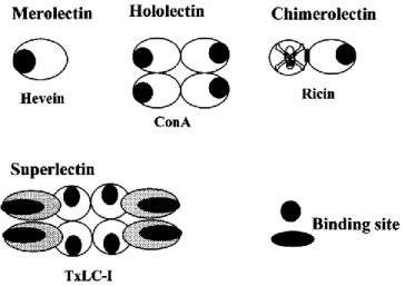
Another division is according to the structure and active center. Lectins are divided in ‘merolectins’, ‘hololectins’, ‘chimerolectins’, and ‘superlectins’ exists (Figure 1):
Merolectins consist of a single carbohydrate-binding domain. Hololectins consist
exclusively of carbohydrate-binding domains but contain at least two such domains that are either identical or very homologous and bind either the same or structurally similar sugar(s) (most plant lectins fall into this category). Chimerolectins are fusion proteins consisting of one or more carbohydrate-binding domain(s) tandemly arrayed to an unrelated domain. Superlectins consist exclusively of at least two carbohydrate binding domains however, unlike the hololectins the carbohydrate binding domains of the superlectins recognize structurally unrelated sugars (Van Damme et al., 1998).
Another commonly used way of differentiating plant lectins is grouping them according to their carbohydrate binding specificity according to which there are six families depending which carbohydrates they preferentially interact with which can be glucose, galactose, mannose, fucose, N-acetylglucosamine, N-acetylgalactosamine or sialic acids,
10 even existing some lectins that bind exclusively with oligosaccharides (Kumar et al., 2012; Walker, 2014; Coelho et al., 2017). The six families are divided (according to their binding specificity) as follows: (1) glucose/mannose; (2) galactose/ N-acetylgalactosamine; (3) N-acetylglucosamine; (4) L-fucose; (5) Sialic acid; (6) Oligosaccharides and complex polysaccharides (Ribeiro, 2008)
Table 1
Taxonomical Distribution of Carbohydrate Domains found in Embryophyta
(Van Damme et al., 2008)
Figure 1. Schematic representation of merolectins, hololectins, chimerolectins, and superlectins
11
3.2. Animal Lectins
Just like plant lectins, animal lectins can be grouped according to several criteria the main of which is the structure and composition of their carbohydrate recognition domain as can be seen in table 2 along with some of their ligand specificities, their subcellular localization and some examples of their functions. It is of value to note that only 4 of the 13 described families of animal lectins are known as intracellular lectins (M-types, P-type, L-type and calnexin family) due to being located in the luminal compartments of the secretory pathway (Gupta, Gupta, & Gupta, 2009; Kumar et al., 2012; Drickamer, 2014).
Table 2
Lectin family, location specificity and functions
12
4. Plant Lectins Structural Features
As were previously mentioned plant lectins can be classified into 12 families according to their carbohydrate recognition domains (CRDs) and respective polypeptide sequencing. Each family is comprised by all the known lectins of related evolutionary CRD structure (in terms of sequence similarity) that are characteristic of said family. The different CRDs are characterized by their own amino acidic sequencing, lectin polypeptide folding, and the structure of the binding site and while CRDs differ in their sequences, they can show reactivity towards similar carbohydrates indicating that specificity is not linked to a particular CRD (Walker, 2014).
Despite the well preserved monomeric unit shared by lectins it is in the quaternary structure, the way that the monomeric units oligomerize, that reside the differences that determine the different carbohydrate binding tendencies and activity that set apart each of the lectin families (Loris, Hamelryck, Bouckaert, & Wyns, 1998).
4.1. Legume lectin Monomer
The legume lectin monomer is structurally well conserved. Studies revealing high similarity in both sequence and structure only existing minor variations in loop and strand length (Lagarda-Diaz, Guzman-Partida, & Vazquez-Moreno, 2017). Approximately 20% of the amino acid residues are invariant in all legume lectins and another 20% are similar with the conserved amino acids including several of those involved in the interaction with the saccharide and almost all the residues that coordinate the metal ions (Ambrosi et al., 2005).
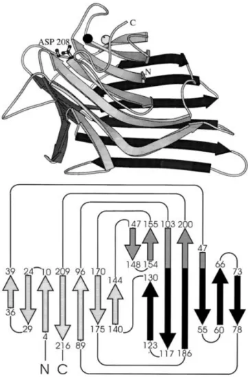
The monomer consists of two large β-pleated sheets that form a scaffold on which the carbohydrate binding region is grafted (Figure 2) (Loris et al., 1998). The architecture of the monomer displays a jerryroll motif, also known as “lectin fold”, that contains a CRD and metal binding sites for divalent cations (tightly bound Ca2+ and a transition metal, usually Mn2+) (Ambrosi et al., 2005). The three-dimensional structure is characterized by three β-sheets that are connected by α turns, β turns and bends. The three β-sheets consist of a 6-stranded back sheet, a 7-stranded front sheet and a smaller 5-stranded sheet that
13 plays a major role in holding the two large sheets together (Lagarda-Diaz et al., 2017; Loris et al., 1998). The main hydrophobic core is located between the back and the front sheet. No α-helix is present and about 50% of the residues are in loop regions with one of these loops curling over the front sheet and resulting in the formation of a second hydrophobic core (Loris et al., 1998).
Figure 2. 3D structure of legume lectin monomer (top) and topology diagram of the legume lectin fold
14
4.2. Quaternary structure
The quaternary structure is responsible for the considerable variation exhibited by legume lectins. This is due to small differences in the aminoacidic sequences at the monomer-monomer interfaces and to different post-translational processing (such as proteolytic cleavage of precursor chain and C- terminal trimming, among other examples) (Loris et al., 1998; Ambrosi et al., 2005).
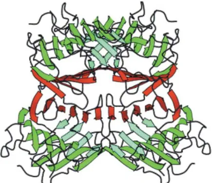
The majority of known legume lectins have tendency to assemble as homodimers or homo-tetramers with most known legume lectins containing a structure termed “canonical legume lectin dimer” which is characterized by anti-parallel side-by-side alignment of the flat six-stranded β-sheets of the two monomers, resulting in the formation of a continuous 12- stranded sheet that extends across the dimer interface (Figure 3) (Ambrosi et al., 2005; Lagarda-Diaz et al., 2017). Some exceptions to this are lectins from coral tree (Erythrina corallodendron) and lectin IV from Griffonia simplicifolia which have more “open” structures (Loris et al., 1998).
As with most legume lectins the pH is a determining factor in the quaternary structure as it can cause the dissociation of tetramers into dimers depending on the conditions (Ambrosi et al., 2005).
15
4.3. Carbohydrate binding site
The advancement of biotechnology in recent years significant progress was made in the molecular and structural understanding of the carbohydrate binding site. It is known that while lectins bind to mono and oligosaccharides, they show specificity for complex sugars and glycoproteins with higher association constants for di-, tri- and tetra- saccharides than for monosaccharides (Ambrosi et al., 2005; Lagarda-Diaz et al., 2017).
The carbohydrate binding sites appear to be preformed as few conformational changes occur upon binding (Ambrosi et al., 2005). In all known plant lectins, the binding of the carbohydrate involves four amino acid residues that are invariant irrespectively of their specificity. These amino acids consist of an aspartic acid, an asparginate, a glycine and an aromatic amino acid or leucine (Ambrosi et al., 2005; Lagarda-Diaz et al., 2017). Despite the well preserved amino acids, the carbohydrate specificity still exists which suggests that amino acid residues from other regions of the pocket are responsible for the specificity.
The sugar-combining site is made up by amino acid residues residing in four different loops (A, B, C and D): the aspartic acid and glycine belong to A and B respectively, whereas the asparagine and the hydrophobic residue are in loop C with additional interactions being attributed to amino acids in loop D which appears to be responsible for carbohydrate specificity (Ambrosi et al., 2005). Loop D is highly variable in terms of length, sequence and conformation with its length, for example, being similar in all mannose-specific lectins (Ambrosi et al., 2005) further suggesting its role in determining the specificity of the lectin families.
Aside from the amino acid residues activity and three-dimensional structure of the binding site, there are two other major components in the carbohydrate binding activity of the legume lectin binding site: water molecules in the CRD and the metal binding
sites for divalent cations (previously mentioned).
The divalent metals, generally Ca2+ and Mn2+, are essential for carbohydrate-binding
16 binding activity but not often directly involved in the binding itself (Lagarda-Diaz et al., 2017).
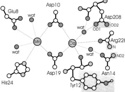
Several studies showed, in different lectins (for example concanavalin A), a loss of said activity when demetallisation essays were performed. As such it is not a surprising fact that the metal binding sites (namely the amino acid residues that interact with the metal ions), first observed in concanavalin A, have been found to be extremely well conserved in all other legume lectin structures (Figure 4) (Loris et al., 1998).
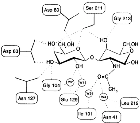
Contacts between the ligand and the protein are often mediated by water molecules with small size and its ability to behave as both hydrogen donor and acceptor make it near-ideal for this function (Ambrosi et al., 2005). In general, water molecules in the carbohydrate-binding region mimic the ligand to a substantial extent not only at the primary site, but also in the regions adjacent to it (Figure 5) (Ambrosi et al., 2005). The position of water molecules in the binding site are well conserved within single species but appear to be poorly conserved in lectins from different species (Loris et al., 1998).
The increase of water mediated H-bonds seems to increase the affinity for complex carbohydrates as opposed to simple sugars (Van Damme et al., 2008). A notable example of water activity in the binding of sugars is the complex of peanut agglutinin with T-antigen disaccharide where a substantial (twenty fold) increase in affinity is observed when compared to the complex of peanut agglutinin and lactose which is entirely due to water mediated protein-carbohydrate interactions (Figure 6) showing that water mediated specificity can be present in carbohydrate recognition (Loris et al., 1998).
Figure 4 Schematic representation of the double metal binding site of concanavalin A (Loris et al., 1998)
17 Figure 5. Schematic representation of protein–carbohydrate interactions in the Peanut agglutinin binding site showing the role of water in the bindingprocess
(Ambrosi et al., 2005)
Figure 6. Schematic diagrams of the binding of lactose a and the T-antigen disaccharide b to peanut agglutinin. The main difference between both complexes are the additional water-mediated interactions in the T-antigen complex (Loris et al., 1998).
18
5. Functions and Bioactivity
Being such a diverse and widespread group of proteins, lectins are involved in several vital processes that ensure the survival of its host. Their functions range from protection of the organism from external threats to maintaining homeostasis through mediation of cell to cell interactions. The communication based on the carbohydrate-lectin interactions is at the focus of biological processes such as transformation, cell growth, cancer metastasis, inflammation and host-pathogen interactions (Majee & Biswas, 2013). Their therapeutic applications are of major importance due to the vast array of intra and inter-cellular processes in which they are a key component.
5.1. Antifungal and Antibacterial Activity
Several studies, have described the role played by lectins in the protection from and elimination of microorganisms. Plant lectins investigated for antifungal potential, mainly against phytopathogenic species, have most reported antifungal effects binding to hyphae, causing inhibition of growth and prevention of spore germination, examples of this are, amongst many others, lectins isolated from Phaseolus vulgaris seeds that were shown to inhibit the growth of Coprinus comatus and Rhizoctonia solani; jackin and
frutackin, two chitin-binding lectins from the genus Artocarpus, demonstrated
inhibition of the germination of Fusarium moniliforme spores (Coelho et al., 2017). It is also worth mentioning, regarding the antifungal properties of lectins, that both human and animal pathogens can be affected, an example of this is lectins extracted from Helianthus
annuus seeds showed the ability to inhibit growth and alter membrane permeability of Candida tropicalis, Candida parapsilosis, Candida albicans, and Pichia membranifaciens while also inducing the production of reactive oxygen species in Candida Tropicalis (Regente et al., 2014).
It is also worth mentioning that lectins, regarding the role in the protection from microorganisms, play a role in the bacterial virulence (like in fungal activity) through the binding of the exogenous lectins to the specific carbohydrates present in the endogenous cells being a factor of significant importance in the recognition and adhesion phase of the infection.
19
5.2. Anti insect and Antiparasitic Activity
In contrast to fungi and bacteria, many plant lectins are moderately to highly toxic for insects and higher animals (Van Damme et al., 2008). The role of protection against external threats played by lectins against insects and parasites is performed through different means than its antibacterial and antifungal activity. Whereas as previously mentioned the antifungal and antibacterial properties of lectins consists mainly in the inhibition of the microorganisms through indirect means such as decreasing its adhesion and recognition and impairing its ability to reproduce and spread, the insect and anti-parasitic effect of lectins is more direct consisting in increasing mortality and delaying development. There are many reported examples such as Arisaema jacquemontii lectin adversely affected the development of Bactrocera cucurbitae larvae (Lam & Ng, 2011); lectins isolated from Coprinopsis cinerea, Aleuria aurantia, and Laccaria bicolor showed larvistatic effect on Haemonchus contortus (Barber’s poleworm), resulting in arresting at L1 phase; and an example of note where lectins isolated from Moringa
oleifera seeds demonstrated larvicidal, ovicidal, and oviposition-stimulant effects on A. aegypti, being considered important candidates for using in control of mosquito
population, including in traps for egg capture (Coelho et al., 2017).
The anti-insect activity of plant lectins is one of its most notable qualities as there have been recent studies that show that insect stimulus induces the production of specific lectins in plants as a means of defense, these lectins are called insect-induced lectins. Interestingly the production of these lectins is not induced by wounding which indicates an insect specific defense mechanism through lectin production (Van Damme et al., 2008).
20
5.3. Antiviral Activity
Due to the high glycoprotein content that is characteristic of the viral envelopes, which are the target of the plant lectins action, this type of application is worthy of note with many possibilities of management in disease therapeutic.
There are many reported plant lectins with antiviral activity. Different lectins have different anti-HIV mechanisms: snowdrop lectin, Concanavalin A and jacalin-related
lectin from banana fruit, among others, exhibited antiretroviral activity inhibiting the
syncytium formation and therefore preventing the entry of the virus into de CD4 cells; extra long autumn purple bean lectin displayed anti-HIV activity by inhibition of reverse transcriptase (Majee & Biswas, 2013); the sea worm (Serpula vermicularis) lectin suppressed the production of viral p24 antigen and cytopathic effect induced by HIV-1 (Lam & Ng, 2011).
5.4. Toxicity
Lectins are considered antinutritional factors due to their inherent toxicity. Due to resistance to digestive enzymes and bacteria they may pose a threat when ingested by animals interacting and binding to the digestive tract of higher animals inhibiting growth and causing diarrhea, vomiting and even death. Their main mechanism of toxicity consists of damaging the cellular membrane, setting off inflammatory processes with internalization, inhibiting protein synthesis or even activating apoptotic pathways.
Because the ability of the lectins to cause intestinal malabsorption is dependent on the presence of enteric bacteria, it has been hypothesized that lectins may also produce toxicity by facilitating bacterial growth in the gastrointestinal tract (Dolan, Matulka, & Burdock, 2010)
The specificity of legume lectins for some typical animal glycans, has further supported the idea that legume lectins play a role in plant defense against insects and/or predator animals.
They have been found to be highly immunogenic to varying degree, inducing inflammatory response and cause GI irritation. In sensitive individuals, lectins can induce
21 severe intestinal damage disrupting digestion, provoke IgG and IgM antibodies
responsible for food allergies and in extreme cases, can cause anemia, which is why
lectin based drug delivery systems should never be used in people with reported food allergies (Majee & Biswas, 2013).
Lectins have been reported to cause damage, including food poisoning by uncooked kidney beans and also hemolytic anemia and jaundice by Mexican fava beans (Kumar et al., 2012).
A noteworthy example is the castor bean lectin ricin (one of the most toxic natural substances known) which is notorious for causing deaths of children, and has been used as an instrument of bioterrorism (Dolan et al., 2010).
5.5. Anticancer activity
The anticancer applications of lectins can be split up into two groups, the diagnostic applications and the therapeutic ones. While the therapeutic applications of lectins are the main focus of this paper it is of note that the role of lectins in diagnostics is of significant importance. They serve as powerful tools in immunological studies and can be employed as immunohistochemistry markers in diagnosis of cancer and profiling of cell surface types due to expression of aberrant glycans on diseased and transformed cells (Majee & Biswas, 2013). As such lectins are frequently used as diagnostic probes and tumour-specific surface markers.
Their anticancer activity per say is related to their activity to specifically bind to the aberrant cells through the identification of the malignant cells surface carbohydrates and then acting as cytotoxins, causing apoptosis in mitochondrial dependent pathway, increasing the content of reactive oxygen species, triggering autophagy or necrosis in cancer cells (Majee & Biswas, 2013). This approach can be improved through the conjugation of said lectins with targeting agents improving their specificity, with lectins also being able to be used as targeting agents. In addition, many anticancer lectins usually possess low cytotoxicity to nontransformed cells.
Hitherto many studies have reported anticancer activity of lectins. European mistletoe demonstrates anticancer properties in, amongst others, Ewing sarcoma (Twardziok et al.,
22 2016), Concanavalin A extracted from Jack bean seeds has been found to induce caspase dependent apoptosis in human melanoma A375 cells (Yau et al., 2015), antiproliferative and cytotoxic effects of the tepary bean lectins on C33-A and Sw480 cells lines has been reported (Valadez-Vega et al., 2011).
It is also worthy of note that, unlike plant lectins, the endogenous animal lectins that we possess play a major role in cell adhesion which shows potential for advanced stage cancer treatment as it can hinder the metastasis process and improve the prognostic by impeding the spreading of the cancer diseases. Lebecin lectin extracted from
Macrovipera lebetina inhibited the integrin-mediated attachment of human breast cancer
cells to fibronectin and fibrinogen (Jebali et al., 2014); Bauhinia forficata seed lectin inhibited adhesion of MCF-7 cells to laminin, collagen I, and fibronectin (Silva et al., 2014).
6. Aberrant glycosylation caused by malignant transformation
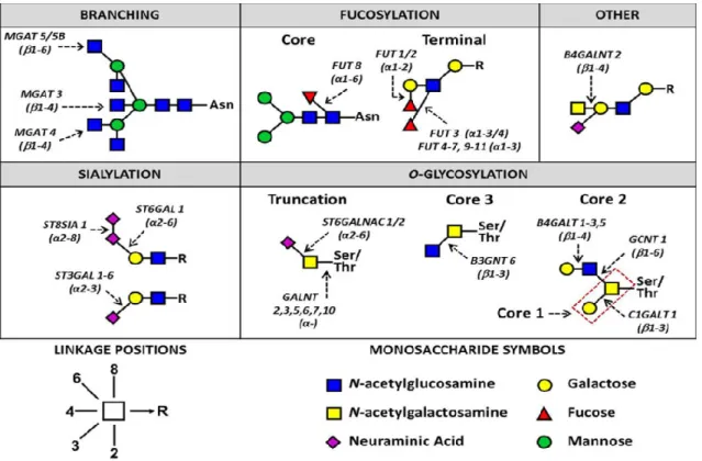
Altered glycosylation is a universal feature of cancer cells, but only certain specific glycan changes are frequently associated with tumors. These include (1) increased β1-6GlcNAc branching of N-glycans; (2) changes in the amount, linkage, and acetylation of sialic acids; (3) truncation of O-glycans, leading to expression of Tn and sialyl Tn antigens; (4) expression of the nonhuman sialic acid N-glycolylneuraminic acid, likely incorporated from dietary sources; (5) expression of sialylated Lewis structures and selectin ligands; (6) altered expression and enhanced shedding of glycosphingolipids; (7) increased expression of galectins and poly-N acetyllactosamines; (8) altered expression of ABH(O) blood-group-related structures; (9) alterations in sulfation of glycosaminoglycans; (10) increased expression of hyaluronan; and (11) loss of expression of GPI lipid anchors (Varki & Freeze, 2009).
However the two main glycan classes found at the cell surface glycoproteins are O-glycans (most common) synthesized in the Golgi complex, and N-O-glycans whose biosynthesis takes place in the endoplasmatic reticulum (with less common forms of protein glycosylation include O-Fucosylation, O-GlucNAcylation and O-Mannosylation), as such the most frequent and relevant glycosylation alterations caused
23 by malignant processes will be the ones affecting both O- and N-glycans with O-glycan alterations being the most important and frequent of the two (Ferreira et al., 2017). The changes in cancer cells surface properties generally manifest through overexpression of aberrant O- and N-glycans exposed at the cell surface (Poiroux, Barre, van Damme, Benoist, & Roug, 2017).
Branching and truncation of both N- and O-glycans, but also sialylation and fucosylation (both core and terminal), appear to be a common theme amongst the cancer-associated changes in glycan structures and can be seen illustrated in Figure 7 (Christiansen et al., 2014).
The most frequent aberrant O-glycans expressed at the surface of cancer cells consist of Tn antigen, T antigen, Lewis a, and Lewis x antigens, and an oncofetal glycotope, the Forssman pentasaccharide antigen which are illustrated in Figure 8 (Poiroux et al., 2017)
With the multitude of different relevant alterations in glycosylation caused by malignant processes the reader is directed to the papers cited above for a further, in depth look at the mechanisms and specific alterations on a molecular level, but also to the works of (Gill, Clausen, & Bard, 2011; Stowell, Ju, & Cummings, 2015; Hanson & Hollingsworth, 2016; Munkley & Elliott, 2016; Dall’Olio & Trinchera, 2017).
Figure 7. N-and O-glycan changes in breast, colorectal, ovarian, liver and melanoma cancers. R indicates N-and O-glycans (Christiansen et al., 2014)
24 Figure 8. Molecular Structure of mutated O-glycans expressed on the cancer
25
7. Anticancer activity mechanism
Legume lectins have been shown to induce programmed cell death in multifactorial ways, affecting many different signaling pathways and a wide range of both pro- and anti- cell death factors.
Many studies have reported the involvement of lectins in the expression of members of the Bcl-2, Autophagy related gene (ATG), and caspase families, as well as p53, ERK, Ras-Raf, and BNIP3 and, being all the mentioned examples integral components of the different programmed cell death pathways, thereby induce both apoptosis and autophagy (Lagarda-Diaz et al., 2017; Yau et al., 2015).
Programmed cell death (PCD) occurs through two main processes: Apoptosis (type I PCD) and Autophagy (type II PCD) with both processes presenting a wide range of different pathways that can be employed some of which are not still fully understood and potentially some not even known. Both processes will be briefly explained bellow and examples of the multitude of lectins and respective PCD pathways can be found in Table 3 as well as Figure 9 which depicts both apoptotic pathways and Figure 10 depicting the autophagic process.
7.1. Apoptosis
Apoptosis, or type I programmed cell death, is a highly regulated and controlled biological process that allows the organism to eliminate defective and unwanted cells allowing for a controlled means of disposal of potentially harmful elements. Apoptosis is characterized by cytoplasmic cell shrinkage, chromatin condensation and DNA fragmentation, and segmentation into apoptotic bodies (called blebbing) and can occur in response to extrinsic and intrinsic stimuli (Fujita et al., 2017).
26
7.1.1. Extrinsic pathway
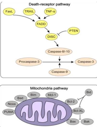
The extrinsic apoptosis pathway (Figure 9), or death-receptor pathway, is triggered by binding of external ligation to cell surface death receptors which then bind to specific ligands (which include Tumor Necrosis Factor (TNF), FAS-Fas Ligand (FasL), TNF-related apoptosis-inducing ligand (TRAIL)), after ligation the intracellular death domains and adapter proteins, such as Fas-Associated protein with Death domain (FADD) and TNF Receptor-1 associated Death Domain protein (TRADD), construct a death inducing signaling complex (DISC) that recruits and activates initiator caspase-8/10 (Estaquier, Vallette, Vayssiere, & Mignotte, 2012; Fujita et al., 2017).
Figure 9. Mechanisms of apoptosis in cancer. Extrinsic pathway (top) and Intrinsic pathway (bottom) (Jiang et al., 2015)
27
7.1.2. Intrinsic pathway
The intrinsic apoptosis pathway (Figure 9), or mitochondrial pathway, is activated by various stress signals, such as severe genetic damage, hypoxia and oxidative stress and is controlled by the Bcl-2 protein family through the regulation of the mitochondrial membrane permeability (Wang, 2014). In response to such stimuli mitochondrial pro-apoptotic proteins, BH3-only members (including Bad, Bik, Bid among others), antagonize anti-apoptotic proteins Bcl-2, Bcl-xL and Mcl-1 creating a pro-apoptotic balance that increases the mitochondrial permeability resulting in the release of specific mitochondrial proteins into the cytosol, such as cytochrome c which forms a complex with Apaf-1, called the apoptosome, which assists in auto-activation of initiator pro-caspase-9 (Jiang et al., 2015; Fujita et al., 2017).
7.2. Autophagy
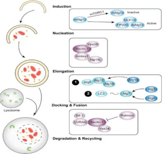
Autophagy is an evolutionary conserved mechanism that disassembles unnecessary or dysfunctional components, through the use of autophagosomes and lysosomes, allowing for the elimination and recycling of cellular components, it constitutes a stress adaptation that avoids cell death (in normal physiological conditions). While there are two main types of autophagy—micro autophagy and macro autophagy—the general term autophagy is commonly used to refer to the process of macro autophagy (Yau et al., 2015): In micro autophagy the lysosome surface directly surrounds and engulfs the cytoplasm and target organelles while in macro autophagy there is a creation of an autophagosome through the isolation of the cytoplasm and creating of a new separate vesicle which then fuses with the lysosome (Li et al., 2011).
In the context of cancer however autophagy can be used by the malignant cells as a physiological mechanism of survival thus becoming a detrimental process to the overall organism (Jiang et al., 2015).
Autophagic programmed cell death (type II PCD), despite the role of autophagy in the survival of the cell, can occur when induced in a massive or prolonged fashion (Lei &
28
Chang, 2009). Autophagy as a process of PCD refers to macro autophagy since it requires the formation of autolysosomes (Lei & Chang, 2009).
There is an increase in the permeability of the autolysosome membrane which originates leakage of catabolic hydrolases, cathepsins and reactive oxygen species (ROS) into the cytosol which can cause mitochondrial membrane increased permeabilization thus activating caspases in an intrinsic apoptosis manner (Figure 10) (Lei & Chang, 2009). Compared to plant lectins involved in apoptosis, a limited number of plant lectins, such as concanavalin A, Mistletoe lectin and P. cyrtonema lectin, have shown activity targeting the autophagic signalling pathways of different types of cancer cells (table 3) (Jiang et al., 2015).
While pro apoptotic factor and pathways are more abundant in cells and more plant lectins have been shown to target such elements, autophagy is nonetheless and important mechanism that should be studied in more detail as it can complement apoptosis and even act as a replacement in treatment of apoptosis resistant cancer cell lines.
Figure 10. Mechanisms of (macro)autophagy in cancer
29 Table 3
Plant lectins targeting apoptotic/autophagic signalling pathways in cancer.
30
8. Biomedical applications
As before mentioned in this work lectins, and particularly plant lectins, have properties that allow their use in healthcare taking advantage of their highly specific interactions and affinities but also with a wide array of possible applications. They take center stage in both the treatment and management of cancer related diseases as the malignant alterations in cells increase the interaction with legume lectins and its specificity.
The use of plant lectins in biomedical applications can be divided into diagnosis and therapeutics.
Table 3 (continued)
31
8.1. Diagnosis
With the knowledge that plant lectins show affinity towards altered cell surface glycans, which are characteristic of aberrant glycosylation caused by cancer diseases, it is not surprising that one of their first uses was as cell surface markers in biochemical assays. The advancement of science, and the means of mitigating plant lectins toxicity, their use grew and expanded with the capability of their use in vivo. Nowadays plant lectins, and also animal endogenous lectins, are used as a major diagnostic, prognostic and therapy management tool in cancer as important biomarkers due to their capability of detecting malignant cells with adequate specificity.
Lectin based serological assays are currently used in clinical practice to detect and quantify glycans in the serum of cancer patients with the measurement of circulating glycoconjugates being used for (a) diagnosis, (b) monitoring of clinical course under therapy, (c) detection of early disease recurrence, and (d) prognosis (Tuccillo et al., 2014).
Several plant lectins have shown their use as T/Tn- specific markers in many cancers (Poiroux et al., 2017). Peanut agglutinin has shown application in identifying hematopoietic cell subpopulations (Lu et al., 2012), CA125 (transmembrane mucin) is one of the best available biomarkers of ovarian cancer (Tuccillo et al., 2014), Parkia pendula lectin has been used as a marker for characterization of meningothelial tumor tissue (Beltrão et al., 2003).
Lectins when used in association with for example fluorescent markers, due to their specificity, can be used to determine the spreading of tumors marking the affected tissues that appear as colored areas in the tests.
32
8.2. Therapeutics
8.2.1. Direct antitumor activity
Plant lectins initial and most obvious use in therapeutics came from its cytotoxic capabilities capable of inducing programmed cell death (apoptosis and/or autophagy) conjugated with their affinity towards malignant cells. A plethora of studies have shown the direct anticancer activity of the many plant lectins on many different cancer cell lines (table 3). However, this type of investigation must continue as new lectins and types of cancer cell lines are discovered as different lectins have different specificities and elicit different reactions while many cancer lines show resistance to several specific lectins.
The most studied and better understood plant lectin is Concanavalin A which shows the ability to induce apoptosis, autophagy and having anti angiogenic capabilities targeting many different pathways of each process, described in (Li et al., 2011), thus having a multitude of applications. Others examples of plant lectins cytotoxic antitumor activity include Polygonatum Odoratum lectin found to induce signs of apoptosis in A549 lung cancer cells without affecting healthy lung cells (Yau et al., 2015), Korean mistletoe
lectin (Viscum album L. coloratum agglutinin) eliciting apoptosis in human
hepatocarcinoma cells (Lyu, Choi, & Park, 2002), Sambucus nigra agglutinin exhibited selectivity towards ovarian carcinoma cells and induced their cell death through mitochondrial disfuction (Chowdhury, Ray, Chatterjee, & Roy, 2017), among others.
There exist many different pathways and, within those pathways, different ways with which plant lectins induce their activity even when belonging to the same family of lectins or acting on the same type of cancer cells which is one of the reasons why their use in cancer related therapeutics is so appealing, if one pathway is inefficient at causing PCD in a certain type of cancer cell line other lectins from the same family or different ones may still be useful.
33
8.2.2. Biological response modifier
Several plant lectins have shown capabilities of modulating the immune system, being powerful mitogens for human lymphocytes aiding in the combat of cancer facilitating the production of host immune cells, with examples including pokeweed mitogen,
concanavalin A and Maclura pumifera agglutinin (Mody, Joshi, & Chaney, 1995).
Other plant lectins have shown the ability to nonspecifically modulate and boost the immune system capabilities through activation of natural killer cells and enhancement of monocyte cell line activity, for example galactose binding mistletoe lectin ML-I, thus inhibiting tumor growth (Mody et al., 1995).
Another important biological response modification which isn’t as relevant in plant lectins but shows great promise in cancer treatment is the regulation of cell adhesion. This process is mainly regulated by animal endogenous lectins such as Galectin-8 (only galectin that appears to have this role from its family) and selectins (of the C-type animal lectin family) making them extremely important and worth mentioning as the adhesion process of cancer is intimately related to its prognostic (Hadari et al., 2000; Nakahara & Raz, 2008). The targeting of the cell adhesion related lectins involved in cancer is an extremely useful tool as, by greatly decreasing or even halting the metastatic process, it both improves the treatment options and their efficiency and also reduces one of the main difficulties of cancer diseases stopping its systemic effects and restricting it to a more treatable level.
8.2.3. Site Specific Drug Delivery Systems
Targeted therapy is most likely the most difficult and rewarding application of lectins in therapy. With the advancement of technology and science, targeted therapy will without a doubt become the most useful and important lectin mediated biomedical application in cancer and possibly in other areas since it provides a way to balance the efficiency of the treatment with the known toxicity that plant lectins have and that is a relevant downside when considering any plant lectin mediated treatment. In addition to using the lectins as the only active agent in a formulation (taking advantage of the lectin specificity), they
34 can also be used in complex drug delivery systems that allow a more specific, prolonged and safe therapeutic usage.
There are two ways in which lectins are used in this field: as carriers for the intended drug, acting as targeting agents, or acting as the active ingredient themselves, being attached to a different targeting agent; the first method frequently applies reverse lectin targeting (the lectin targets the cell surface carbohydrates), while the second method often uses direct lectin targeting (where the targeting agent has affinity towards endogenous cell lectins) (Gupta et al., 2009).
The controlled deliverance techniques (liposomes, nanosuspensions, bioadhesive systems) provide an adequate release rate and duration, producing the desired effect and reducing the toxic effects of plant lectins but have a main disadvantage of non-specificity to substrate while lectin mediated bioadhesion allows direct binding to target cells establishing specific interactions (Coelho et al., 2017).
The plant lectins specificity towards mutated cells makes them an ideal targeting agent allowing for an effective delivery of the drug to the intended tissues and reducing systemic side effects often related to cancer therapy due to the difficulty of finding an effective targeting agent. Chemotherapy and Radiotherapy are fitting examples of this with patients experiencing severe side effects associated with the lack of effective means with which to target the affected tissues.
8.2.3.1 Nanoparticle systems
The use of nanoparticle drug delivery systems is an expanding field that shows great promise as it increases the half-life of drugs, reduces its side effects and increases its specificity and efficacy by both inhibiting the drugs deliverance to healthy cells and directly conducting the drugs to the tumor site and promoting its accumulation there (Bahrami et al., 2017). There are several different types of nanoparticle systems such as
Nanoconstructed lipid carriers, dendrimers and liposomes to name a few (Bahrami et
al., 2017).
Nanoparticle internalization in cancer cells occurs predominantly through its endocytosis in target cells, which can be further classified as phagocytosis or pinocytosis.
35 Glycotargeting is the main application of lectin and lectin based therapy in the nano drug delivery system field due to the lectin high biocompatibility and specific cell surface carbohydrate recognition with the main criteria of choice of ligand in the drug delivery system being the affinity level of the carbohydrates for their receptors and the expression levels of said receptors in the target cells (Bahrami et al., 2017; Malekzad et al., 2017).
Long circulating lectin conjugated paclitaxel loaded magnetic nanoparticles have shown to increase uptake in Bcr-Abl positive chronic myelogenous leukemia (Singh, Dilnawaz, & Sahoo, 2011), wheat germ agglutinin and Ulex europaeus agglutinin showed high specifity towards human urinary carcinoma 5637 cells, which allowed their targeting of bladder cancer cells (Plattner, Wagner, Ratzinger, Gabor, & Wirth, 2008), Concanavalin A was associated with clarithromycin in an mucoadhesive microparticles complex against Helicobacter pylori improving adhesion (Adebisi & Conway, 2014), among many other reported studies (table 4).
Plants lectins also have been shown to be able to be used in site specific drug delivery systems for pulmonary, epithelial and ocular tissues (Majee & Biswas, 2013).
Drug delivery to gut epithelium requires specificity for epithelial cells and stability at low pHs which is why plant lectins are considered such good reagents for this purpose, with their propensity to interact with mucins present on the absorptive epithelial cells leading to bioadhesion, with Peanut agglutinin and Wheat germ agglutinin having been used in such studies (Bahrami et al., 2017; Lavín de Juan et al., 2017; Malekzad et al., 2017). It is also worth noting that nasal mucosa is also covered in M-cells being the equivalent of the gastrointestinal tract and as such, also possessing the same potential for targeted drug delivery mediated by lectins with studies in rats using Griffonia
simplicifolia I isolectin-B4 and isolectin B4 from Bandeiraea simplicifolia 1 (seen in
table 4) having shown promise (Lavín de Juan et al., 2017).
Pulmonary drug delivery activity of wheat germ agglutinin, Concanavalin A and soybean agglutinin has also been reported in functionalized liposomes causing increase
in uptake of the drug delivery systems (Gupta et al., 2009; Lavín de Juan et al., 2017; Majee & Biswas, 2013).
Lectins tend to bind on corneal and conjunctival surfaces presenting suitable bioadhesive properties for ocular drug delivery, which is promising since tearing, mucin secretion and blinking all contribute to the difficult ocular drug absorption (Majee &
36 Biswas, 2013). Studies in rats and rabits using Solanum tuberosum lectin and wheat germ agglutinin have shown promise increasing the binding of the drug delivery systems (Lavín de Juan et al., 2017)
Table 4
Examples of lectins used in drug targeting (tissue specific)
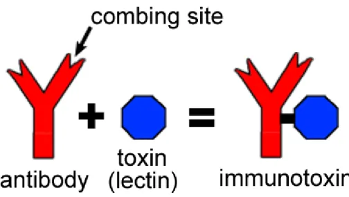
37 8.2.3.2 Immunotoxins
Immunotoxins are a specific type of nanoparticle drug delivery systems that most commonly couple a toxic unit with an antibody (or antibody related) unit allowing for extremely high specificity (Lavín de Juan et al., 2017).
The lectins used in this type of system are usually ribosome inactivating lectins (also known as type 2 ribosome inactivating proteins) consisting of polypeptide lectin chain (B chain) connected by a disulfide bridge with a ribosome inactivating chain (A chain), with the A chain exhibiting the ribosomal specificity and being responsible for the protein synthesis inhibition in mammals and as such, the immunotoxin formulations that include these lectins are composed only by these A-chain that is extracted from said lectins (Lavín de Juan et al., 2017).
Lectins used in such systems include Abrin (isolated from Abrus precatorius), which showed increase in the activity of systems in a specific manner through targeting of the gonadotropin releasing hormone receptor (GnRH- R) molecule which is overexpressed in breast carcinoma cells (Gadadhar & Karande, 2013), and Maackia amurensis seed lectin that showed results in targeting podoplanin (which is transmembrane receptor that promotes tumor cell mobility) and inducing mitochondrial membrane permeability and consequential cell death (Ochoa-Alvarez et al., 2015).
38
9. Conclusions
As was explored throughout this work, lectin have major potential applications and used in a plethora of areas, however their applications in healthcare, and specifically in cancer related diseases, shows the most promise. With the growing control over infectious diseases combined with the general improvement of living conditions and healthcare, life expectancy has suffered a dramatic increase and as such diseases of chronic, genetic and age-related character have become the main focus of studies as they now pose a bigger problem to our quality of life. Cancer, now more than ever, is in the spotlight as with longer lifespans, with its previous relatively slow onset of symptoms and deterioration of health condition has now become more apparent.
Plant lectin use has always been limited by our knowledge of them and technological means for both their study and application. As science evolves, and our techniques and understanding advance along with it, lectin use will without a doubt become as ubiquitous as their presence in living organisms has shown to be.
As our structural understanding of lectins grows, so do the applications with which to use them. The more studies are conducted, the more unknown specific interactions will be found and mechanisms of action, through both poorly understood and completely new pathways, will become available to be utilized and put to proper use.
The studying and application of lectins has increased at an astounding rate in the last few decades. Ever since the 1960s, with the realization that these readily available proteins are invaluable tools for the study of carbohydrates both simple and complex, in solution and on cell surfaces, as well as for cell characterization, this field has been extensively explored and studied. We have progressed from the mere knowledge that plant lectins have anticancer properties, among many others, to being able to create nanodrug delivery systems that utilize them.
While plant lectin toxicity will always be a concern in any form on in vivo application, the drastic advancement in every area of our life that technology has provided in the last century has shown that studies in this field will surely bear fruit in the near future and that further investment in this area is required and will surely pay dividends.
39
References
Adebisi, A. O., & Conway, B. R. (2014). Lectin-conjugated microspheres for eradication of Helicobacter pylori infection and interaction with mucus. International Journal of Pharmaceutics, 470(1–2), 28–40. https://doi.org/10.1016/j.ijpharm.2014.04.070 Ambrosi, M., Cameron, N. R., & Davis, B. G. (2005). Lectins: tools for the molecular
understanding of the glycocode. Organic & Biomolecular Chemistry, 3(9), 1593. https://doi.org/10.1039/b414350g
Bahrami, B., Hojjat-Farsangi, M., Mohammadi, H., Anvari, E., Ghalamfarsa, G., Yousefi, M., & Jadidi-Niaragh, F. (2017). Nanoparticles and targeted drug delivery in cancer therapy. Immunology Letters, 190(April), 64–83. https://doi.org/10.1016/j.imlet.2017.07.015 Beltrão, E. I. C., Medeiros, P. L., Rodrigues, O. G., Figueredo-Silva, J., Valença, M. M.,
Coelho, L. C. B. B., & Carvalho, L. B. (2003). Parkia pendula lectin as histochemistry marker for meningothelial tumour. European Journal of Histochemistry : EJH, 47(2), 139–42. Retrieved from http://www.ncbi.nlm.nih.gov/pubmed/12777210
Chowdhury, S. R., Ray, U., Chatterjee, B. P., & Roy, S. S. (2017). Targeted apoptosis in ovarian cancer cells through mitochondrial dysfunction in response to Sambucus nigra agglutinin. Cell Death & Disease, 8(5), e2762. https://doi.org/10.1038/cddis.2017.77 Christiansen, M. N., Chik, J., Lee, L., Anugraham, M., Abrahams, J. L., & Packer, N. H. (2014).
Cell surface protein glycosylation in cancer. Proteomics, 14(4–5), 525–546. https://doi.org/10.1002/pmic.201300387
Coelho, L. C. B. B., Silva, P. M. D. S., Lima, V. L. de M., Pontual, E. V., Paiva, P. M. G., Napoleão, T. H., & Correia, M. T. D. S. (2017). Lectins, Interconnecting Proteins with Biotechnological/Pharmacological and Therapeutic Applications. Evidence-Based Complementary and Alternative Medicine : eCAM, 2017, 1594074.
https://doi.org/10.1155/2017/1594074
Dall’Olio, F., & Trinchera, M. (2017). Epigenetic Bases of Aberrant Glycosylation in Cancer. International Journal of Molecular Sciences, 18(5). https://doi.org/10.3390/ijms18050998 Dolan, L. C., Matulka, R. A., & Burdock, G. A. (2010). Naturally occurring food toxins. Toxins,
2(9), 2289–332. https://doi.org/10.3390/toxins2092289
40 http://www.imperial.ac.uk/research/animallectins/ctld/lectins.html
Estaquier, J., Vallette, F., Vayssiere, J., & Mignotte, B. (2012). Advances in Mitochondrial Medicine, 942, 157–183. https://doi.org/10.1007/978-94-007-2869-1
Ferreira, J. A., Magalhães, A., Gomes, J., Peixoto, A., Gaiteiro, C., Fernandes, E., … Reis, C. A. (2017). Protein glycosylation in gastric and colorectal cancers: Toward cancer detection and targeted therapeutics. Cancer Letters, 387, 32–45.
https://doi.org/10.1016/j.canlet.2016.01.044
Fu, L. L., Zhou, C. C., Yao, S., Yu, J. Y., Liu, B., & Bao, J. K. (2011). Plant lectins: Targeting programmed cell death pathways as antitumor agents. International Journal of
Biochemistry and Cell Biology, 43(10), 1442–1449. https://doi.org/10.1016/j.biocel.2011.07.004
Fujita, K., Iwama, H., Oura, K., Tadokoro, T., Samukawa, E., Sakamoto, T., … Masaki, T. (2017). Cancer Therapy Due to Apoptosis: Galectin-9. International Journal of Molecular Sciences, 18(1). https://doi.org/10.3390/ijms18010074
Gadadhar, S., & Karande, A. A. (2013). Abrin immunotoxin: targeted cytotoxicity and intracellular trafficking pathway. PloS One, 8(3), e58304.
https://doi.org/10.1371/journal.pone.0058304
Gill, D. J., Clausen, H., & Bard, F. (2011). Location, location, location: New insights into O-GalNAc protein glycosylation. Trends in Cell Biology, 21(3), 149–158.
https://doi.org/10.1016/j.tcb.2010.11.004
Gupta, A., Gupta, R. K., & Gupta, G. S. (2009). Targeting cells for drug and gene delivery: Emerging applications of mannans and mannan binding lectins. Journal of Scientific & Industrial Research, 68, 465–483. Retrieved from
http://nopr.niscair.res.in/bitstream/123456789/4323/1/JSIR 68(6) 465-483.pdf
Hadari, Y. R., Arbel-Goren, R., Levy, Y., Amsterdam, A., Alon, R., Zakut, R., & Zick, Y. (2000). Galectin-8 binding to integrins inhibits cell adhesion and induces apoptosis. Journal of Cell Science, 113 ( Pt 13), 2385–97. Retrieved from
http://www.ncbi.nlm.nih.gov/pubmed/10852818
Hanson, R. L., & Hollingsworth, M. A. (2016). Functional Consequences of Differential O-glycosylation of MUC1, MUC4, and MUC16 (Downstream Effects on Signaling). Biomolecules, 6(3). https://doi.org/10.3390/biom6030034
Jebali, J., Fakhfekh, E., Morgen, M., Srairi-Abid, N., Majdoub, H., Gargouri, A., … Sarray, S. (2014). Lebecin, a new C-type lectin like protein from Macrovipera lebetina venom with
41 anti-tumor activity against the breast cancer cell line MDA-MB231. Toxicon, 86, 16–27. https://doi.org/10.1016/j.toxicon.2014.04.010
Jiang, Q. L., Zhang, S., Tian, M., Zhang, S. Y., Xie, T., Chen, D. Y., … Jiang, X. (2015). Plant lectins, from ancient sugar-binding proteins to emerging anti-cancer drugs in apoptosis and autophagy. Cell Proliferation, 48(1), 17–28. https://doi.org/10.1111/cpr.12155
Kumar, Kk., Reddy, Gs., Reddy, B., Shekar, Pc., Sumanthi, J., & Chandra, Kl. P. (2012). Biological role of lectins: A review. Journal of Orofacial Sciences, 4(1), 20. https://doi.org/10.4103/0975-8844.99883
Lagarda-Diaz, I., Guzman-Partida, A. M., & Vazquez-Moreno, L. (2017). Legume Lectins: Proteins with Diverse Applications. International Journal of Molecular Sciences, 18(6). https://doi.org/10.3390/ijms18061242
Lam, S. K., & Ng, T. B. (2011). Lectins: Production and practical applications. Applied
Microbiology and Biotechnology, 89(1), 45–55. https://doi.org/10.1007/s00253-010-2892-9
Lavín de Juan, L., García Recio, V., Jiménez López, P., Girbés Juan, T., Cordoba-Diaz, M., & Cordoba-Diaz, D. (2017). Pharmaceutical applications of lectins. Journal of Drug Delivery Science and Technology. https://doi.org/10.1016/j.jddst.2017.05.018
Lei, H.-Y., & Chang, C.-P. (2009). Lectin of Concanavalin A as an anti-hepatoma therapeutic agent. Journal of Biomedical Science, 16(1), 10. https://doi.org/10.1186/1423-0127-16-10 Li, W. wen, Yu, J. ying, Xu, H. long, & Bao, J. ku. (2011). Concanavalin A: A potential
anti-neoplastic agent targeting apoptosis, autophagy and anti-angiogenesis for cancer
therapeutics. Biochemical and Biophysical Research Communications, 414(2), 282–286. https://doi.org/10.1016/j.bbrc.2011.09.072
Loris, R., Hamelryck, T., Bouckaert, J., & Wyns, L. (1998). Legume lectin structure.
Biochimica et Biophysica Acta - Protein Structure and Molecular Enzymology, 1383(1), 9–36. https://doi.org/10.1016/S0167-4838(97)00182-9
Lu, Q., Li, N., Luo, J., Yu, M., Huang, Y., Wu, X., … Li, G. (2012). Pinellia pedatisecta agglutinin interacts with the methylosome and induces cancer cell death. Oncogenesis, 1(10), e29. https://doi.org/10.1038/oncsis.2012.30
Lyu, S. Y., Choi, S. H., & Park, W. B. (2002). Korean mistletoe lectin-induced apoptosis in hepatocarcinoma cells is associated with inhibition of telomerase via mitochondrial controlled pathway independent of p53. Archives of Pharmacal Research, 25(1), 93–101. Retrieved from http://www.ncbi.nlm.nih.gov/pubmed/11885700
42 Majee, S. B., & Biswas, G. R. (2013). Exploring plant lectins in diagnosis , prophylaxis and
therapy. Journal of Medicinal Plants Research, 7(47), 3444–3451. https://doi.org/10.5897/JMPR2013.5289
Malekzad, H., Mirshekari, H., Sahandi Zangabad, P., Moosavi Basri, S. M., Baniasadi, F., Sharifi Aghdam, M., … Hamblin, M. R. (2017). Plant protein-based hydrophobic fine and ultrafine carrier particles in drug delivery systems. Critical Reviews in Biotechnology, 8551(April), 1–21. https://doi.org/10.1080/07388551.2017.1312267
Mody, R., Joshi, S. H. antaram, & Chaney, W. (1995). Use of lectins as diagnostic and therapeutic tools for cancer. Journal of Pharmacological and Toxicological Methods, 33(1), 1–10. https://doi.org/10.1016/1056-8719(94)00052-6
Munkley, J., & Elliott, D. J. (2016). Hallmarks of glycosylation in cancer. Oncotarget, 7(23), 35478–89. https://doi.org/10.18632/oncotarget.8155
Nakahara, S., & Raz, A. (2008). Biological modulation by lectins and their ligands in tumor progression and metastasis. Anti-Cancer Agents in Medicinal Chemistry, 8(1), 22–36. Retrieved from http://www.ncbi.nlm.nih.gov/pubmed/18220503
Ochoa-Alvarez, J. A., Krishnan, H., Pastorino, J. G., Nevel, E., Kephart, D., Lee, J. J., … Goldberg, G. S. (2015). Antibody and lectin target podoplanin to inhibit oral squamous carcinoma cell migration and viability by distinct mechanisms. Oncotarget, 6(11), 9045– 9060. https://doi.org/10.18632/oncotarget.3515
Plattner, V. E., Wagner, M., Ratzinger, G., Gabor, F., & Wirth, M. (2008). TarPlattner, V. E., Wagner, M., Ratzinger, G., Gabor, F., & Wirth, M. (2008). Targeted drug delivery: binding and uptake of plant lectins using human 5637 bladder cancer cells. European Journal of Pharmaceutics and Biopharmaceutics : Official Journal of A. European Journal of Pharmaceutics and Biopharmaceutics : Official Journal of Arbeitsgemeinschaft Fur Pharmazeutische Verfahrenstechnik e.V, 70(2), 572–6.
https://doi.org/10.1016/j.ejpb.2008.06.004
Poiroux, G., Barre, A., van Damme, E., Benoist, H., & Roug?, P. (2017). Plant Lectins Targeting O-Glycans at the Cell Surface as Tools for Cancer Diagnosis, Prognosis and Therapy. International Journal of Molecular Sciences, 18(6), 1232.
https://doi.org/10.3390/ijms18061232
Regente, M., Taveira, G. B., Pinedo, M., Elizalde, M. M., Ticchi, A. J., Diz, M. S. S., … Gomes, V. M. (2014). A sunflower lectin with antifungal properties and putative medical mycology applications. Current Microbiology, 69(1), 88–95.