(Annals of the Brazilian Academy of Sciences) ISSN 0001-3765
www.scielo.br/aabc
Neurochemical phenotype and birthdating of specific cell populations
in the chick retina
KARIN DA COSTA CALAZA1 and PATRICIA F. GARDINO2
1Departamento de Neurobiologia, Instituto de Biologia, Programa de Pós-Graduação em Neurociências
Universidade Federal Fluminense, Av. Outeiro São João Batista, s/n, 24020-140 Niterói, RJ, Brasil
2Laboratório de Neurobiologia da Retina, Instituto de Biofísica Carlos Chagas Filho
Programa de Pós-Graduação em Ciências Biológicas (Biofísica), Universidade Federal do Rio de Janeiro Av. Carlos Chagas Filho 373, bloco C, sala C1-031, 21941-902 Rio de Janeiro, RJ, Brasil
Manuscript received on March 18, 2009; accepted for publication on November 16, 2009
ABSTRACT
The chick embryo is one of the most traditional models in developing neuroscience and its visual system has been one of the most exhaustively studied. The retina has been used as a model for studying the development of the nervous system. Here, we describe the morphological features that characterize each stage of the retina development and studies of the neurogenesis period of some specific neurochemical subpopulations of retinal cells by using a combination of immunohistochemistry and autoradiography of tritiated-thymidine. It could be concluded that the proliferation period of dopaminergic, GABAergic, cholinoceptive and GABAceptive cells does not follow a common rule of the neurogenesis. In addition, some specific neurochemical cell groups can have a restrict proliferation period when compared to the total cell population.
Key words:developing, neurogenesis, neurotransmitter systems, ontogenesis, proliferation.
INTRODUCTION
Cells in the chick retina are generated during specific pe-riods in a regulated profile, and some cell populations overlap the periods of their neurogenesis, as occurs in other species (Marquardt and Gruss 2002, Martins and Pearson 2008). The chick retina has been widely used as a model for studying the development of the nervous system, particularly because: (a) it is a tissue where the access is simplified by the fact that it is located out of the neuro axis, although it is part of the central ner-vous system; (b) it has a pattern of synaptic organization and a development similar to other central structures; (c) it shows a highly ordered histological organization; (d) it has almost all, if not all, neurotransmitters found in the brain; (e) pre- and post-synaptic components of
Correspondence to: Karin da Costa Calaza E-mail: karin@vm.uff.br
the neurotransmitter circuitries are present in the tis-sue. Since the diversity of neurotransmitters is extremely high, and as the beginning of the expression of the ma-chinery responsible for the synthesis, release and recog-nition of many neurotransmitters is not synchronous, dif-ferent neurochemical phenotypes may be generated dur-ing the same proliferatdur-ing periods or only durdur-ing a re-stricted developmental interval. Thus, it is very impor-tant to associate the histogenesis of this tissue with the neurogenesis of specific cellular types.
NEUROCHEMICAL DIVERSITY OF CELL TYPES IN THE CHICK RETINA
al. 1990). In particular, the heterogeneous population of amacrine cells employs a wide spectrum of synaptic transduction modulators, including substance P, dopa-mine, enkephalin, vasoactive intestinal polypeptide, ade-nosine, glucagon, somatostatin, serotonin, avian pancre-atic polypeptide, acetylcholine, neurotensin, neuropep-tide Y, corticotrophin releasing factor, glutamate and GABA (Kiyama et al. 1985, Fukuda et al. 1981, Spira et al. 1987, Hokoç et al. 1990, Paes de Carvalho and de Mello 1982, de Carvalho et al. 1992, Gardino et al. 1993, Kalloniatis and Fletcher 1993, Thoreson and Witkovs-ky 1999, Sun and Crossland 2000).
The retina has two basic circuitries for light ana-lysis: one formed by photoreceptors, bipolar and gan-glion cells that represent the vertical pathway, and the other formed by horizontal and amacrine cells, called lateral pathway, which modulates the vertical pathway of visual information. At a glance, the retina seems to have simplified circuitries because of the small number of neuronal types composing the tissue (only five in the chick retinal tissue: photoreceptors, horizontal, bipolar, amacrine and ganglion cells) and one main glial type: Müller cells. However, it is not that simple. In general, morphological, electrophysiological and neurochemical parameters are used to classify the many cell subtypes of the retina. Each cell type subdivides into subtypes that make specific connections to other specific cells con-tributing to different functions of the retina. Even in the chick, both types of photoreceptors (cones and rods) are necessarily functional in different light conditions. There are at least four distinct subtypes of horizontal cells, and it has been suggested that there may be more than thirty different types of amacrine cells (Fischer et al. 1998b, 1999, 2005, 2006, 2007, Masland 2001, 2004).
HISTOGENESIS OF THE CHICK RETINA
The eyes arise from the posterior part of the forebrain, the diencephalon, while the optic tecta derive from the dorsal portion of the midbrain. An invagination of the primary optic vesicle leads to a bilaminated structure, the eye cup or the secondary optic vesicle. The neu-ral retina derives from the thickened inner layer of this primordial eye, while the outer layer, in contact with ag-gregating mesenchyme that will later form the sclera and the choroid coat, also gives rise to the retinal pigmented
epithelium (RPE). At the beginning of retinal develop-ment all cells are multipotent (Hyer et al. 1998), and even embryonic day 4.5 (E4.5) epithelial cells can be induced to transdifferentiate into neural phenotypes (Coulombre and Coulombre 1965).
Proliferation of RPE ceases early while stem cells continue to divide in the presumptive neural retina. The basic patterns of mitosis, vertical cellular migration and differentiation resemble the processes found throughout the early neural tube (Mey and Thanos 2000). The pat-tern of mitotic activity and, consequently, the maturation of the retina, follow a central to peripheral and temporal to nasal gradient (Rager et al. 1993).
After the report of the embryological origin of the neural retina, morphological features that characterize each embryonic stage established by Hamburger and Hamilton (1951; referred as H-H) will be revised as follow.
EMBRYONIC DAY3-5 (E5 = H-HSTAGE25)
align just next to the prospective ganglion cell layer be-fore migrating back again to their final laminar position in the external part of the future inner nuclear layer. The bi-directional migration occurs between Hamburger and Hamilton stages 24 and 33, which are equivalent to em-bryonic days 4.5 and 8 (Edqvist and Hallböök 2004).
Finally, in this period, a very high density of apop-totic nuclei appears in the neuroepithelium of the central retina representing the cell death related to early prolif-erative retinal stages (Chavarría et al. 2007).
EMBRYONIC DAY6-7 (H-HSTAGES29-31)
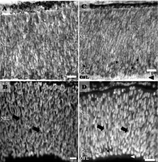
Because of the intense early proliferation of ganglion cells cited above, at E6 the ganglion cell layer (GCL) is constituted of two or three rows of cell bodies (Fig. 1C-D) extending over most central retina (Coulombre 1955). A clear segregation of retinal strata begins at about E6 with the appearance of the GCL, OFL and NBL (Spence and Robson 1989, Meller and Tetzlaff 1976). The first axonal process from RGC begins to arrive at the optic tectum at E6 (Delong and Coulombre 1965, LaVail and Cowan 1971). In this period, it has been described a low incidence of cell death in both NBL and RGC (Chavarría et al. 2007).
EMBRYONIC DAY8 (H-HSTAGE34)
At E8, very distinguishable from early retinas, the inner plexiform layer (IPL) appears in the central region of the retina (Fig. 2A) even though the GCL still appears with three rows of cell bodies. Furthermore, Müller fibers and the outer limiting membrane are found throughout the neural retina as can be observed with 2M6, a glial marker specific for chick retina (Schlosshauer et al. 1991) (Fig. 2B-C). The two plexiform layers of the mature retina result from neurite growth and dendritic branching that takes place during all the embryonic life.
EMBRYONIC DAY9-10 (H-HSTAGES35-36)
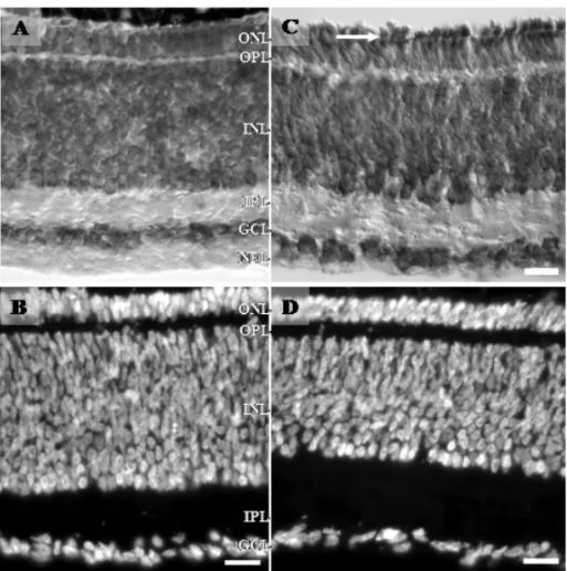
The retina of animals on the 9thday of incubation is very typical because the outer plexiform layer (OPL) makes its appearance (Fig. 2D-E). Besides that, between E9 (Fig. 2D-E) and E10 (Fig. 2F-G), displaced amacrine cells occupy a transitory intraplexiform position in the IPL, forming a discontinuous one-cell-thick layer (Fig. 2D-G; Génis-Gálvez et al. 1977). These authors
sug-gested that the unusual position of the displaced ama-crine cells in the IPL could be related to a growth in the thickness of the IPL between the displaced amacrine and the ganglion cells during the 9th and 10th days of incubation. This would lead to their appearance in an intraplexiform position. Then, the ulterior rapid elonga-tion of a cytoplasmic stalk between the bushy arboriza-tion and the soma explains the apparent disappearance of those cells in that intraplexiform position, and the displaced amacrine cells become mixed with the gan-glion cells. Thus, as we can see in Figure 2 (D-E), on the 9thday of incubation, the cell bodies are in the center of the IPL and, on the 10th day, the nucleus adopts a posi-tion nearer the GCL (Fig. 2F-G). Finally, it is not before E9 that photoreceptor neuroblasts show a round shaped apical process beyond the outer limiting membrane into the subretinal space (Fig. 2D-E), and the extension of inner segments of photoreceptors is the first indication of their morphogenesis (Olson 1979, Meller 1984apud
Mey and Thanos 2000).
At E10-E11, the number of ganglion cell axons is estimated at 4 million, but 40% of them will be elimi-nated up to E18 and, then, remain constant (Rager and Rager 1978). Probably because of the peak of cell death period, at E9, GCL does not appear with three rows of cell bodies anymore (Fig. 2E; Cook et al. 1998, Chavar-ría et al. 2007).
EMBRYONIC DAY11-13 (H-HSTAGES37-39)
devel-Fig. 1 – Photomicrographs of retinal sections from E5 (A and B) and E6 (C and D). Retinal sections stained for cresyl violet (A and C) and DAPI (B and D). In C, arrows point to round shaped cells of the future ganglion cell layer (GCL), and black arrowhead shows the optic fiber layer (OFL) region. In B and D, elongated (thick arrows) and round shaped nucleus (thin arrows) in separate layers; neuroblastic (NBL) and presumptive ganglion cell layer (GCL) can be visualized. In D, white arrowhead points to the OFL. Calibration bars = 20µm.
opment. The first receptor potentials are detected only after E13, and with the progress of the embryonic age, their increasing amplitude could be correlated with the appearance of photoreceptor outer segments that begin on the 15thday (Meller and Tetzlaff 1976, Hanawa et al. 1976).
EMBRYONIC DAY14-16 (STAGEH-H 40-42)
Around E14, horizontal cells undergo some morpholog-ical changes and, from one of their main dendrites, a single axon arises of these cells. With Golgi silver im-pregnation, synaptic spines can be distinguished 2 days later on dendrites and axon terminals (Génis-Galvéz et al. 1981). In parallel, oligodendrocyte precursors are observed in the retina around E14 and one day later oligodendrocyte processes multiply and begin to envelop thicker axons. However, as at E18, only 3% of the ax-ons of the optic nerve are ensheathed, and most of the
myelination takes place during the early postnatal period (Rager 1976).
It is on the 15th day that rod and cone outer seg-ments appear (Fig. 3C, white arrow), and both photo-receptor types begin a very rapid increase in length. Al-though the extension of RGCs spine-like process to the IPL begins after E7, the morphological development of these cells is completed around E16. From this age, the retina tissue would only increase in complexity and thickness.
LATE DEVELOPMENT TO POST-HATCH
photore-Fig. 2 – Photomicrographs of retinal sections from E8-E10 animals. In A, retinal section from E8 stained for cresyl violet (A) and immunoreactivity to 2M6, a specific Müller cell marker of the chick retina (B and C). In A, a clear segregation between the GCL and the inner nuclear layer (INL) is observed by the presence of the presumptive inner plexiform layer (IPL). In B and C, white arrows point to Müller cells endfeet, and soma cells take the middle region of INL (white rectangle). At E9 (D and E) and E10 (F and G), retinal sections stained for cresyl violet (D and F) and DAPI (E and G). At E9, the future outer plexiform layer (OPL) is present, turning the retina into a three nuclear layer tissue. The displaced amacrine cells in the characteristic intraplexiform position in the IPL, forming a discontinuous one-cell-thick layer in both ages (E9 and E10, white rectangles and arrows). Note the difference of displaced amacrine cells position in E9 and E10. OLM, outer limiting membrane. Calibration bars = 20µm.
ceptors. The chick retina has a specialized area (Ehrlich 1981), called area centralis, functionally similar to the primate fovea with the highest visual acuity enriched in red and green cones and no rods (Cepko 1996). During the development, red and green opsins are expressed first (E14), then rhodopsin follows (E15), and finally the blue and violet opsins (E16) (Bruhn and Cepko 1996). The development of rod and cone photoreceptors is similar, although rod outer segments are formed earlier. First, synapses between bipolar cells and photorecep-tors are not observed in the OPL before E17 (Hughes and LaVelle 1974, Meller 1984apudMey and Thanos 2000). In accordance to that, the normal electrical re-sponse to light stimulation develops between E17 and P3 (Hanawa et al. 1976). In fact, the electroretinogram shows a mature profile only at E19. At that time, 2 days before hatching, light evokes beak clapping and move-ments of the eyelids (Oppenheim 1968, Rager 1979apud
Mey and Thanos 2000). Concerning to the cell death,
none TUNEL-positive nuclei are seen in the GCL and INL at E16 and E18, respectively (Chavarría et al. 2007). Finally, in post-hatched retinas, the outer limiting membrane (OLM) (Fig. 4A, thick arrow) and some cells in the middle of IPL, called intraplexiform cells (Fig. 4B-C), can also be visualized.
NEUROGENESIS OF RETINAL CELLS
As discussed above, retinal neuroblasts leave the cell cycle in a specific order with respect to their prospective cell type: the first to be generated are retinal ganglion cells, followed by photoreceptors, then amacrine cells, horizontal cells and bipolar cells, which are the last neu-rons to leave the cell cycle in the chick retina. The pro-duction of photoreceptors continues over a longer pe-riod (Prada et al. 1991, Fekete et al. 1994, Meller 1984
apudMey and Thanos 2000). Those specific sequences
Fig. 3 – Photomicrographs of retinal sections from E13 (A and B) and E15 (C and D) stained for cresyl violet (A and C) and DAPI (B and D). In C, an arrow points to the outer segments of photoreceptors. Note the round shaped cells in the innermost regions of the INL and the growing of the plexiform layers. GCL is almost constituted by a one cell row (C and D). Calibration bars = 20µm.
Pearson 2008). In addition, the neurogenesis sequence occurs in a stereotypical order, but shows a strong over-lap (Kahn 1973, Spence and Robson 1989, Prada et al. 1991). However, a question remains to be clarified: would there be a specific temporal order for neurogene-sis of different neurochemical subpopulations of a spe-cific cell type in the chick retina? Efforts to answer this question have been made through studies involving the neurogenesis of dopaminergic, GABAergic, choli-noceptive and GABAceptive cells using the association of immunohistochemical and autoradiography of triti-ated thymidine techniques in the chick retina. The main methodology to evaluate neurogenesis is the cumulative approach, developed by Fujita and Horii (1963). Origi-nally, it was described with the use of [3H]-thymidine as a tracer. Nowadays, this problem can also be approached with the use of BrdU (Farah 2004). Either 3H-thymidine or BrdU have to be applied for a prolonged period to label all the cells that go through S-phase after tracer admin-istration. So, for neurogenesis in avian retina, the tracer is injected oncein ovoon different embryonic days (E1-E11), and the tissue is dissected at the end of the incuba-tion period (around E19) when the retina is considered to be morphologically mature and the programmed cell death is finished. After that, retinas are analyzed for the presence of the tracer, and two populations are found: labeled cells (those that passed through S-phase while the tracer was available) and unlabeled cells (those that completed their terminal S-phase before the initiation of the cumulative labeling).
BIRTH DATING SPECIFIC RETINAL CELLS
Several reports have addressed the neurogenesis period of specific neurochemical subpopulations of retinal cells (Gardino et al. 1993, 1996, da Costa Calaza et al. 2000, Barros et al. 2003) by using a combination of immuno-histochemistry and the cumulative method (Fujita and Horii 1963). As the [3H]-thymidine injectedin ovo re-mains available to be incorporated into the DNA for several days, immunolabeled cells without [3H]-thymi-dine, i.e. without autoradiographical silver grains, are judged to have been generated prior the time of [3H]-thymidine injection. On the other hand, double-labeled cells (immunolabeled cells showing autoradiographical silver grains) are considered to be in mitotic division and,
therefore, are judged to have not been generated at the time of the [3H]-thymidine injection.
DOPAMINERGIC AMACRINE CELLS
hydroxy-lase phenotype of retinal dopaminergic cells within this narrow window of development. Finally, the neurogene-sis period of dopaminergic cells in the chick retina ends when the width of the IPL rapidly increases (Coulom-bre 1955), and the TH immunoreactive cells are first detected after amacrine migration to their final position (Prada et al. 1987). In addition, the first detection of TH positive cells is temporally coincident with some of the above cited events of the chick retina development, for example the beginning of synaptogenesis in the IPL and the maximum activation of dopamine-dependent adeny-lyl cyclase activity (Ventura et al. 1984). Nevertheless, the factors responsible for dopaminergic specific cell proliferation remain unsolved.
2 3 4 5 6 7 8 9 10 11
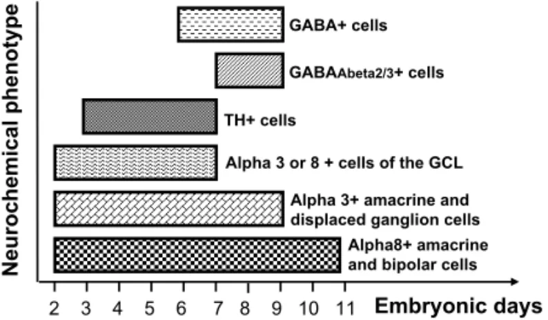
Fig. 5 – Schematic histogram of neurogenesis periods of different neu-rochemical cell subpopulations Note that subpopulations of amacrine cells, for example, expressing different phenotypes, can have different neurogenesis periods (compare alpha8+ amacrine cells and TH+ cells).
CHOLINOCEPTIVE CELLS
The chick retina contains a prominent cholinergic sys-tem, which includes three populations of cholinergic amacrine cells that could be identified by the expres-sion of choline acetyltransferase (ChAT), the enzyme responsible for the synthesis of acethylcoline (ACh) (Millar et al. 1985, Spira et al. 1987). Both ChAT and acetylcholinesterase (AchE), the enzyme that metabo-lizes ACh, are found very early in the developing tissue and are also present in the mature retina (Shen et al. 1956, Spira et al. 1987). These cholinergic markers are found in amacrine cells in the INL (cholinergic cell type I and III) and in displaced amacrine in the GCL (cholin-ergic cell type II) since E6.5 (ChAT) and E4.5 (AChE). Other aspects of the cholinergic system, such as
mod-ulation of choline acetyltransferase activity and regu-lation of vesicular acetylcholine transporter (Loureiro-dos-Santos et al. 2001, 2002, Prado et al. 2002), have also been studied.
Concerning cholinoceptive aspects, nicotinic and muscarinic acetylcholine receptors are expressed in the chick retina (Vogel and Nirenberg 1976, Large et al. 1985, Hamassaki-Britto et al. 1994, Fischer et al. 1998a, da Costa Calaza et al. 2000). Muscarinic acetylcholine receptors modulate cell cycle and calcium signaling in the chick retinal ventricular zone (Pearson et al. 2002, Syed et al. 2004). Acting through nicotinic acetylcholine receptors, ACh seems to play important roles in neurite outgrowth of ganglion cells, dendritic filopodia motil-ity and remodeling during synaptogenesis, and devel-opment of spontaneous rhythmic activity in retinal gan-glion cells during the period in which their connectivity pattern is shaped (Wong et al. 1998, Wong and Wong 2001). Therefore, the neurogenesis pattern of the cholin-ergic system is an important parameter considered to es-tablish the temporal relationship between histogenesis and definition of functional phenotypes.
Alpha 3 subunit nicotinic receptors are observed in amacrines, displaced ganglion cells, and cells in the ganglion cell layer. Alpha 8 subunits are seen in ama-crine and bipolar cells, as well as in cells in the ganglion cell layer (Hamassaki-Britto et al. 1994). The first cells that exhibit either alpha 3 or alpha 8 immunolabeling are almost certainly presumptive ganglion cells. There-after, the expression of both subunits are seen in ama-crine neurons, followed by alpha 8 subunit in bipolar cells and alpha 3 in displaced ganglion cells in the in-ner nuclear layer. These results reveal not only different developmental patterns of cells containing alpha 3 and alpha 8 nAChR subunits, but also indicate that both sub-units are expressed in the chick retina before ChAT and well before retinal synaptogenesis (Araki et al. 1982). In addition, despite the fact that nAChRs alpha 3 and alpha 8 subunits have different time courses of develop-ment in the embryonic chick retina, it could also be sug-gested that the birth of cholinoceptive cells followed the neurogenesis time course of specific cell types.
al. 1996). Alpha 3-immunoreactive cells in the INL (amacrine and displaced ganglion cells) leave the cell cycle from E2 through E9. The alpha 8-positive cells in the ganglion cell layer are born between E1 and E7, and those in the inner nuclear layer (amacrine and bipo-lar cells) from E2 through E11. At least one difference was noticed between the neurogenesis of alpha 3- and al-pha 8-positive neurons in the chick retina (Fig. 5). The cells in the ganglion cell layer that stained for alpha 3 begin to leave the cell cycle almost one day later than the alpha 8-positive neurons do. Furthermore, until E4, the alpha 3-positive cells were observed to leave the cell cycle at a slower rate than the one of alpha 8-positive cells. However, the expression of the alpha 3 subunit begins almost one day before (Hamassaki-Britto et al. 1994). These results suggest that the factors controlling the expression of alpha 3 and alpha 8 nAChR subunits in the chick retina appear to be relatively independent of those that control cell genesis. Indeed, the ontogene-sis of alpha 3 and alpha 8 nAChR subunits in the chick retina is not related in any simple way to the neurogenesis of neurons bearing those receptor subunits. The neuro-genesis rates of alpha 3- and alpha 8-positive amacrine cells also appear to differ slightly, although in this case no difference in their ontogenesis has been noticed in a previous study (Hamassaki-Britto et al. 1994). Finally, previously reported data suggest that the time of birth of cholinoceptive neurons in the chick retina follows the general pattern of cell generation in this tissue (Gardino et al. 1996).
GABAERGIC ANDGABACEPTIVECELLS
L-Glutamate (L-Glu) andγ-aminobutyric acid (GABA) are widely recognized as mediators of excitatory and inhibitory neurotransmissions, respectively, in the cen-tral nervous system, including the retina of many ver-tebrate species (Mosinger et al. 1986, Yazulla 1986, Massey and Redburn 1987, Thoreson and Witkovsky 1999). The GABAergic system is composed of a large population of cells, mainly constituted of amacrine neu-rons, with many important functions in the development and physiology of the retina (Catsicas and Mobbs 2001, Barnstable 1993, Ge et al. 2007, Luján et al. 2005, Ser-nagor et al. 2003, Tachibana and Kaneko 1988, Yazulla 1986). In fact, GABA is visualized in horizontal and
amacrine cells and in cells in the GCL, most of them containing glutamate, probably used as a precursor for GABA synthesis (Sun and Crossland 2000). Neurons expressing GABA in the chick retina appear at E6 and, at this period, GABA is mainly synthesized from pu-trescine (de Mello et al. 1976, Hokoç et al. 1990). The classical enzyme that synthesizes GABA from glutamate (glutamic decarboxylase, GAD) first appears only at E11 (Hokoç et al. 1990). After this stage, GABA is mainly synthesized by the classical pathway involving GAD.
As observed in several regions of the CNS (Ben-Ari 2002 for review), retinal GABA, acting on iono-tropic receptors, also seems to be a depolarizing media-tor at the beginning of the development (Yamashita and Fukuda 1993, Catsicas and Mobbs 2001). At E3, GABA is able to induce calcium influx by its depolarizing ac-tion (Yamashita and Fukuda 1993). Depolarizaac-tion by GABA reaches a peak at E8 and undergoes a slight de-crease around E12 (Catsicas and Mobbs 2001). Finally, at E14, GABA no more induces calcium influx and prob-ably assumes its classical inhibitory function (Catsicas and Mobbs 2001).
Concerning to the neurogenesis pattern, the profile of GABAergic amacrine and horizontal cells showed a very slow proliferation rate up to E6, when only 20% of GAD-positive cells were considered generated. Then, between E6 and E9, 80% of GAD expressing neurons are born (da Costa Calaza et al. 2000) (Fig. 5). Birth dates of GABAergic neurons were also determined in the rat retina by using BrdU or thymidine as markers (Lee et al. 1999, Silveira et al. 2007). In both cases, the rate of GABAergic cell generation peaked at E18 in both central and peripheral sectors of the retina, and displayed a homogenous generation rate from E14 to PN4, ending after that.
Costa Calaza et al. 2000), and long before GAD ex-pression (Hokoç et al. 1990). Considering GABA as a neurotrophic molecule and knowing its inhibitory effect on DNA synthesis (LoTurco et al. 1995), GABA could carry specific signals that are need to arrest the mitotic cycle of GAD immunoreactive cells. Therefore, it could constitute a factor acting to control the proliferation of its own population. However, one cannot rule out the possibility of other substances influencing this process, and this possibility remains to be confirmed.
During the development, GABA functions are apparently mediated by ionotropic GABA receptors. GABAAβ2-3, an important subunit of these receptors, is expressed in amacrine cells and in cells in the GCL (Barros et al. 2003). At E9, GABAAβ2-3 immunoreac-tivity was restricted to the inner plexiform layer, and the first cell bodies immunoreactive to GABAAβ2-3 were seen at E14. Thereafter, the number of cell bod-ies and the intensity of GABAAβ2-3 immunoreactivity increased until the adult pattern is established. Inter-estingly, GABAAβ2-3amacrine positive cells are born in the same pattern as GAD positive cells, late in the neu-rogenesis period of the overall amacrine cell popula-tion (E7-E9) (Fig. 5).
CONCLUSION: NO REGULAR PLAN FOR BIRTH!
One interesting point that could arise from the data dis-cussed above is that the neurogenesis period of the to-tal cell population of the retina is not necessarily the same for different neurochemical cell subpopulations. Thus, although the neurogenesis of the total population of amacrine cells in chick retina takes place mainly from E2 to E9 (Fujita and Horii 1963, Kahn 1974, Spence and Robson 1989), subpopulations of amacrine cells express-ing different phenotypes, like dopaminergic amacrine cells for example, can have distinct periods of neuro-genesis, i.e. from E3 to E7 (Gardino et al. 1993, 1996, da Costa Calaza et al. 2000). By studying the ontoge-nesis and the neurogeontoge-nesis of different retinal cell sub-populations, with distinct phenotypes, one could think that, for some of them, such as for the GABAergic sys-tem, there is a temporal window during the develop-ment, during which specific signal(s), responsible for their decision to leave the cell cycle, must be given. For the GABAergic system, it appears that there are
im-portant signals around E6, which determine the end of the proliferation period. We cannot exclude the collabo-ration of progenitor cells competent to respond to some signal, which is present before E6 at their retinal mi-croenvironment. However, for a full comprehension of the signals that modulate the neurogenesis of each spe-cific neurochemical system, further experiments must be done. If there is not a common plan for the birth of the overall population of retinal cell, we can conclude that, among the great diversity of neurochemical cell types in the retina, there may be several genetic or epigenetic sig-nals, factors, molecules, or even a combination of these, which are responsible not only for phenotype definition, but also for the establishment of the end of the mitotic cycle for each neurochemical cell type. The identifica-tion of signals and the determinaidentifica-tion of the proliferaidentifica-tion periods of each cell type of the retina could be important for future therapeutic interventions using stem cells.
ACKNOWLEDGMENTS
This work was supported by funds from Conselho Nacional de Desenvolvimento Científico e Tecnológico (CNPq), Institutos Nacionais de Ciência e Tecnologia/ CNPq/ Instituto Nacional de Neurociência Translacional, Fundação Carlos Chagas Filho de Amparo à Pesquisa do Estado do Rio de Janeiro (FAPERJ) and Programa de Apoio a Núcleos de Excelência (PRONEX), Pro-reitoria de Pesquisa, Pós-Graduação e Inovação (PROPPi)/UFF. We thank Prof. Fernando G. de Mello for the scientific reviews and Prof. Maria Christina F. de Mello for the manuscript revision. We also thank Prof. B. Schlosshauer for gently providing the mouse monoclonal antibody 2M6.
RESUMO
imuno-histoquímica e autoradiografia de timidina-tritiada. Conclui-se que o período de proliferação das células dopaminérgicas, GABAérgicas, colinoceptivas e GABAceptivas não segue uma regra comum. Além disso, alguns grupos celulares neuroqui-micamente distintos podem ter um período de proliferação mais restrito quando comparado ao da população total destas células.
Palavras-chave: desenvolvimento, neurogênese, sistemas
neurotransmissores, ontogênese, proliferação.
REFERENCES
ARAKIM, IDECANDSAITOT. 1982. Ultrastructural local-ization of acetylcholinesterase activity in the developing chick retina. Acta Histochem Cytochem 15: 242–255. ARAKIM, FUKADAY, SHICHIDAYANDYOSHIZAWAT.
1990. Localization of iodopsin in the chick retina during in vivoandin vitrocone differentiation. Invest Ophthal-mol Visual Sci 31: 1466–1473.
BARNSTABLECJ. 1993. Glutamate and GABA in retinal circuitry. Curr Opin Neurobiol 3: 520–525.
BARROSPH, CALAZAKDA CANDGARDINO PF. 2003. GABA (Aβ2-3) immunoreactive cells in the developing chick retina. Int J Dev Neurosci 21: 35–40.
BEN-ARI Y. 2002. Excitatory actions of GABA during de-velopment: the nature of the nurture. Nat Neurosci 3: 728–739.
BLASINA MF, FARIA AC, GARDINO PF, HOKOC JN,DE MELLO FGANDDAJASF. 2000. Evidence for a non-cholinergic function of acetylcholinesterase during devel-opment of chicken retina as shown by fasciculin and other enzyme inhibitors. J Cell Tissue Res 299: 173–184. BORBA JC, HENZE IP, SILVEIRA MS, KUBRUSLY
RC, GARDINOPF,DEMELLOMC, HOKOÇJNAND DE MELLOFG. 2005. Pituitary adenylate cyclase-activating polypeptide (PACAP) can act as determinant of the tyrosine hydroxylase phenotype of dopaminergic cells during retina development. Brain Res Dev Brain Res 156: 193–201.
BRUHN S AND CEPKO CL. 1996. Development of the pattern of photoreceptors in the chick retina. J Neurosci 16: 1430–1439.
CATSICASMANDMOBBSP. 2001. GABAB receptors regu-late chick retinal calcium waves. J Neurosci 21: 897–910. CEPKOCL. 1996. The patterning and onset of opsin expres-sion in vertebrate retinae Curr Opin Neurobiol 6: 542–546. CHAVARRÍAT, VALENCIANOAI, MAYORDOMO R, EGEA J, COMELLAJX, HALLBÖÖK F,DEPABLOF AND DE
LAROSAEJ. 2007. Differential, age-dependent MEK-ERK and PI3K-Akt activation by insulin acting as a sur-vival factor during embryonic retinal development. Dev Neurobiol 67: 1777–1788.
COOKB, PORTERA-CAILLIAUCANDADLERR. 1998. De-velopmental neuronal death is not a universal phenomenon among cell types in the chick embryo retina. J Comp Neu-rol 396: 12–19.
COULOMBREAJ. 1955. Correlations of structural and bio-chemical changes in the developing retina of the chick. Am J Anat 96: 153–189.
COULOMBREJL ANDCOULOMBRE AJ. 1965. Regenera-tion of neural retina from the pigmented epithelium in the chick embryo. Dev Biol 12: 79–92.
DA COSTA CALAZA K, HOKOÇ JN AND GARDINO PF. 2000. Neurogenesis of GABAergic cells in the chick ret-ina. Int J Devl Neuroscience 18: 721–726.
DECARVALHORP, BRAASKM, ADLER RANDSNYDER SH. 1992. Developmental regulation of adenosine A1 re-ceptors, uptake sites and endogenous adenosine in the chick retina. Brain Res Dev Brain Res 70: 87–95. DEMELLOFG, BACHRACHUANDNIRENBERGM. 1976.
Ornithine and glutamic acid decarboxylase activities in the developing chick retina. J Neurochem 27: 847–851. DELONGGRANDCOULOMBREAJ. 1965. Development of
the retinotectal topographic projection in the chick em-bryo. Exp Neurol 13: 351–363.
DUPREEJL ANDBIGBEE JW. 1996. Acetylcholinesterase inhibitor treatment delays recovery from axotomy in cultured dorsal root ganglion neurons. J Neurocytol 25: 439–454.
EDQVISTPHANDHALLBÖÖKF. 2004. Newborn horizontal cells migrate bi-directionally across the neuroepithelium during retinal development. Development 131: 1343– 1351.
EHINGERB. 1978. Biogenic Monoamines and aminoacids as retinal neurotransmitters. In: COOLSJ ANDSMITH EL (Eds), Frontiers in Visual Science. Berlin-Heidelberg-New York Springer-Verlag, p. 42–53.
EHRLICHD. 1981. Regional specialization of the chick retina as revealed by the size and density of neurons in the gan-glion cell layer. J Comp Neurol 195: 643–657.
EHRLICHDANDMORGANIG. 1980. Kainic acid destroys displaced amacrine cells in posthatch chicken retina. Neurosci Lett 17: 43–48.
FARAHMH. 2004. Cumulative labeling of embryonic mouse neural retina with bromodeoxyuridine supplied by an os-motic minipump. J Neurosci Meth 134: 169–178. FEKETE DM, PEREZ-MIGUELSANZ J, RYDER EF AND
CEPKOCL. 1994. Clonal analysis in the chicken retina reveals tangential dispersion of clonally related cells. Dev Biol 166: 666–682.
FISCHER AJ, MCKINNON LA, NATHANSON NM AND STELL WK. 1998a. Identification and localization of muscarinic acetylcholine receptors in the ocular tissues of the chick. J Comp Neurol 392: 273–84.
FISCHER AJ, SELTNER RL, POON J AND STELL WK. 1998b. Immunocytochemical characterization of quis-qualic acid- and N-methyl-D-aspartate-induced excitotox-icity in the retina of chicks. J Comp Neurol 393: 1–15. FISCHERAJ, WALLMAN J, MERTZ JR ANDSTELL WK.
1999. Localization of retinoid binding proteins, retinoid receptors, and retinaldehyde dehydrogenase in the chick eye. J Neurocytol 28: 597–609.
FISCHERAJ, OMARG, WALTONNA, VERRILL TAAND UNSONCG. 2005. Glucagon-expressing neurons within the retina regulate the proliferation of neural progenitors in the circumferential marginal zone of the avian eye. J Neurosci 25: 10157–10166.
FISCHERAJ, SKORUPAD, SCHONBERGDLANDWALTON NA. 2006. Characterization of glucagon-expressing neu-rons in the chicken retina. J Comp Neurol 496: 479–494. FISCHERAJ, STANKEJJ, ALOISIOG, HOYHANDSTELL WK. 2007. Heterogeneity of horizontal cells in the chicken retina. J Comp Neurol 500: 1154–1171. FUJITASANDHORIIM. 1963. Analysis of cytogenesis in
chick retina by tritiated thymidine autoradiography. Arch histol Jpn 23: 359–366.
FUKUDA M ET AL. 1981. Localization of vasoactive in-testinal polypeptide and neurotensin immunoreactivities in the avian retina. Curr Eye Res 1: 115–118.
GARDINO PF, DOS SANTOS RM ANDHOKOÇ JN. 1993. Histogenesis and topographical distribution of tyrosine hydroxylase immunoreactive amacrine cells in the devel-oping chick retina. Dev Brain Res 72: 226–236. GARDINO PF, CALAZA KC, HAMASSAKI-BRITTO DE,
LINDSTROM JM, BRITTO LRANDHOKOÇ JN. 1996. Neurogenesis of cholinoceptive neurons in the chick ret-ina. Brain Res Dev Brain Res 95: 205–212.
GE S, PRADHAN DA, MING GL AND SONG H. 2007. GABA sets the tempo for activity-dependent adult neuro-genesis. Trends Neurosci 30: 1–8.
GÉNIS-GÁLVEZ JM, PULELLESL ANDPRADA C. 1977. Inverted displaced retinal amacrine cells and their em-bryonic development in the chick retina. Exp Neurol 56: 151–157.
GÉNIS-GÁLVEZJM, GARCIA-LOMASV, PRADAFAAND ARMENGOLJA. 1981. Developmental study of axon for-mation in the horizontal neurons of the retina of the chick embryo. Anat Embryol 161: 305–317.
GUIMARÃESMZ, HOKOÇJN, DUVOISINR, REISRAAND DEMELLOFG. 2001. Dopaminergic retinal cell differ-entiation in culture modulation by forskolin and dopamine. Eur J Neurosci 13: 1931–1937.
HAMASSAKI-BRITTODE, GARDINOPF, HOKOÇJN, KEY -SER KT, KARTEN HJ, LINDSTROM JM ANDBRITTO LR. 1994. Differential development of alpha-bungaro-toxin-sensitive and alpha-bungarotoxin-insensitive nico-tinic acetylcholine receptors in the chick retina. J Comp Neurol 347: 161–170.
HAMBURGER V ANDHAMILTON HL. 1951. A series of normal stages in the development of the chick embryo. J Morphol 88: 49–92.
HANAWAJ, TAKAHASHIKANDKAWAMOTON. 1976. A correlation of embryogenesis of visual cells and early re-ceptor potential in the developing retina. Exp Eye Res 23: 587–594.
HERING HANDKRÖGERS. 1996. Formation of synaptic specializations in the inner plexiform layer of the devel-oping chick retina. J Comp Neurol 375: 393–405. HOKOÇ JN, VENTURA ALM, GARDINO PF AND DE
MELLOFG. 1990. Developmental immunoreactivity for GABA and GAD in the avian retina: possible alternative pathway for GABA synthesis. Brain Res 532: 197–202. HUGHESWFANDLAVELLEA. 1974. On the synaptogenic
sequence in the chick retina. Anat Rec 179: 297–302. HYERJT, MIMATANDMIKAWA T. 1998. FGF1 patterns
the optic vesicle by directing the placement of the neural retina domain. Development 125: 869–877.
KAHNAJ. 1973. Ganglion cell formation in the chick neural retina. Brain Res 63: 285–290.
KAHNAJ. 1974. An autoradiographic analysis of the time of appearance of neurons in the developing chick neural retina. Dev Biol 38: 30–40.
KALLONIATISMANDFLETCHEREL. 1993. Immunocyto-chemical localization of the amino acid neurotransmitters in the chicken retina. J Comp Neurol 336: 174–193. KIYAMA H, KATAYAMA-KUMOI Y, KIMMEL J, STEIN
1985. Three dimensional analysis of retinal neuropeptides and amine in the chick. Brain Res Bull 15: 155–165. LANKFORD KL, DE MELLO FG AND KLEIN WL. 1987.
A transient embryonic dopamine receptor inhibits growth cone motility and neurite outgrowth in a subset of avian retina neurons. Neurosci Lett 75: 169–174.
LANKFORD KL, DEMELLO FG ANDKLEIN WL. 1988. D1-type dopamine receptors inhibit growth cone motil-ity in cultured retina neuronsevidence that neurotransmit-ters act as morphogenic growth regulators in the develop-ing central nervous system. Proc Nat Acad Sci USA 85: 2839–2843.
LARGE TH, RAUH JJ, DE MELLO FG AND KLEIN WL. 1985. Two molecular weight forms of muscarinic acetylcholine receptors in the avian central nervous sys-temswitch in predominant form during differentiation of synapses. Proc Natl Acad Sci USA 82: 8785–8789. LAVAILJHANDCOWANWM. 1971. The development of
the chick optic tectum. I. Normal morphology and cyto-architectonic development. Brain Res 28: 391–419. LEEMY, SHINSL, HANSHANDCHUNMH. 1999. The
birth dates of GABA immunorreactive amacrine cells in rat retina. Exp Brain Res 128: 309–314.
LIPTONSA, FROSCHMP, PHILLIPSMD, TAUCKDLAND AIZENMANE. 1988. Nicotinic antagonists enhance pro-cess outgrowth by rat retinal ganglion cells in culture. Sci-ence 239: 1293–1296.
LOTURCO JJ, OWENS DF, HEATH MJS, DAVIES MBE ANDKRIEGSTEINAR. 1995. GABA and glutamate de-polarize cortical progenitor cells and inhibit DNA synthe-sis. Neuron 15: 1287–1298.
LOUREIRO-DOS-SANTOS NE, REIS RA, KUBRUSLYRC, DEALMEIDAOM, GARDINOPF,DEMELLOMCAND DEMELLOFG. 2001. Inhibition of choline acetyltrans-ferase by excitatory amino acids as a possible mechanism for cholinergic dysfunction in the central nervous system. J Neurochem 77: 1136–1144.
LOUREIRO-DOS-SANTOS NE, PRADO MA, REIS RA, GARDINO PF, DE MELLO MC AND DE MELLO FG. 2002. Regulation of vesicular acetylcholine transporter by the activation of excitatory amino acid receptors in the avian retina. Cell Mol Neurobiol 22: 727–740.
LUJÁNR, SHIGEMOTO RANDLÓPEZ-BENDITO G. 2005. Glutamate and GABA receptor signalling in the develop-ing brain. Neuroscience 130: 567–580.
MARQUARDTTANDGRUSSP. 2002. Generating neuronal diversity in the retina one for nearly all. Trends Neurosci 25: 32–38.
MARTINS RA AND PEARSON RA. 2008. Control of cell proliferation by neurotransmitters in the developing verte-brate retina. Brain Res 1192: 37–60.
MASLANDRH. 2001. Neuronal diversity in the retina. Curr Opin Neurobiol 11: 431–436.
MASLANDRH. 2004. Neuronal cell types. Curr Biol 14: R497–500.
MASSEYSCANDREDBURND. 1987. Transmitter circuits in the vertebrate retina. Prog Neurobiol 28: 55–96. MELLERKANDTETZLAFFW. 1976. Scanning electron
mi-croscopic studies on the development of the chick retina. Cell Tissue Res 170: 145–159.
MEYJANDTHANOSS. 2000. Development of the visual sys-tem of the chick. I. Cell differentiation and histogenesis. Brain Res Brain Res Rev 32: 343–379.
MILLAR T, ISHIMOTO I, JOHNSON CD, EPSTEIN ML, CHUBBIWANDMORGANIG. 1985. Cholinergic and acetylcholinesterase-containing neurons of the chicken retina. Neurosci Lett 61: 311–316.
MITROFANISJ, VIGNYAANDSTONEJ. 1988. Distribution of cathecolaminergic cells in the retina of rat, guinea-pig, cat and rabbitindependence of ganglion cell distribution. J Comp Neurol 26: 71–14.
MOSINGERJL, YAZULLASANDSTUDHOLMEKM. 1986. GABA-like immunoreactivity in the vertebrate retina species comparison. Exp Eye Res 42: 631–644.
OKANO T, KOJIMA D, FUKADA Y, SHICHIDA Y AND YOSHIZAWAT. 1992. Primary structures of chicken cone visual pigments: vertebrate rhodopsins have evolved out of cone visual pigments. Proc Nat Acad Sci USA 89: 5932–5936.
OLSONMD. 1979. Scanning electron microscopy of devel-oping photoreceptors in the chick retina. Anat Rec 193: 432–438.
OPPENHEIMRW. 1968. Light responsivity in chick and duck embryos just prior to hatching. Anim Behav 16: 276–280. PAES DE CARVALHO R AND DE MELLO FG. 1982. Adenosine-elicited accumulation of adenosine 3′, 5′ -cyc-lic monophosphate in the chick embryo retina. J Neu-rochem 38: 493–500.
PEARSON R, CATSICAS M, BECKER D AND MOBBS P. 2002. Purinergic and muscarinic modulation of the cell cycle and calcium signaling in the chick retinal ventricu-lar zone. J Neurosci 22: 7569–7579.
PRADA C, PUGA J, PÉREZ-MÉNDEZ L, LÓPEZ R AND RAMÍREZG. 1991. Spatial and temporal patterns of neu-rogenesis in the chick retina. Eur J Neurosci 3: 559–569. PRADO MA, REIS RA, PRADO VF, DE MELLO MC, GOMEZMVAND DEMELLO FG. 2002. Regulation of acetylcholine synthesis and storage. Neurochem Int 41: 291–299.
RAGERG. 1976. Morphogenesis and physiogenesis of the tectal connection in the chicken. II. The retino-tectal synapses. Proc R Soc Lond B Biol Sci 192: 353– 370.
RAGERGANDRAGERU. 1978. Systems-matching by de-generation. I. A quantitative electron microscopic study of the generation and degeneration of retinal ganglion cells in the chicken. Exp Brain Res 33: 65–78.
RAGERU, RAGERGANDFREIB. 1993. Central retinal area is not the site where ganglion cells are generated first. J Comp Neurol 334: 529–544.
REISRA, VENTURAAL, KUBRUSLYRC,DEMELLOMC AND DEMELLOFG. 2007. Dopaminergic signaling in the developing retina. Brain Res Rev 54: 181–188. ROBINSONSR. 1988. Cell death in the inner and outer
nu-clear layers of the developing cat retina. J Comp Neurol 267: 507–515.
SCHLOSSHAUER B, GRAUERD, DTTINGDANDVANSE -LOWJ. 1991. Expression of a novelMuller gliaspecific antigen during development and after optic nerve lesion. Development 111: 789–799.
SERNAGOR E, YOUNG C ANDEGLEN SJ. 2003. Devel-opmental modulation of retinal wave dynamics shedding light on the GABA saga. J Neurosci 23: 7621–7629. SHENSC, GREENFIELDPANDBOELLEJ. 1956.
Localiza-tion of acetylcholinesterase in chick retina during histoge-nesis. J Comp Neurol 106: 433–461.
SILVEIRA ACD, GARDINO PF, BEVILAQUA MCN AND HOKOÇJN. 2007. Neurogenesis of GABAergic cells in the retina of malnourished rats. Int J Dev Neurosci 25: 325–333.
SPENCESGANDROBSON JA. 1989. An autoradiographic analysis of neurogenesis in the chick retinain vitroandin vivo. Neuroscience 32: 801–812.
SPIRAAW, MILLARTJ, ISHIMOTOI, EPSTEINML, JOHN -SONCD, DAHLJLANDMORGANIG. 1987. Localiza-tion of choline acetyltransferase like immunoreactivity in the embryonic chick retina. J Comp Neurol 260: 526–538.
SUNHANDCROSSLANDWJ. 2000. Quantitative assessment of localization and colocalization of glutamate, aspartate, glycine and GABA immunoreactivity in the chick retina. Anat Rec 260: 158–179.
SYEDMM, LEES, HESANDZHOUZJ. 2004. Spontaneous waves in the ventricular zone of developing mammalian retina. J Neurophysiol 91: 1999–2009.
TACHIBANA M AND KANEKO A. 1988. Retinal bipolar cells receive negative feedback input from GABAergic amacrine cells. Vis Neurosci 12: 297–305.
THORESONWBANDWITKOVSKYP. 1999. Glutamate re-ceptors and circuits in the vertebrate retina. Prog Ret Eye Res 18: 765–810.
TUNGNN, MORGANIGANDEHRLICHD. 1990. A quanti-tative analysis of the effects of excitatory neurotoxins on retinal ganglion cells in the chick. Vis Neurosci 4: 217– 223.
VENTURA ALM, KLEIN WLAND DEMELLO FG. 1984. Differential ontogenesis of D1 and D2 dopaminergic re-ceptors in the chick embryo retina. Dev Brain Res 12: 217–223.
VOGELZANDNIRENBERGM. 1976. Localization of acetyl-choline receptors during synaptogenesis in retina. Proc Natl Acad Sci USA 73: 1806–1810.
WONGWTANDWONGRO. 2001. Changing specificity of neurotransmitter regulation of rapid dendritic remodeling during synaptogenesis. Nature Neurosci 4: 351–352. WONGWT, SANESJR ANDWONGRO. 1998.
Develop-mentally regulated spontaneous activity in the embryonic chick retina. J Neurosci 18: 8839–8852.
WUDKANDCEPKOCL. 1993. Development of dopamin-ergic neurons is insensitive to optic nerve section in the neonatal rat retina. Brain Res Dev Brain Res 742: 53–60. YAMASHITA MANDFUKUDAY. 1993. Calcium channels and GABA receptors in the early embryonic chick retina. J Neurobiol 24: 1600–1614.