Comparison of optical coherence tomographic findings between Behcet
disease patients with and without ocular involvement and healthy subjects
Comparação de achados de tomograia de coerência óptica em pacientes com doença de Behcet
com e sem envolvimento ocular e indivíduos saudáveis
Ayse sevgi KArAdAg1, BurAK Bilgin2, Merih BAnu soylu3
Submitted for publication: September 26, 2016 Accepted for publication: November 18, 2016
1 Department of Ophthalmology, School of Medicine, Adıyaman University, Adıyaman, Turkey. 2 Department of Ophthalmology, Medical Park Hospital, Gaziantep, Turkey.
3 Department of Ophthalmology, School of Medicine, Cukurova University, Adana, Turkey.
Funding: No specific financial support was available for this study.
Disclosure of potential conflicts of interest: None of the authors have any potential conflict of interest to disclose.
Corresponding author: Burak Bilgin. Medical Park Hospital/Gaziantep, Mücahitler, 52063. Sk. No: 2, Şehitkamil/Gaziantep 27584 - Turkey - E-mail: [email protected]
Approved by the following research ethics committee: Adiyaman University (#2016/2-14). ABSTRACT
Purpose: We aimed to compare the retinal nerve fiber layer, ganglion cell layer, inner plexiform layer, and the choroid thickness between patients with Behcet disease and healthy subjects by using spectral domain optical coherence tomo-graphy (SD-OCT ).
Methods: Ninety eyes of 45 healthy subjects and 104 eyes of 52 patients with Behcet disease were included in this study. Rheumatoid factor and C-reactive protein levels were measured by blood testing in the patients.
Results: The mean thickness of the retinal nerve fiber layer, ganglion cells layer, and inner plexiform layer were significantly lower in patients with Behcet’s disease than in the healthy subjects. The mean choroidal thickness was significantly higher in the patients than in the healthy subjects.
Conclusions: SD-OCT was a useful and non-invasive tool for the detection of retinal nerve degeneration and choroidal changes in patients with Behcet disease even in the absence of ocular involvement.
Keywords: Retinal degeneration; tomography, optical coherence; Behcet disease/ complications
RESUMO
Objetivo: Comparar a camada de fibras nervosas da retina, a camada de células ganglionares, a camada plexiforme interna e a espessura coróide entre os pacientes com doença de Behçet e indivíduos saudáveis usando tomografia de coerência óptica (OCT ) de domínio espectral (SD).
Métodos: Noventa olhos de 45 indivíduos saudáveis e 104 olhos de 52 pacientes com doença de Behcet foram incluídos no estudo. O fator reumatoide e os níveis de proteína C-reativa foram medidos por exames de sangue em pacientes com doença de Behcet. Resultados: As médias de espessura da camada de fibras nervosas da retina, da camada de células ganglionares e da camada plexiforme interna dos pacientes com Doença de Behcet foram significativamente menores do que o grupo controle. As medidas de espessura coróide média dos pacientes com doença de Behcet foram significativamente mais elevadas do que o grupo controle.
Conclusões: Tomografia de coerência óptica é uma ferramenta útil e não invasiva para acompanhar a degeneração nervosa retiniana e as alterações coroidais em pacientes com doença de Behcet, mesmo sem envolvimento ocular.
Descritores: Degeneração retiniana; Tomografia de coerência óptica; Doença de Behçet/complicações
INTRODUCTION
Behcet disease (BD) is an immune-mediated systemic occlusive vasculopathy for which the etiology and pathogenesis have not been fully clarified. The major symptoms include oral aphthous ulcers and skin and ocular lesions(1). Ocular involvement, which is seen in
60-80% of patients, is characterized by anterior uveitis, posterior uvei-tis, or panuveitis with retinal vasculitis. In particular, posterior seg ment involvement can significantly affect visual acuity(2-4).
In addition to clinical fundus fluorescein angiography, indocya-nine green angiography ultrasound biomicroscopy, and optical coherence tomography (OCT) can be used to evaluate the posterior segment(5,6).
OCT entered clinical use in the late 1990s and made it possible to examine the retinal layers in detail. In 2002, with the introduction of the Stratus OCT instrument, more detailed and high-quality images of the retina could be obtained. By 2006, parallel to developments in OCT technology, spectral domain (SD) OCT began to be used clinically, which made even more detailed evaluations of the retina possible with higher resolution, speed, and contrast(7-10). In this clinical
study, we compared the retinal nerve fiber layer (RNFL), ganglion cell layer (GCL), inner plexiform layer (IPL), and choroid thickness (CT) between patients with BD and healthy subjects by using SD-OCT. Additionally, we compared the OCT parameters, including macular volume (MV), between patients with and without ocular involvement.
METHODS
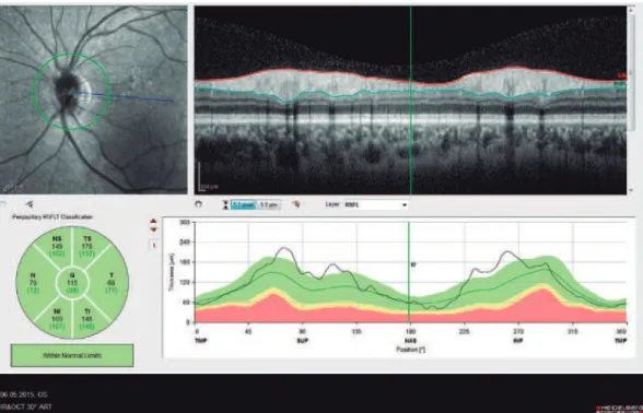
subjects was performed. All measurements were performed by the same experienced examiner (ASK) in the morning. RNFL, GCL, IPL, and CT were measured and recorded. The new Spectralis segmentation software was used to obtain RNFL, GCL, and IPL. The SD-OCT choroid layer, RNFL, GCL, and IPL imaging of a normal eye are shown in figures 1, 2, 3, and 4, respectively.
The horizontal cross-sectional view running through the center of the fovea was used to evaluate the central foveal choroidal thick-ness and 500-µm nasal and 500-µm temporal choroidal thickthick-ness to the foveal center. The average of these three location measurements were calculated and compared. The choroidal thickness was manua-lly measured from the outer line of the retinal pigment epithelium to
the inner border of the sclera using the device’s software. A choroidal thicknesss calculation for each subject was performed by two of the co-authors and then averaged for analysis.
In the patients with BD, MV measurements were taken by SD-OCT. Additionally, the rheumatoid factor (RF) and C-reactive protein (CRP) levels were measured by blood testing in the patients with BD. The exclusion criteria included history of ocular surgery, glaucoma, retinal and macular pathology, diabetes, and uveitis cau-sed by other etiologies. Statistical analysis was performed by using Statistical Package for Social Sciences (SPSS) 21.0 for windows (SPSS Inc., Chicago, IL, USA).
Figure 1. The parafoveal choroidal thickness measurement of one of the patients.
RESULTS
Of the 52 patients with BD, 34 (65.4%) were female and 18 (34.6%) were male. Of 45 healthy subjects in the control group, 23 (51.1%) were female and 22 (48.9%) were male. The mean ages of the BD patients and control group were 37.03 ± 10.57 and 39.55 ± 15.94, respectively. There were no statistically significant differences in sex and age between the patients and control group (p=0.154 and
p=0.356, respectively). Of the 52 patients with BD, 25 (48.1%) had ocular involvement and 27 (51.9%) had no ocular involvement. Of the 25 patients with ocular involvement, 4 had an acute uveitis attack and 21 had chronic uveitis.
A comparison of the mean RNFL, choroid, GCL, and IPL thickness measurements between the 2 groups is presented in table 1. The mean RNFL thickness was significantly lower for the patients with BD
Figure 3. Spectral domain optical coherence tomography ganglion cell layer (GCL) imaging of a normal eye.
than for the control group (p=0.026). The mean choroid thickness measurements were significantly higher in the patients with BD than in the control group (p=0.00). The GCL and IPL thickness measure-ments were significantly lower in the patients with BD than in the control group (p=0.00).
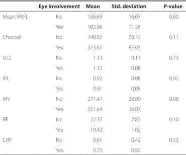
A comparison of the mean RNFL, choroid, GCL, IPL, and MV thickness measurements between the BD patients with ocular in-volvement and without ocular inin-volvement is presented in table 2. There were no statistically significant differences in CT, GCL, IPL, RNFL, and MV measurements (p=0.11, p=0.73, p=0.92, p=0.19, and p=0.08, respectively) between the BD patients with and without ocular invol-vement. There were also no statistically significant differences in the CRP and RF levels (p=0.533 and p=0.102, respectively) between the BD patients with and without ocular involvement.
Correlation analyses results are shown in table 3. There was a significant negative correlation between CRP-GCL and CRP-IPL (p=0.006 and p=0.036, respectively). There was also a significant ne-gative correlation between RF-GCL and RF-IPL (p=0.00 and p=0.00, respectively).
DISCUSSION
Behcet disease is an idiopathic, polysymptomatic, chronic, and recurrent systemic vasculitis(1). Although the disease occurs worldwide,
its incidence increases from the Mediterranean and Middle East, eastward to Japan(11). It is more common in males, with a male:female
ratio of 2:1, but this ratio is reversed in the US and European coun-tries(11). In our BD group, there were more female patients than male
patients. Studies have shown that HLA-B5 and HLA-B51 are related to BD(12). In the disease etiology, toxic exposure, infective agents,
genetic factors, chromosomal anomalies, IL-8 polimorphism, T-cell CD-4+ type 1 polarization, and auto-immunity have been conside-red as factors(13-15). However, there is insufficient evidence to support
any of these etiological factors. Although the cause of the disease is unknown, histopathological studies have shown that accumulation of immuno-complexes in blood vessel walls activates the comple-ment system and results in vasculitis, which causes tissue damage(16).
In our study, of the 52 patients with BD, 27 (51.9%) had ocular involvement and 25 (48.1%) did not. Of the 27 patients with BD and ocular involvement, 4 (7.7%) patients had acute uveitis attacks and 21 (40.4%) had chronic uveitis. BD usually affects both eyes asymme-trically but rarely, unilateral involvement may be seen. In 80% of the cases with unilateral involvement, the other eye becomes involved within 5 years. Unilateral involvement is more likely associated with HLA-B27+ uveitis. In 20% of the patients with BD, the eye is the first organ to be affected. The rate of eye involvement in patients with BD varies between 50% and 90%. Eye involvement is usually seen after 2 years of oral involvement, but it may take years in some cases. BD can affect the anterior and posterior segments of the eyes separa-tely or jointly(17). Recently, with SD-OCT, image resolution, imaging
speed, and sensitivity has improved, and high-quality 3-dimensional images that show all the retinal layers in detail can be obtained(18-20).
Consequently, ocular involvement in patients with BD has become an important tool for follow-up.
The retina, which consists of receptors, ganglion cells, glial cells, and axons, is accepted as an extension of the brain by many anato-mists. In this aspect, it is thought that the retina is a clearly visible part of the brain. Although the retina does not contain myelin, its ganglion cell neurons and axons make it an ideal model of neuronal tissue. Considering the hypothesis above, in this study, we used OCT to investigate RNFL to show possible degeneration in the retinal nerves of patients with BD without neurological involvement and compared the results with those of healthy subjects. In our study, the mean RNFL thickness was significantly lower for the patients with BD than for the control group. In the literature, there are a few studies about RNFL thickness measurements made by SD-OCT in BD patients with and without ocular involvement. Oray M et al. reported that there was no significant difference in RNFL thickness between BD patients with and without ocular involvement(21). Consistent with
this previous study, we did not find a statistically significant difference in RNFL measurements between BD patients with and without ocular involvement.
Table 1. Comparison of OCT parameters between the control group and Behcet disease-patient group
Group Mean Std. deviation P-value
Mean RNFL Control group 107.22 8.92 0.026
BD group 104.63 13.92
Mean choroid Control group 245.48 28.48 0.000
BD group 330.29 78.92
GCL Control group 1.20 0.04 0.000
BD group 1.14 0.10
IPL Control group 0.96 0.05 0.000
BD group 0.92 0.07
RNFL= retinal nerve fiber layer; GCL= ganglion cell layer; IPL= inner plexiform layer; BD= Behcet disease.
Table 2. Comparison of OCT parameters, serum RF, and CRP levels beween Behcet disease patients with and without ocular involvement
Eye involvement Mean Std. deviation P-value
Mean RNFL No 106.69 16.07 0.80
Yes 102.96 11.55
Choroid No 340.02 70.31 0.11
Yes 313.67 85.03
GCL No 1.13 00.11 0.73
Yes 1.12 00.08
IPL No 00.92 00.08 0.92
Yes 00.91 00.05
MV No 271.47 28.80 0.08
Yes 261.64 26.07
RF No 022.37 07.82 0.10
Yes 19.42 01.02
CRP No 00.61 00.42 0.53
Yes 00.75 00.92
RNFL= retinal nerve fiber layer; GCL= ganglion cell layer; IPL= inner plexiform layer; MV= macular volume; RF= romatoid factor; CRP= C-reactive protein.
Table 3. Correlation between OCT parameters and inlammatory markers in patients with Behcet disease
GCL thickness IPL thickness RNFL thickness Choroid thickness Macular volume
CRP r -0.435 -0.342 -0.036 -0.112 -0.030
p -0.006 -0.036 -0.832 -0.511 -0.859
RF r -0.564 -0.468 -0.205 -0.103 -0.126
p -0.000 -0.003 -0.216 -0.544 -0.457
To our knowledge, no study has reported GCL and IPL thickness measurements in patients with BD. In our study, GCL and IPL thick-ness measurements were significantly lower in the patients with BD than in the control group (p=0.00 and p=0.00, respectively). In the patients with BD, there were no statistically significant differences in GCL and IPL measurements between the patients with and without ocular involvement. Some studies have reported that fundus exami-nation and photographs can detect RNFL damage following the loss of 50% of ganglion cells(22,23). Measuring GCL and IPL thickness is an
important and easy way to detect ganglion cell damage in the early phase during follow-up.
The choroid is one of the most vascularized tissues in the human body; it has important roles in the perfusion of outer retinal layers, thermo-regulation of the retina, maintenance of the anatomical position of the retina, removal of residues, and secretion of growth factors(24). In our study, the mean choroid thickness was significantly
higher in the patients with BD than in the control group (p=0.00). Kim et al. reported that subfoveal choroidal thickness measured by enhanced depth imaging -OCT was higher in patients with BD with ocular involvement than in the healthy control group(25). In the same
study, subfoveal choroid thickness was higher in the patients with BD with ocular involvement than in the patients with BD without ocular involvement. Additionally, subfoveal choroid thickness was higher in the affected eye than in the other eye in the patients with BD with unilateral ocular involvement. In our study, there was no statistically significant difference in choroid thickness between the BD patients with and without ocular involvement (p=0.11). It has been shown that leakage from vascular structures is responsible for the thickening of the choroid(25). In studies on patients with BD with inactive
pos-terior segment involvement, subfoveal choroidal thickness and retinal thickness were significantly thinner than in those of a healthy population. These studies reported that failure of the choroid to support oxygen and the metabolites in the retina, caused by choroidal atrophy and affected choroidal circulation secondary to recurrent posterior uveitis, were the main reasons for the decrease in subfoveal choroidal and retinal thickness(26-27).
It has been shown that pro-inflammatory mediators, such as IL-1, IL-1RA, IL-2, IL-6, IL-8, IL-12, IL-15, IL-17, IL-18, T cells, and neutrophil hyper-activation are etiological factors(28-30). Although increases in
CRP and RF levels, which are indicators of inflammation, can be ex-pected, we could not find a statistically significant difference in the CRP and RF levels between the BD patients with and without ocular involvement (p=0.533 and p=0.102, respectively). However, there was a significant negative correlation between CRP-GCL and CRP-IPL. As inflammatory indices increase, GCL and IPL decrease significantly, which may be indicative of the toxic effects of the inflammatory process on retinal nerve layers in patients with BD.
In conclusion, BD itself, even without neurological involvement or eye involvement, may independently cause retinal nerve degene-ration, such as a decrease in GCL and IPL and an increase in choroidal thickness. OCT makes it possible to examine the retinal layers in detail. In this aspect, OCT is a useful, inexpensive, and non-invasive tool for detecting retinal nerve degeneration and choroidal changes in patients with BD, even without ocular involvement.
REFERENCES
1. Behcet H. Uber rezidivierende, aphhose, durch ein virus verursachte Geschwure am Munde, am Auge und an Genitalien. Dermatol Wochenschr. 1937;105:1152-7. 2. Tugal-Tutkun I, Onal S, Altan-Yaycioglu R, Huseyin Altunbas H, Urgancioglu M. Uveitis
in Behçet disease: An analysis of 880 patients. Am J Ophthalmol. 2004;138(3):373-80. 3. Kitaichi N, Miyazaki A, Stanford MR, Ohno S, Stanford MR, Chams H. Ocular features of
Behcet’s disease: An international collaborative study. Br J Ophthalmol. 2007;91(12): 1579-82.
4. Ozdal PC, Ortac S, Taskintuna I, Firat E. Posterior segment involvement in ocular Behçet’s disease. Eur J Ophthalmol. 2002;12(5):424-31.
5. Atmaca LS, Sonmez PA. Fluorescein and indocyanine green angiography findings in Behçet’s disease. Br J Ophthalmol. 2003;87(12):1466-8.
6. Klaeger AJ, Tran VT, Hiroz CA, Morisod L, Herbort CP. Use of ultrasound biomicroscopy, indocyanine green angiography and HLA-B51 testing as adjunct methods in Behçet’s uveitis. Int Ophthalmol. 2004;25(1):47-63.
7. Fujimoto JG, Pitris C, Boppart SA, Brezinski ME. Optical coherence tomography: an emerging technology for biomedical imaging and optical biopsy. Neoplasia. 2000; 2(1-2):9- 25.
8. Fujimoto JG. Optical coherence tomography for ultrahigh resolution in vivo imaging. Nat Biotechnol. 2003;21(11):1361-7.
9. Gupta V, Gupta P, Singh R, Dogra MR, Gupta A. Spectral-domain Cirrus high definition optical coherence tomography is better than time-domain Stratus optical coherence tomography for evaluation of macular pathologic features in uveitis. Am J Ophthalmol. 2008;145(6):1018-22.
10. Wolf S, Wolf-Schnurrbusch U. Spectral-domain optical coherence tomography use in macular diseases: a review. Ophthalmologica. 2010;224(6):333-40.
11. Mishima S, Masuda K, Izawa Y, Mochizuki M, Namba K. The eighth Frederick H. Verhoeff Lecture. presented by saiichi mishima, MD Behcet’s disease in Japan: ophthalmologic aspects. Trans Am Ophthalmol Soc. 1979;77:225-79.
12. Yazici H, Chamberlain MA, Schreuder I, D‘Amaro J, Muftuoglu M. HLA antigens in Behcet’s disease: a reappraisal by a comparative study of Turkish and British patients. Ann Rheum Dis. 1980;39(4):344-8.
13. Ishigatsubo Y, Samukawa S. [Behcet’s disease from the aspect of autoinflammatory disease]. Nihon Rinsho Meneki Gakkai Kaishi. 2011;34(5):408-19. Japanese 14. Ilhan F, Demir T, Turkcuoglu P, Turgut B, Demir N, Gödekmerdan A. Th1 polarization
of the immune response in uveitis in Behcet’s disease. Can J Ophthalmol. 2008;43(1): 105-8.
15. Durmazlar SP, Ulkar GB, Eskioglu F, Tatlican S, Mert A, Akgul A. Significance of serum interleukin-8 levels in patients with Behcet’s disease: high levels may indicate vascular involvement. Int J Dermatol. 2009;48(3):259-64.
16. Burton-Kee JE, Mowbray JF, Lehner T. Different cross-reacting circulating immu-ne complexes in Behcet’s syndrome and recurrent oral ulcers. J Lab Clin Med. 1981;97(4):559-67.
17. Turkcuoglu P. Behcet’s disease and retinal vasculitis. Ret-Vit. 2012;20:130-5. 18. Van Velthoven MEJ, Faber DJ, Verbraak FD, van Leeuwen TG, de Smet MD. Recent
developments in optical coherence tomography for imaging the retina. Prog Retin Eye Res. 2007;26(1):57-77.
19. Nassif N, Cense B, Park BH, Pierce M, Yun S, Bouma B, et al. In vivo human retinal imaging by ultra-high speed spectral domain optical coherence tomography. Opt Express. 2004;12(3):367-76.
20. Schmidt-Erfurth U, Leitgeb RA, Michels S, Povazay B, Sacu S, Hermann B, et al. Three-dimensional ultrahigh resolution optical coherence tomography of macular diseases. Invest Ophthalmol Vis Sci. 2005;46(9):3393-402.
21. Oray M, Onal S, Bayraktar S, Izgi B, Tugal-Tutkun I. Nonglaucomatous localized retinal nerve fiber layer defects in Behçet uveitis. Am J Ophthalmol. 2015;159(3):475-81 22. Honrubia F, Calonge, B. Evaluation of the nerve fi ber layer and peripapillary atrophy
in ocular hypertension. Int Ophthalmol. 1989;13(1-2):57-62.
23. Quigley HA, Addicks EM. Quantitative studies of retinal nerve fi ber layer defects. Arch Ophthalmol. 1982;100(5):807-14.
24. Parver LM. Temperature modulating action of choroidal blood flow. Eye (Lond). 1991;5 (Pt 2):181-5.
25. Kim M, Kim H, Kwon HJ, Kim SS, Koh HJ, Lee SC. Choroidal thickness in Behcet’s üveitis: an enhanced depth imaging optical coherence tomography and its association with angiographic changes. Invest Ophthalmol Vis Sci. 2013;54(9):6033-9.
26. Coskun E1, Gurler B, Pehlivan Y, Kisacik B, Okumus S, Yayuspayı R, et al. Enhanced depth imaging optical coherence tomography findings in Behcet disease. Ocul Immunol Inflamm. 2013;21(6):440-5.
27. Takeuchi M, Iwasaki T, Kezuka T, Usui Y, Okunuki Y, Sakai J, et al. Functional and mor-phological changes in the eyes of Behçet’s patients with uveitis. Acta Ophthalmol. 2010;88(2):257-62.
28. Hamzaoui K, Hamzaoui A, Guemira F, Bessioud M, Hamza M, Ayed K. Cytokine profile in Behçet’s disease patients. Scand J Rheumatol. 2002;31(4):205-10.
29. Akdeniz N, Esrefoglu M, Keles MS, Karakuzu A, Atasoy M. Serum interleukin-2, inter-leukin-6, tumour necrosis factor-alpha and nitric oxide levels in patients with Behcet’s disease. Ann Acad Med Singapore. 2004;33(5):596-9.