Antioxidant, cytotoxic and antimutagenic activities of 7-epi-clusianone obtained
from pericarp of
Garcinia brasiliensis
Luciano Bruno Carvalho-Silva
a,⁎
, Maysa do Vale Oliveira
a, Vanessa Silva Gontijo
b,
Williana F. Oliveira
b, Priscilla B.M.C. Derogis
b, Paulo C. Stringheta
c, Tanus J. Nagem
d,
Maísa R.P.L. Brigagão
b, Marcelo H. dos Santos
baFaculty of Nutrition, Federal University of Alfenas, MG (Unifal-MG), Brazil bFaculty of Pharmaceutical Sciences, Unifal-MG, Brazil
cFood Technology Department, Federal University of Viçosa, MG, Brazil d
Federal University of Ouro Preto, MG, Brazil
a b s t r a c t
a r t i c l e
i n f o
Article history:
Received 5 April 2011 Accepted 5 March 2012
Keywords:
Medicinal plants Antioxidant Micronucleus test Mutations
Rheedia brasiliensis
7-Epi-clusianone
This paper describes the investigation of the cytotoxic and antioxidant activities andin vivomutagenic/ antimutagenic potential of different concentrations of the hexane extract (EHP) and isolated molecule 7-epi-clusianone (MI) ofRheedia brasiliensis. Thein vitroantioxidant activity of MI was investigated by mon-itoring the reduction of radical scavenging and metal chelating activity of DPPH (1,1-diphenyl-2-picryl-hydrazil). Cytotoxic activity was assessed by measuring the mortality of brine shrimp in the presence and absence of the compounds. The mutagenic, antimutagenic and cytotoxic effects of these compounds were evaluated by a micronucleus test. During the antioxidant activity assessment, the 7-epi-clusianone was significantly higher than that of EHP at all concentrations in three assays. From the results obtained with the assessment of cytotoxic activity, all samples had a mortality rate (LC50b100 mg/mL) lower than the pos-itive control (thymol). The results of the micronucleus test revealed that MI at 5, 10 and 15 mg/kg b.w. is antimutagenic. In conclusion, these results suggest that in the future, the EHP and MI could be used as prophylactic agents in cancer prevention.
1. Introduction
Medicinal plants have been used since ancient times as medicines for the treatment of diseases and still play a key role in world health. The chemical diversity of plants has made them one of the main sources of the isolation of bioactive organic compounds (Frutuoso et al., 2007). Several plants are now being used in part or as a whole to treat many diseases. The active components of these plants are now being investigated, extracted and developed into drugs with little or no negative effects or contraindication (Oluyemi, Okwuonu, Baxter, & Oyesola, 2007).
Rheedia, a genus of the Clusiaceae family, is commonly used in folk medicine to treat innumerous disorders including constipation, rheu-matism, inflammation and pain. Plants of this genus are rich in bifl avo-noids, benzophenones,flavonoids, xanthones, triterpenes and steroids (Brandão et al., 2008; Corrêa, 1978).Santos, Nagem, Oliveira, and Braz-Filho (1999)isolated the compound 7-epi-clusianone from the
fruits ofRheedia brasiliensis, and its structure was identified using different methods of organic analysis.
Antioxidant substances, such as phenolic compounds,flavonoids, tocopherol and ascorbic acid, appear in many fruits and vegetables (Clerici & Carvalho-Silva, 2011; Suzuki et al., 2004). A diet of natural foods with antioxidant compounds can protect the human body from oxidative stress and associated chronic diseases induced by en-dogenous and exogenous factors (Morganti, 2009). The antioxidant capacity of plant foods is derived from the cumulative synergistic action of a wide variety of antioxidants such as vitamins C and E; polyphenols, mainly phenolic acids andflavonoids; carotenoids; ter-penoids; Maillard compounds and trace minerals. These antioxidants appear to play a role in the prevention of oxidative stress-related dis-eases and in the reduction of total mortality associated with diets rich in plant foods, particularly fruits and vegetables (Bazzano et al., 2002; Brighenti et al., 2005; Pérez-Jiménez et al., 2008; Pitsavos et al., 2005; Trichopoulou, Costacou, Bamia, & Trichopoulos, 2003). The consump-tion of diets that are rich in phenolic content is associated with a de-creased risk of cardiovascular diseases and certain cancers. These health effects have been partially attributed to the presence of pheno-lic compounds in plants and exotic fruits dietary that may exert their effects as a result of their antioxidant properties (Guo, Wei, Sun, Hou, & Fan, 2011), being necessary mainly to promote a consumption of
–
⁎ Corresponding author at: Faculty of Nutrition, Gabriel Monteiro da Silva Street, 700, 37130-000, Alfenas-MG, Brazil. Tel.: +55 3532991106; fax: +55 3532991067.
E-mail address:luciano@unifal-mg.edu.br(L.B. Carvalho-Silva).
doi:10.1016/j.foodres.2012.03.003
Contents lists available atSciVerse ScienceDirect
Food Research International
j o u r n a l h o m e p a g e : w w w . e l s e v i e r . c o m / l o c a t e / f o o d r e s
2012 Elsevier Ltd.Open access under the Elsevier OA license.
©
exotic fruits (a rich source of natural antioxidants) as a supplement to everyday human diet (Dembitsky et al., 2011; Saviranta et al., 2011). Genetic toxicology studies have given rise to a number of testing procedures, bothin vitroandin vivo, that are designed to assess the effects of chemicals on genetic mechanisms and the potential risk of these compounds to organisms, including humans. Studies on the mechanisms by which adverse genetic effects are mediated and epi-demiological studies on the frequency of chemical exposure-related effects are equally important. Thus far, it is clear that information on three levels of mutation,e.g., gene, chromosomal and cellular appara-tus required for chromosome segregation, is necessary to provide broad coverage of the mutagenic and presumably carcinogenic poten-tial of a chemical or radiation (Oluyemi et al., 2007).
In vivoevaluation of the micronucleus frequency is the primary test in a battery of genotoxicity tests and is recommended by regula-tory agencies worldwide to be conducted as part of product safety assessments (Krishna & Makoto, 2000). The micronucleus test has many advantages including the following: reliable identification of cells that have completed only one nuclear division, sensitivity and precision, quickness and simplicity, the ability to screen large num-bers of cells and good reproducibility. Micronuclei which appear in the cytoplasm of divided cells as small additional nuclei result from chromosome fragments or whole chromosomes that are left behind during mitotic division. Thus, the presence of micronuclei is an indi-cation of exposure to clastogenic and/or aneugenic agents (Ramírez, Surrallés, Puerto, Creus, & Marcos, 1999).
In this context, the current study evaluated the mutagenic and antimutagenic effects of Rheedia brasiliensis in the bone marrow cells of mice through the micronucleus test and measurement of the antioxidant activity of the EHP of R. brasiliensis and its isolated benzophenone.
2. Materials and methods
2.1. Plant material
The fruits ofR. brasiliensis(Mart.) were collected at the campus of the Federal University of Viçosa-MG, Brazil in February (summer) of 2010. Botanical identification was performed in the Horto Botânico of the Federal University of Viçosa by Dr. João Augusto Alves Meira Neto. A voucher specimen (number VIC2604) was deposited at the Herbarium of Federal University of Viçosa.
2.2. Sample preparation, extraction and isolation procedures
To obtain an extract rich in 7-epi-clusianone, the pericarp were dried at 40 °C in a forced air oven for 48 h, powdered into powder (1 kg) and extracted with n-hexane in a Soxhlet apparatus over 24 h. The solvent was removed under reduced pressure and then dried with a spray dryer (BÜCHI Mini Spray Dryer B-290). The total yield of theR. brasiliensishexane extract (EHP) was 8.5%. To isolate the bioactive compound, EHP was separated by chromatography on
a silica gel (230–400 mesh) column (8×100 cm) and eluted with
crescent polarity mixtures of (n-hexane, n-hexane-ethyl acetate (95:5), n-hexane-ethyl acetate (80:20), n-hexane-ethyl acetate (50:50), n-hexane-ethyl acetate (20:80) and ethanol) to give 25 frac-tions. These fractions were pooled into four groups according to their similarities after the analysis using thin layer chromatography (TLC) and compared to the standard 7-epiclusianone previously isolated from hexane extracts of the fruit of R. brasiliensis. Fractions 4–10
were chromatographed on a silica gel (230–400 mesh) column
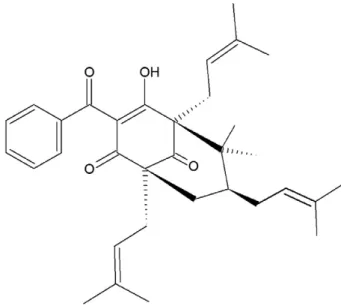
(8×100 cm) eluted with crescent polarity mixtures of n-hexane/ ethyl-acetate and ethyl-acetate/ethanol to purify the prenylated ben-zophenone 7-epiclusianone with a yield of 5% (Fig. 1). The bioactive
compound was identified as the prenylated benzophenone,
7-epi-clusianone, using spectrometric techniques (IR, UV, MS and1H and
13C NMR). The data were compared with those veri
fied in a previous study that investigated the chemical structure of this compound with an authentic sample analyzed by chromatography (Derogis et al., 2008; Yen, Chang, & Duh, 2005).
2.3. Materials and chemicals
Ascorbic acid, 2,2-diphenyl-1-picrylhydrazyl (DPPH), butylated hydroxytoluene (BHT), trichloroacetic acid (TCA), ethylenediamine tet-raacetic acid (EDTA), ferricchloride (FeCl3), ferrozine (3-(2-pyridyl)-5.6-bis(4-phenyl-sulfonic acid)-1,2,4-triazine), potassium ferricyanide (K3Fe(CN)6), dimethylsulfoxide (DMSO), horse heart ferricytochrome ctype VI (Cytc), H2O2(35%), 2-thiobarbituric acid (TBA) and sodium ascorbate were purchased from VETEC (Rio de Janeiro, Brazil). All other chemicals and solvents utilized in this study were of analytical grade. Column chromatography was conducted over silica gel using gra-dient solvents. The studies were performed using compounds isolated from the extracts of theRheediaspecies.
2.4. General instrumentation methods
Melting points were determined on a Mettler melting point apparatus (FP 80 HT). UV spectra were determined on a Shimadzu U-2000 spectrophotometer. Infrared spectra were determined using
KBr discs in a Schimadzu/IR-408 spectrophotometer. 1H and 13C
NMR spectra were determined on a Bruker spectrometer equipped with a 5 mL1H and13C probe operating at 400.1 and 100.6 MHz, re-spectively, with TMS as the internal standard. Mass spectra were de-termined using a gas chromatography–mass spectrometry (GC–MS), using a Schimadzu GCMS-QP5050A spectrometer connected to an ion detector operating in Electron Impact mode at 70 eV. Optical rota-tion was determined using a Perkin-Elmer-241 spectrophotometer.
2.5. Antioxidant activity
2.5.1. DPPH free radical-scavenging property
The antioxidant property of benzophenone and the extract to scavenge DPPH free radicals was measured according to the method described by Yen et al. (2005). The 1,1-diphenyl-2-picrylhydrazyl (DPPH) radical was diluted in ethanol (0.5 mmol L−1). One microliter of DPPH solution was added to a 4.0 mL aliquot of the samples, previ-ously dissolved in ethanol, yieldingfinal concentrations of 800, 400, 200, 100, 50 and 25μg/mL. Each mixture was shaken and maintained
for 30 min at room temperature in the dark. Ascorbic acid and BHT at the same concentrations of the samples, dissolved in ethanol, were used as standard compounds. DPPH solution (1.0 mL) in ethanol (4.0 mL) served as control. Absorbances of the resulting solutions were measured using a UV/VIS spectrophotometer at 517 nm (Shimada, Fujikawa, Yahara, & Nakamura, 1992), and the percent in-hibition was determined by comparison with an ethanol treated con-trol group. The activities of the drugs were calculated as follows: percent scavenging capacity = [(∆A517(control)− ∆A517(samples)) / ∆A517(control)] 100. Ascorbic acid and BHT were used for compari-son. For each concentration tested, three samples were assayed.
2.5.2. Reducing power assay
The reducing power of each compound was determined according to the method published byYildirim, Mavi, and Kara (2001). Different concentrations of samples in ethanol (1 mL), 2.5 mL of 0.2 M phos-phate buffer at pH 6.6 and 2.5 mL of 1% potassium ferricyanide were mixed and then incubated at 50 °C for 30 min. Afterwards, an aliquot (2.5 mL) of 10% trichloroacetic acid was added to the mixture. From each of the above mixtures, an aliquot (2.5 mL) was diluted with 2.5 mL distilled water and mixed with 0.5 mL of 0.1% ferric chloride in a test tube. After 10 min of reaction, the absorbance was measured at 700 nm. An increased absorbance of the reaction mixture indicated a high reducing power. The ascorbic acid and BHT standards were used as positive controls. The reducing capability of samples was expressed as a percentage of action in which the ascorbic acid and BHT absorbance were compared to the same concentrations of the
samples (800, 400, 200, 100, 50 and 25μg/mL). The values are
presented as the mean of triplicate analyses.
2.5.3. Evaluation of the chelating activity of Fe2+
The chelating activity of Fe2+ was measured according to the
method published by Tang, Kerry, Sheehan, and Buckley (2002)
with modifications. Briefly, 1 mL of the samples in solution at dif-ferent concentrations (800, 400, 200, 100, 50 and 25μg/mL) was mixed with 3.7 mL of a hydroalcoholic solution, 0.1 mL of a 2 mM FeSO4 (Fe2+) solution and 0.2 mL of 5 mmol L−1 ferrozine. After 20 min of reaction, the absorbance was measured at 562 nm. The control contained all the reaction reagents except the samples or the positive control. The standard employed was EDTA. A low absorbance in-dicated chelating activity. The chelating activity of the Fe2+(% CA) was calculated using the following equation: %CA=[(∆A562(control)-∆A517 (samples))/∆A562(control)]100. The values are presented as the mean of triplicate analyses. The EC50value was the effective concentration able to chelate 50% of Fe2+.
2.5.4. Assay of toxicity against brine shrimp
Lethality towards brine shrimp was assayed using procedures previously reported by (Meyer, Ferrigni, Jacobson, Nichols, & Mclaughein, 1982), with modifications. Brine shrimp encysted eggs (10 mg) were incubated in 500 mL of seawater under artificial light at 28 °C, pH 7–8. After incubation for 24 h, nauplii were collected with a Pasteur pipette and kept for an additional 24 h under the same conditions to reach the metanauplii stage. The samples to be assayed were dissolved in 1% DMSO (dimethyl sulfoxide) yielding
final concentrations of 100, 50, 25 and 12.5μg/mL in seawater. About 10 nauplii were added to each set of tubes containing the sam-ples. Controls containing 1% DMSO in seawater were included in each experiment. After 48 h, the number of survivors was recorded, and the lethal dose 50% (LD50value) and 95% confidence intervals were calculated by Probit analysis. The repeatability of the method was evaluated using at leastfive replicates of each concentration of each sample.
2.6. In vivo mutagenic test in mice
2.6.1. Animals
Three-week-old male Swiss mice weighing 25 g were obtained from the Central Animal Facility of the Federal University of Alfenas. The animals were housed in wire-topped opaque polycarbonate cages and maintained under constant room conditions on a 12 h light/dark schedule. The room temperature was 20 ± 2 °C, and the hu-midity was maintained at 50%. Commercial food pellets and water were providedad libitum. The animals were allowed to habituate to the housing facilities for at least 1 week before the experiments began. All experiments were conducted in accordance with the Decla-ration of Helsinki on the welfare of experimental animals and with the approval of the Ethics Committee of the Federal University of Alfenas (#239/2009).
2.6.2. Micronucleus test
Animals were segregated into 14 groups. Group 1: EHP 50 mg/kg bw + CPA; Group 2: EHP 50 mg/kg bw + NaCl; Group 3: EHP 100 mg/kg bw + CPA; Group 4: EHP 100 mg/kg bw + NaCl; Group 5: EHP 200 mg/kg bw + CPA; Group 6: EHP 200 mg/kg bw + NaCl; Group 7: 7-epi-clusianone isolated from the hexane extract of the pericarp (MI) 5 mg/kg bw + CPA; Group 8: MI 5 mg/kg bw + NaCl; Group 9: MI 10 mg/kg bw + CPA; Group 10: MI 10 mg/kg bw + NaCl; Group 11: MI 15 mg/kg bw+CPA; Group 12: MI 15 mg/kg bw+ NaCl; Group 13: CPA (negative control) and Group 14: NaCl (negative control). The test substances were dissolved in water and Tween 40 (5%) and administered by gavage daily over 15 days in 150μL doses at concentrations of 50, 100 and 200 mg/kg body weight of hexane extract and at concentrations of 5, 10 and 15 mg/kg body weight of 7-epi-clusianone. Negative and positive controls received only the vehicle. Half of the groups received intraperitoneal injections of 50 mg/kg body weight of cyclophosphamide 24 h before the euthanasia, and the other half received injections of NaCl. Both femur bones were excised, and their bone marrow wasflushed into test tubes using a syringe containing bovine fetal serum. The percentage of reduction in the frequency of CP-induced DNA damage was calculated as follows: % reduction= [(y_A)−(y_B)/(y_A)−(y_C)]100.
Where A = positive control group treated with CP; B = group fed with the EHP and MI + CP; and C = negative control group.
All animals were euthanized 24 h after treatment by cervical dislo-cations under ether anesthesia.
For the conventional assessment of micronucleus frequencies, two slides for each animal were prepared according to the method of
MacGregor et al. (1987). Briefly, femurs were dissected and cleaned
of any adhering muscle, and bone marrow cells wereflushed with
fetal calf serum into a centrifuge tube. The cells were stained with Leishman stain and centrifuged at 2000 rpm for 5 min, and the super-natant was removed. The slides were coded, and the cells were blind-ly scored by light microscopy at 1000 magnification. The frequency of micronucleated polychromatic erythrocytes (MNPCE) in individual mice was used as the experimental unit with variability (standard deviation) based on differences among animals within the same group. The polychromatic erythrocytes/normochromatic erythrocytes (PCE/NCE) ratio was also determined on a total of 1000 erythrocytes counted.
2.7. Statistical evaluation
In order to analyze the mutagenic activity of EHP and MI, it was compared the MNPCE frequencies obtained for the treated groups and the negative control group using ANOVA, followed by a multiple comparison procedure (Tukey test). To analyze EHP and MI antimuta-genicity, it was compared the MNPCE frequencies observed in the treated groups and the positive control group by ANOVA followed by the Tukey test. To evaluate the cytotoxicity of EHP and MI, the polychromatic erythrocytes/normochromatic erythrocytes ratio (PCE/NCE) of all treated groups was compared to the result obtained in the mutagenic effect evaluation for the negative control group, and the results found in the antimutagenic effect evaluation for the posi-tive control, using chi-square test; p values of 0.05 or less were con-sidered statistically significant.
3. Results and discussion
3.1. Antioxidant activity
3.1.1. Radical scavenging activity
The radical scavenging activity of DPPH, like the scavenging activ-ity of other substances, is considered to be a quick and valid method that enables simple evaluation of the antioxidant activity because DPPH is a stable radical that needs not to be generated (Gülçin, Alici, & Cesur, 2005; Sánchez-Moreno, 2002). The method is based on the reduction of DPPH to the corresponding hydrazine by its reac-tion with hydrogen donors. In a methanol or ethanol solureac-tion, the radical is purple in color. Upon reduction to the hydrazine, the solu-tion turns yellow, and this transformasolu-tion is characterized by a de-crease in absorbance that can be monitored spectrophotometrically at 517 nm (Guo et al., 2011). In the current study, different concentra-tions and different reaction times were used; therefore, for prelimi-nary evaluation of the antioxidant activity of the EHP and the 7-epiclusianone they were previously tested at concentrations of 200μg/mL and 400μg/mL for the kinetic behavior of samples; and thus, the best time for reading was determined. They were obtained to determine the time required to reach a plateau of scavenging action for all the compounds. The ascorbic acid reaches the plateau al-most in thefirst minute, as described similarly byAruoma, Halliwell, & Williamson, 1997. As BHT has been described as being of slow kinetics depending on the in-use concentration, its action varied from 40 min at 200μg/mL to 30 min at 400μg/mL. The samples at 200μg/mL showed no significant difference in different times; how-ever, at 400μg/mL, they seemed to have reached a plateau of reaction. Considering the behavior of standards and samples, it was deter-mined that an optimal time of 30 min should be used to determine the scavenging activity of different compounds at different con-centrations. No difference in scavenging activity was observed for ascorbic acid in the concentrations tested. BHT and ascorbic acid at 200μg/mL were 4.88 and 23.8-fold more active than MI, respectively. Once confirmed the existence of the antioxidant capacity of sam-ples, different concentrations of EHP and MI were evaluated in a test dose dependency. At concentrations of 25, 50, 100, 200, 400 and 800μg/L, the scavenging activities ranged between 0 and 33.6 ± 1.06% for 7-epi-clusianone and between 0 and 31.1 ± 1.31% for EHP (Table 1). The scavenging capacity of 7-epi-clusianone was statistically higher than that of EHP at all concentrations (pb0.5). 7-Epi-clusianone (with EC50>800μg/L–EC50= 1267.4 ± 4.35μ g/mL-estimated) and EHP (with EC50>800μg/L–EC50=1714.3±3.52μg/ mL-estimated) exhibited weak radical-scavenging activities, which were 34 and 46 times less active than BHT (EC50=37.2±0.47μg/mL) and one hundred eighty-four and two hundred forty-eight times less
active than ascorbic acid (EC50=6.9±0.25μg/mL), respectively.
Extracts or compounds with scavenging activity are believed to in-hibit lipid peroxidation by stabilizing transition metals. Accordingly, it is suggested that the low-to-moderate chelating effect of
7-epi-clusianone would be at least partially beneficial in protecting against oxidative damage (Sun, Zhang, Lu, Zhang, & Zhang, 2011).
The number and configuration of the hydroxyl groups of H donors in phenolic compounds are important for radical scavenging activity (Soobrattee, Neergheen, Luximon-Ramma, Aruoma, & Bahorun, 2005). It has been reported for theflavonoids that the presence of the 3-OH group of ring C and the catechol group of ring B are essential for the scavenging activity, as described for quercetin (Rice-Evans, 1995) and biflavonoids isolated from pine Araucaria angustifolia (Yamaguchi, Vassão, Kato, & Mascio, 2005). In nemorosone, a poly-isoprenylated benzophenone isolated from propolis, the study by
Piccinelli et al. (2005), showed an EC50for the DPPH radical scaveng-ing activity equal to 22.2 ± 0.04μg/mL. MI showed a scavenging ca-pacity 37.26 times smaller than that of nemorosone. The possible reason for this difference is the presence of two hydroxyl groups in ring B of the nemorosone structure that, along withflavonoid struc-ture, is essential for antioxidant activity.
3.1.2. Reducing power assay
In the reducing power assay, the presence of reducing substances (antioxidants) resulted in the reduction of the Fe+3/ferricyanide complex to the ferrous form by the donation of an electron (Sahreen, Khan, & Khan, 2010). The reducing capability of samples was expressed in terms of absorbance of action in comparison to BHT and ascorbic acid reducing power at concentrations of 25, 50, 100, 200, 400 and 800μg/L. Higher reducing power resulted in greater blue complex formation and consequently in greater absorbance measured spectrophotometrically at 700 nm. At all concentrations, 7-epi-clusianone and EHP exhibited weak relative reducing power in comparison to the standards, acid ascorbic and BHT, and did not show any significant complex formation. Despite the low activity, in-creased concentration of these compounds resulted in an inin-creased reducing power. However, the most active sample was the MI sample, which showed a complex formation approximately 11 times lower than BHT and 19 times lower than ascorbic acid (Table 1).
3.1.3. Evaluation of the chelating activity of Fe2+
Iron is an important metal that can stimulate lipid peroxidation via the Fenton reaction and accelerate the peroxidation of the lipid decomposition pathway of hydroperoxides into peroxyl and alkoxyl radicals that can abstract hydrogen and perpetuate the chain reaction of lipid peroxidation (Gülçin et al., 2005). The chelating activity of Fe2+ions in the samples and standards were determined according toGu et al., 2010. To evaluate the chelating activity, a pink color change was observed with ferrozine, a chromogenic reagent, in accor-dance with the amount of Fe2+available in solution that was mea-sured by a spectrophotometer at 562 nm (Nićiforovićet al., 2010).
In the presence of a chelating agent, smaller numbers of Fe2+ions will be available for complex formation with ferrozine, resulting in decreased absorbance (Table 1). In this test, EDTA was used as the Table 1
Scavenging of free radical DPPH, reducing power and chelating activity ions Fe2+
eval-uation and kinetic reaction against DPPH of the standards and the samples (EHP and 7-epi-clusianone) ofGarcinia brasiliensis.
Samples Antioxidant activitya
Radical scavenging Reducing power assay Chelating activity of Fe2+
EC50(μg/mL) ± SD EC50(μg/mL) ± SD EC50(μg/mL) ± SD
EHP >800 >800 >800
MI >800 >800 >800
AAb 6.9 ± 0.25 42.73 ± 0.89
– BHTb 37.2 ± 0.47 72.85 ± 3.37
–
EDTAb – – 32.56 ± 2.26
aEach value is expressed as mean ± standard deviation (n= 3).
b AA (ascorbic acid), BHT (butylated hydroxytoluene) and EDTA (ethylenediamine
standard chelating agent. The enhancement of chelatable ions in the sample and the number of available ions to react with ferrozine in-duced lower absorbance, which resulted in the chelating property at concentrations of 25, 50, 100, 200, 400 and 800μg/L analyzed. Com-pared to the standard antioxidant EDTA (EC50= 32.56 ± 2.26μg/mL), the benzophenone 7-epi-clusianone (with EC50> 800μg/L–EC50= 1003.45 ± 5.45μg/mL estimated) and EHP (with EC50> 800μg/L–
EC50= 1003.45± 5.45μg/mL estimated) exhibited weak chelating
activity of Fe2+. Such activity varies according to the sample concentration.
3.1.4. Assay of toxicity against brine shrimp
According to the literature (Meyer et al., 1982), when assessing the toxicity of plant extracts and compounds isolated with bioassays in brine shrimp, a LC50value of less than 1000μg/mL is considered to be bioactive. Therefore, we found that for the samples analyzed in this study, all of them demonstrated satisfactory biological activity, as all samples had a lower mortality rate (LC50b100 mg/mL) than the positive control (thymol) (Table 2). We also showed that the in-creased mortality of brine shrimp nautilus was proportional to the concentration increase, resulting in a linearity of the dose–effect rela-tionship for the samples. Among the samples, the EHP showed the lowest toxicity in percentage of survivors at the concentrations ana-lyzed and showed no statistical difference.
3.1.5. In vivo mutagenic test in mice
During the experiment, the animals were weighed, and the con-sumption of rations was controlled. The results are shown in
Table 3, suggesting that there was no significant variation of body weight and ration consumption between experimental groups (pb0.05) during the study period. These results indicate that the con-sumption of EHP and MI, in different concentrations, did not interfere with animal development and growth.
The frequency of micronuclei (MN) after administration of EHP and MI in polychromatic erythrocytes (MNPCEs) of bone marrow in mice is presented inTable 4. The ratio of PCE:NCE from CP and treated groups was not significantly different from negative control group (p > 0.05), indicating that EHP and MI did not present cytotoxic properties in mice bone marrow cells at any doses tested. The PCE: NCE relationship, also shown inTable 4.
Among the methods ofin vivogenetic toxicity investigation, the micronucleus test has been largely used in bone marrow of mice and accepted by regulatory agencies and the scientific community. This test detects genomic alterations and/or damage in mitosis. Although genetic toxicity is not a carcinogenicity measurement, this is often associated with the appearance of cancer as there is a positive correlation between enhanced frequency of micronuclei and the
appearance of tumors in mice and humans (Ramírez et al., 1999;
Vinod, Tiwari, & Meshram, 2011).
Cyclophosphamide, a mutagenic substance of bone marrow, has been used as a positive control substance in many assay systems because cyclophosphamide and its metabolites can bind DNA, causing damage that may result in chromosome breaks, micronucleus for-mation and cell death (Ahmadi, Hosseinimehr, Naghshvar, Hajir, & Ghahremani, 2008). In our experiment, it showed a statistically Table 2
Distribution of the results of the cytotoxic activity of the hexanic extract of the pericarp (EHP) and of the 7-epi-clusianone (MI) on brine shrimp.a
Concentration (μg/mL) % EHP % MI % TIMOL
100.0 13.3 ± 1.37d 20.0 ±1.15d 100.0
50.0 33.3 ± 3.33c 40.0 ±2.31c 100.0
25.0 50.0 ± 2.38b 50.0 ±2.88b 100.0
12.5 60.0 ± 6.10a 66.6 ±6.61a 100.0
aEach value is expressed as mean ± standard deviation (n = 3). Means with
differ-ent letters within a column are significantly different (one-way ANOVA and Tukey test, pb0.05).
Table 3
Mean and standard deviation of body weight and gain of weight in mice during experiment.a
Group/treatment Number of animals Initial weight (g)b Finish weight (g)b Gain weight (g)b,c Total consumption (g)
(G1) EHP 50 + CPA 8 23.40 ± 0.99 25.74 ±2.79 2.34 ± 2.57 463.44
(G2) EHP 50 + NaCl 8 22.33 ± 2.39 27.35 ±2.64 5.02 ± 0.88 522.16
(G3) EHP 100 + CPA 8 23.97 ± 2.08 27.04 ±2.22 2.93 ± 1.08 508.75
(G4) EHP 100 + NaCl 8 23.76 ± 2.20 28.07 ±2.14 4.30 ± 1.95 587.18
(G5) EHP 200 + CPA 8 20.46 ± 4.10 26.83 ±3.55 5.64 ± 3.17 543.09
(G6) EHP 200 + NaCl 8 22.79 ± 2.63 24.65 ±2.61 1.86 ± 1.71 503.10
(G7) MI 5 + CPA 8 22.60 ± 1.31 27.05 ±2.16 4.45 ± 1.55 624.53
(G8) MI 5 + NaCl 8 21.59 ± 3.56 27.63 ±3.83 5.56 ± 1.56 638.19
(G9) MI 10 + CPA 8 23.56 ± 2.53 27.96 ±2.41 4.40 ± 3.83 628.21
(G10) MI 10 + NaCl 8 23.25 ± 2.38 27.94 ±3.46 4.69 ± 1.67 663.61
(G11) MI 15 + CPA 8 22.35 ± 2.58 27.60 ±2.82 5.25 ± 1.65 649.07
(G12) MI 15 + NaCl 8 23.39 ± 2.48 26.81 ±2.75 3.42 ± 2.42 642.65
(G13) CPA 8 24.93 ± 1.98 27.26 ±2.40 2.33 ± 2.30 639.34
(G14) NaCl 8 22.90 ± 2.17 27.03 ±2.90 4.13 ± 1.84 662.33
There was no significant variation of body weight and ration consumption between experimental groups by Tukey test (pb0.05).
aAbbreviations used: EHP, hexanic extract of pericarp ofRheedia brasiliensis; MI, molecule 7-epiclusianone isolated of hexanic extract of pericarp ofRheedia brasiliensis; CPA,
Cyclophosphamide.
b Means ±standard deviation of three determinations. c Relative the two weeks experimentals.
Table 4
Frequency of erythrocytes polychromatics micronucleuds (MNPCEs) of bone marrow cells of Swiss mice in experimental groups treated with EHP and MI.
Groups/treatments Number of PCEs analyzed
MNPCEs Relation
PCE/NCE No. Mean +
SD (%)⁎ % Reduction
(G1) EHP 50 + CPA 8000 164 2.05 ± 0.18b 09 0.76 ± 0.17
(G2) EHP 50 + NaCl 8000 067 0.84 ± 0.20d 22 1.07 ± 0.22
(G3) EHP 100 + CPA 7000 131 1.87 ± 0.10b 27 0.86 ± 0.21
(G4) EHP 100 + NaCl 8000 064 0.80 ± 0.15d 26 1.54 ± 0.17
(G5) EHP 200 + CPA 7000 189 2.70 ± 0.12a
– 0.84 ± 0.21 (G6) EHP 200 + NaCl 7000 073 1.04 ± 0.12d 15 1.17 ± 0.31
(G7) MI 5 + CPA 8000 153 1.91 ± 0.16b 15 0.95 ± 0.31
(G8) MI 5 + NaCl 8000 078 0.98 ± 0.10d 09 1.32 ± 0.20
(G9) MI 10 + CPA 8000 155 1.94 ± 0.25b 14 1.16 ± 0.21
(G10) MI 10 + NaCl 8000 085 1.06 ± 0.18d 01 1.11 ± 0.46
(G11) MI 15 + CPA 8000 128 1.60 ± 0.10c 29 0.94 ± 0.39
(G12) MI 15 + NaCl 7000 055 0.79 ± 0.22d 36 1.24 ± 0.10
(G13) CPA 8000 180 2.25 ± 0.20b
– 1.12 ± 0.18 (G14) NaCl 8000 086 1.08 ± 0.17d – 1.23 ± 0.28
significant induction of MNPCE. The CPA positive control (vehicle +
CPA) was shown to be efficient in the induction of chromosomal
damage in immature erythrocytes (PCEs) because the frequency of MNPCEs present was statistically superior to the negative control.
In this study, it was found that the MNPCE frequency in groups treated with the three different hexane extract concentrations and the three different isolated molecule concentrations + NaCl was sig-nificant and equal to that of the negative control group. In mice trea-ted with EHP (50, 100 and 200 mg/kg b.w.) + NaCl and MI (5, 10 and 15 mg/kg b.w.)+ NaCl, the number of MNPCEs decreased compared to the negative control group, which indicates that the substances evaluated are not causing mutagenic effects because DNA damage was not induced (micronuclei). The MNPCE frequency was less than or equal to the negative control.
Ramos et al. (2003)evaluated antimutagenicity infive extracts, in-cludingRheedia aristata. This plant displayed a positive response in at least two of the former biochemical testing systems withEscherichia colireversion upon oxidative damage induced bytert-butyl hydroper-oxide as a model to test suppression of the mutagenic response. A 25–50% decrease in the revertant count was observed whenE. coli was treated with the extracts ofR. aristatain the concentration rang-ing from 2.5 to 10 mg/plate, even in the absence of TBH, which indi-cates some toxicity by the species assayed.
The antimutagenic activity was tested by observing the number of MNPCEs in mice treated with EHP (50 and 100 mg/kg bw) + CPA and MI (5, 10 and 15 mg/kg b.w.)+ CPA when compared to the positive control group. The number of MNPCEs decreased, indicating that the substances analyzed had antimutagenic effects, excluding the group of major concentrations of EHP (200 mg/kg b.w.), which showed an increase in the number of MNPCEs. This increase in mutation rate can indicate a synergism with the CPA, as the mutagenicity test does not show mutagenic effects. The group with the higher dosage of MI administered was statistically different from the positive con-trol group and along with the decrease in the number of MNPCEs, demonstrating a major protection against mutagenic effects.
Many researchers report that high consumption of fruits is associ-ated with the Incidence of degenerative diseases, including cancer (Gerber et al., 2002; Rufino et al., 2010). However, few studies have demonstrated such effects with exotic fruits. For the fruits of the Clusiaceae family, noin vivomutagenicity studies were found.
Based on the results obtained, it can be noted that both the EHP and MI showed low antioxidant activity, but showed antimutagenic activity.Negi, Jayaprakasha, and Jena (2003)evaluated the antioxi-dant and antimutagenic activities of pomegranate peel extracts. In this study, researchers found that the water extract showed low anti-oxidant activity, but very strong antimutagenic activity. In this con-text, it is not always noted that antioxidant values are determinants of the antimutagenic potential of products. A study bySanta-Cecília et al. (2012)with MI has shown that, even as low antioxidant activity, the MI was able to down regulate inflammatory phagocyte superox-ide anion release through a mechanism controlled by protein tyrosine phosphorylation and by the direct stimulation of the protein kinase C.
4. Conclusions
The present study, reveals that the antioxidant properties of the hexanic extract (EHP) and 7-epi-clusianone (MI) that were investi-gated using three different antioxidant assays; (i) DPPH radical scav-enging, (ii) reducing power and (iii) metal chelation, exhibited lower activities in the concentrations analyzed. Moreover, antioxidant capacities in all assays were concentration dependent. In addition, in the assay of toxicity against brine shrimp the samples presented satisfactory activity, since they all showed a lower mortality rate (LC50b100μg/mL) compared to the positive control (thymol). Con-sidering that the increased mortality of brine shrimp nautilus was proportional to the concentration increase, resulting in a linearity of
dose–effect relationship for the samples. Among the samples, the hexanic extract showed the lowest toxicity in percentage of survivors at the concentrations analyzed compared to the benzophenone. The EHP and MI did not show mutagenic effect in mice. EHP (50 and 100 mg/kg bw) and MI (higher dosage) demonstrated protection against mutagenic effects. Possibly, the antimutagenic and antioxi-dant activities shown by 7-epi-clusianone might contribute to an anticarcinogenic effect and cytotoxicity to antitumor activity, and the study of new drugs of natural origin as prophylactic agents in the cancer prevention.
Acknowledgments
The authors would like to thank CNPq, CAPES, FINEP and FAPEMIG for theirfinancial support.
References
Ahmadi, A., Hosseinimehr, S. J., Naghshvar, F., Hajir, E., & Ghahremani, M. (2008). Chemo protective effects of hesperidin against genotoxicity induced by cyclophos-phamide in mice bone marrow cells.Archives of Pharmacal Research,31, 794–797. Aruoma, O. I., Halliwell, B., & Williamson, M. (1997).In vitromethods for characterizing potential prooxidant and antioxidant actions of nonnutritive substance in plant foods. In O. I. Aruoma & S. L. Cuppett (Eds.), Urbana: AOCS Press.
Bazzano, L. A., He, J., Ogden, L. G., Loria, C. M., Vupputuri, S., Myers, L., et al. (2002). Fruit and vegetable intake and the risk of cardiovascular disease in US adults: Thefirst National Health and Nutrition Examination Survey Epidemiologic Follow-up Study.American Journal of Clinical Nutrition,76, 93–99.
Brandão, M. G. L., Zanetti, N. N. S., Oliveira, P., Grael, C. F. F., Santos, A. C. P., & Monte-Mór, R. L. M. (2008). Brazilian medicinal plants described by 19th century European naturalists and in the Official Pharmacopeia.Journal of Ethnopharmacol-ogy,120, 141–148.
Brighenti, F., Valtuenã, S., Pellegrini, N., Ardigo, D., Del Rio, D., Salvatore, S., et al. (2005). Total antioxidant capacity of the diet is inversely and independently related to plasma concentrations of high sensitive C-reactive protein in adult Italian subjects.
British Journal of Nutrition,93, 619–625.
Clerici, M. T. P. S., & Carvalho-Silva, L. B. (2011). Nutritional bioactive compounds and technological aspects of minor fruits grown in Brazil.Food Research International,
44, 1658–1670.
Corrêa, M. P. (1978).Dicionário das plantas úteis do Brasil e das plantas exóticas cultiva-das.Rio de Janeiro: Imprensa Nacional.
Dembitsky, V. M., Poovarodom, S., Leontowicz, H., Leontowicz, M., Vearasilp, S., Trakhtenberg, S., et al. (2011). The multiple nutrition properties of some exotic fruits: Biological activity and active metabolites.Food Research International,44, 1671–1701.
Derogis, P. B., Martins, F. T., De Souza, T. C., Moreira, M. E., Souza Filho, J. D., Doriguetto, A. C., et al. (2008). Complete assignment of the 1H and 13C NMR spectra of garci-niaphenone and keto-enol equilibrium statements for prenylated benzophenones.
Magnetic Resonance in Chemistry,46, 278–282.
Frutuoso, V. S., Monteiro, M. M., Amendoeira, F. C., Almeida, A. L. F., Nascimento, D. D., Bérenger, A. L. R., et al. (2007). Analgesic and anti-inflammatory activity of the aqueous extract of Rheedia longifoliaPlanch & Triana. Memórias do Instituto Oswaldo Cruz,102, 91–96.
Gerber, M., Boutron-Ruault, M. C., Hercberg, S., Riboli, E., Scalbert, A., & Siess, M. H. (2002). Food and cancer: State of the art about the protective effect of fruits and vegetables.Bulletin du Cancer,89, 293–312.
Gu, F. L., Kim, J. M., Abbas, S., Zhang, X. M., Xia, S. Q., & Chen, Z. X. (2010). Structure and antioxidant activity of high molecular weight Maillard reaction products from casein–glucose.Food Chemistry,120, 505–551.
Gülçin,İ., Alici, H. A., & Cesur, M. (2005). Determination ofin vitroantioxidant and radical scavenging activities of propofol.Chemical & Pharmaceutical Bulletin,53(n.3281), 281–285.
Guo, T., Wei, L., Sun, J., Hou, C., & Fan, L. (2011). Antioxidant activities of extract and fractions fromTuber indicumCooke & Masse.Food Chemistry,127, 1634–1640. Krishna, G., & Makoto, H. (2000). In vivo rodent micronucleus assay: Protocol, conduct
and data interpretation.Mutation Research,455, 155–166.
MacGregor, J. T., Heddle, J. A., Hite, M., Margolin, B. H., Ramel, C., Salamone, M. F., et al. (1987). Guidelines for the conduct of micronucleus assay in mammalian bone marrow erythrocytes.Mutation Research,189, 103–112.
Meyer, N. B., Ferrigni, N. R., Jacobson, J. E., Nichols, D. E., & Mclaughein, J. L. (1982). Brine shrimp: A convenient general bioassay for active plant constituents.Planta Medica,45, 31–34.
Morganti, P. (2009). The photoprotective activity of nutraceuticals.Clinics in Dermatol-ogy,27, 166–174.
Negi, P. S., Jayaprakasha, G. K., & Jena, B. S. (2003). Antioxidant and antimutagenic activities of pomegranate peel extracts.Food Chemistry,80, 393–397.
Nićiforović, N., Mihailović, V., Masković, P., Solujić, S., Stojković, A., Pavlović, D., et al. (2010). Antioxidant activity of selected plant species; potential new sources of natural antioxidants.Food and Chemical Toxicology,48, 3125–3313.
Oluyemi, K. A., Okwuonu, U. C., Baxter, D. G., & Oyesola, T. O. (2007). Toxic effects of methanolic extract ofAspilia africanaleaf on the estrous cycle and uterine tissues of Wistar rats.International Journal of Morphology,25, 609–614.
Pérez-Jiménez, J., Arranz, S., Tabernero, M., Díaz-Rubio, M. E., Serrano, J., Goñi, I., et al. (2008). Updated methodology to determine antioxidant capacity in plant foods, oils and beverages: Extraction, measurement and expression of results.Food Research International,41, 274–285.
Piccinelli, A. L., Cuesta-Rubio, O., Chica, M. B., Mahmood, N., Pagano, B., Pavone, M., et al. (2005). Structural revision of clusianone and 7-epi-clusianone and anti-HIV activity of polyisoprenylated benzophenones.Tetrahedron,61, 8206–8211. Pitsavos, C., Panagiotakos, D. B., Tzima, N., Chrysohoou, C., Economou, M., Zampelas, A.,
et al. (2005). Adherence to the Mediterranean diet is associated with total anti-oxidant capacity in healthy adults: The ATTICA study.American Journal of Clinical Nutrition,82, 694–699.
Ramírez, M. J., Surrallés, J., Puerto, S., Creus, A., & Marcos, R. (1999).Mutation Research,
440, 163–169.
Ramos, A., Visozo, A., Piloto, J., Garcıa, A., Rodrıguez, C. A., & Rivero, R. (2003). Screening of antimutagenicity via antioxidant activity in Cuban medicinal plants.Journal of Ethnopharmacology,87, 241–246.
Rice-Evans, C. (1995).Plant polyphenols: Free radical scavenger or chain-breaking anti-oxidants? Free radicals and oxidative stress: Environment, drugs and food additives
(pp. 103–116). London, England: Portland Press.
Rufino, M. S. M., Alves, R. E., Brito, E. S., Brito, J. P. P., Saura-Calixto, F., & Mancini-Filho, J. (2010). Bioactive compounds and antioxidant capacities of 18 non-traditional tropical fruits from Brazil.Food Chemistry,121, 996–1002.
Sahreen, S., Khan, M. R., & Khan, R. A. (2010). Evaluation of antioxidant activities of various solvent extracts ofCarissa opacafruits.Food Chemistry,122, 1205–1211. Sánchez-Moreno, C. (2002). Review: Methods used to evaluate the free radical
scavenging activity in foods and biological systems.Food Science and Technology International,8, 121–137.
Santa-Cecília, F. V., Santos, G. B., Fuzissaki, C. N., Derogis, P. B. M. C., Freitas, L. A. S., Gontijo, V. S., et al. (2012). 7-Epiclusianone, the natural prenylated benzophenone, inhibits superoxide anions in the neutrophil respiratory burst.Journal of Medicinal Food,15, 200–205.
Santos, M. H., Nagem, T. J., Oliveira, T. T., & Braz-Filho, R. (1999). 7-Epiclusianone, the new tetraprenylated benzophenone and others chemical constituents from the fruits ofRheedia gardneriana.Química Nova,22, 654–660.
Saviranta, N. M. M., Veeroos, L., Granlund, L. J., Hassinen, V. H., Kaarniranta, K., & Karjalainen, R. O. (2011). Plantflavonol quercetin and isoflavone biochanin A differentially induce protection against oxidative stress and inflammation in ARPE-19 cells.Food Research International,44, 109–113.
Shimada, K., Fujikawa, K., Yahara, K., & Nakamura, T. (1992). Antioxidative properties of xanthan on the autoxidation of soybean oil in cyclodextrin emulsion.Journal of Agricultural and Food Chemistry,40, 945–948.
Soobrattee, M. A., Neergheen, V. A., Luximon-Ramma, A., Aruoma, O. I., & Bahorun, T. (2005). Phenolics as potential antioxidant therapeutic agents: Mechanism and actions.Mutation Research,579, 200–213.
Sun, L., Zhang, J., Lu, X., Zhang, L., & Zhang, Y. (2011). Evaluation to the antioxidant activity of totalflavonoids extract from persimmon (Diospyros kaki L.) leaves.
Food and Chemical Toxicology,49, 2689–2696.
Suzuki, N., Fujimura, A., Nagai, T., Mizumoto, I., Itami, T., Hatate, H., et al. (2004). Anti-oxidative activity of animal and vegetable dietaryfibers.Bio Factors,21, 329–333. Tang, S. Z., Kerry, J. P., Sheehan, D., & Buckley, D. J. (2002). Antioxidative mechanisms of
tea catechins in chicken meat systems.Food Chemistry,76, 45–51.
Trichopoulou, A., Costacou, T., Bamia, C., & Trichopoulos, D. (2003). Adherence to a Mediterranean diet and survival in a Greek population.The New England Journal of Medicine,348, 2599–2608.
Vinod, V., Tiwari, P. K., & Meshram, G. P. (2011). Evaluation of mutagenic and anti-mutagenic activities of neem (Azadirachta indica) seed oil in the in vitro Ames Salmonella/microsome assay and in vivo mouse bone marrow micronucleus test.
Journal of Ethnopharmacology,134, 931–937.
Yamaguchi, L. F., Vassão, D. G., Kato, M. J., & Mascio, P. (2005). Biflavonoids from brazil-ian pineAraucária angustifóliaas potentials protective agents against DNA damage and lipoperoxidation.Phytochemistry,66, 2238–2247.
Yen, W. J., Chang, L. W., & Duh, P. D. (2005). Antioxidant activity of peanut seed testa and its antioxidative component, ethyl protocatechuate.Food Science and Technol-ogy,38, 193–200.
Yildirim, A., Mavi, A., & Kara, A. A. (2001). Determination of antioxidant and antimicro-bial activities ofRumex crispusL. extracts.Journal of Agricultural and Food Chemistry,