REVISTA CIENTÍFICA DE MEDICINA VETERINÁRIA - ISSN 1679-7353 Ano XV - Número 31 –
JULHO de 2018 – Periódico Semestral
1 Departamento de Cirurgia. Faculdade de Medicina Veterinária e Zootecnia. Universidade de São Paulo (USP). São Paulo, SP, Brasil. *Author contact. Erick Eduardo Silveira. Av. Prof. Dr. Orlando Marques de Paiva, 87. Cidade Universitária, CEP 05508-270, São Paulo, SP. Brasil. Phone number: +55-11-30911316
2 Universidade de Sorocaba (UNISO). Sorocaba, SP, Brasil.
3 Instituto de Ciências da Saúde da Universidade Paulista. (UNIP). São Paulo, SP, Brasil.
4 Departamento de Anatomia, Patologia e Clínica Veterinária. Escola de Medicina Veterinária e Zootecnia. Universidade Federal da Bahia. Salvador, BA, Brasil.
DESCRIPTION OF TERMINAL RAMIFICATIONS OF THE ABDOMINAL AORTA IN DOGS (Canis familiaris)
Erick Eduardo SILVEIRA¹*
Amilton Cesar dos SANTOS1
Helen B. ABUD²
Rodrigo Filippi PRAZERES3
Caio BIASI4
ABSTRACT
Current study describes possible alterations of the terminal branches of dogs´ abdominal aorta to determine their origin and compare them with other species. Thirty specimens of dogs (race not defined), 20 females and 10 males, varying in age and size, were used. The corpses were retrieved from previous practical classes at the veterinary anatomy laboratory of the Universidade de Sorocaba (UNISO), Sorocaba, Brazil. The dogs´ femoral artery was dissected and catheterized by 10% formaldehyde injection and submerged for at least 48 hours in formalin-fixed vats for fixation. All animals were submitted to abdominal cavity access through the abdominal and lateral abdominal wall and later removed from the abdominal viscera to visualize the abdominal aorta and its terminal branches. Most of the animals under analysis presented, right and left external iliac artery, right and left internal iliac artery and a median sacral artery as terminal branches of the abdominal aorta, whereas 9 dissected specimens showed a common trunk with internal iliac artery and medial sacral artery. Results show that there are variations among mammals with regard to the origins of terminal branches of the abdominal aorta.
Keywords: External iliac artery. Internal iliac artery. Median sacral artery.
RESUMO
Esse estudo teve o objetivo de descrever as possíveis alterações dos ramos terminais da aorta abdominal dos cães determinando a sua origem e comparando com as demais espécies. Foram utilizados 30 exemplares de cães SRD com idade e portes variados, sendo destes 20 fêmeas e 10 machos, todos os cadáveres provenientes de aulas práticas anteriormente ministradas no laboratório de anatomia veterinária da universidade de Sorocaba- UNISO. Os cadáveres tiveram sua artéria femoral dissecada em seguida cateterizada para injeção de formaldeído a 10% e posteriormente foram mantidos submersos por no mínimo 48 horas em cubas com formol para fixação. Todos os animais foram submetidos ao acesso da cavidade abdominal através da retirada da parede abdominal ventral e lateral e posteriormente retirada das vísceras abdominais para visualização da aorta abdominal e seus ramos terminais. A maioria dos animais estudados apresentou como ramos terminais da aorta abdominal, artéria ilíaca externa direita e esquerda, artéria ilíaca interna direita e esquerda e uma artéria sacral mediana, enquanto 9 espécimes dissecados apresentaram um tronco comum onde surgiu a artéria ilíaca interna e artéria sacral mediana, concluindo-se que existem variações das origens dos ramos terminais da aorta abdominal entre os mamíferos. Palavras chave: Artéria ilíaca externa. Artéria ilíaca interna. Artéria Sacral mediana.
INTRODUCTION
Knowledge on the anatomy of the abdominal aorta and its arterial ramifications is highly relevant for radiological and surgical practice in animals used as experimental models (XAVIER-SILVA et al., 2011). It is also important for veterinarians so that they could interpret certain diseases such as thromboembolism and aneurism of the abdominal aorta.
Anatomic variations of the vascular system are identified modifications when compared to an established pattern (AVEDILLO et al., 2016). Although variations may be described as an anomaly, the relationships between vascular variations and congenital malformations of the vessels are absent. Consequently, variations do not have any effect on the functioning of the organ in normal circumstances (WINSLOW, 1883; LIPPERT; PABST, 1985).
A dissection of anatomical models was conducted, highlighting the final branches of the abdominal aorta to better understand the characteristics of the terminal ramification of dogs´ abdominal aorta and apply data to technical and surgical practices and define the most common anatomic patterns.
MATERIAL AND METHODS
Current analysis was conducted at the laboratory of veterinarian anatomy of the Universidade de Sorocaba (UNISO), Sorocaba, Brazil. Study was authorized by Institutional Ethical Committee.
Thirty dogs, 20 females and 10 males, with no defined race, varying in age and size, were used in current study. Specimens, which had been donated to the institution, were retrieved from laboratory practice in veterinarian anatomy classes. Femoral artery was dissected and catheterized with 40X12 needles, perfused in a solution of formaldehyde 10% and maintained up to 48 hours submerged in vats with an aqueous formaldehyde solution for fixing.
The animals were dissected with surgery instruments and the abdominal cavity was reached by removing the ventral and lateral abdominal wall. Abdominal viscera were removed to visualize the cavity, abdominal aorta and its terminal ramifications.
Description of the ramifications was based on terminology according to the
Nomina Anatomica Veterinaria (INTERNATIONAL COMMITTEE ON
VETERINARY GROSS ANATOMICAL NOMENCLATURE, 2012).
RESULTS
Dissection of the anatomic specimens revealed the terminal segment of the abdominal aorta close to the lumbar quadratus and the psoas minor muscle, contacting the ventral surface of the lumbar and sacral vertebrae and in syntopy with the caudal vena cava.
In most animals (21 times or 70%, 16 females and 5 males), the abdominal aorta ended in the ramification of the external right and left iliac artery, right and left internal iliac artery and median sacral artery.
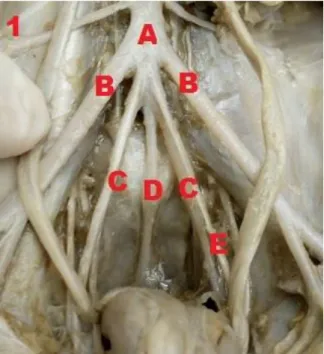
In the cases mentioned above, the external iliac artery emerged from the lateral face of the abdominal aorta at L5 and L6 and from the cranial side to the origin of the internal iliac artery and had significantly greater caliper than the internal iliac artery. The internal iliac arteries showed a medial pathway with regard to the external iliac arteries. The median sacral artery in the 21 specimens rose from the dorsal face of the abdominal aorta and moved towards the caudal segment (Figure 1).
Less frequently (9 times or 30%, in 4 females and 5 males), the end of the abdominal aorta in the dissected specimens occurred through ramification in the right and left external iliac artery and a single trunk from where the right and left internal iliac artery and median sacral artery emerged. Whereas the animals´ internal iliac arteries emerged from the lateral face of the single trunk, median sacral artery rose from the dorsal face of the single trunk (Figure 2).
The right and left umbilical artery, emerging at the initial portion of the ventral face of the internal iliac artery of all dissected specimens was also detected.
Figure 1- Ventral cross-section with abdominal aorta (A) giving rise to right and left
external iliac artery (B), right and left internal iliac artery (C), median sacral artery (D) and left umbilical artery (E).
Figure 2- Ventral cross-section with abdominal aorta (A) giving rise to the right and
left external iliac artery (B), right and left internal iliac artery (C), median sacral artery (D) and right and left umbilical artery (E), common trunk (*). (Bar = 30 mm).
DISCUSSION
A common iliac artery as the end branch of the abdominal aorta has been reported in skunks and nutrias, respectively by Silva and Martins (2004) and by Culau et al. (2008). It has also been described in rabbits by Daólio et al. (2011) and Bavaresco et al. (2012), but current research did not corroborate above data.
A research by Macedo and coworkers (2013) showed a left common trunk prior to its ramifications in external and internal iliac arteries in a female anteater. It was not reported in dogs in current research.
The external iliac artery was detected by Silva e Martins (2004) in skunks; by Culau et al. (2008) in nutrias; by Daólio et al. (2011) and Bavaresco et al. (2012) in rabbits, as a branch of the common iliac artery, and by Silva et al. (2011) in the squirrel monkey; by Geraldo et al. (2013) in cats; by Silva et al. (2014) in guinea pigs; Pinheiro et al. (2014) in the ocelot; Biihrer et al. (2015) in the raccoon. The external iliac artery was described as a direct branch of the abdominal aorta emerging from the lateral face.
The internal iliac artery was also described in other mammals as smaller when compared to the external iliac one. The internal iliac artery was described as a branch of the external iliac artery by Silva and Martins (2004) in the skunk; by Silva et al. (2011) in squirrel monkeys; by Silva et al. (2014) in guinea pigs; by Faria et al. (2016) in the night monkey (Aotus azarae infulatus). The internal iliac artery was described as a branch of the common iliac artery by Daólio et al. (2011) and Bavaresco et al. (2012) and by Culau et al. (2008) in nutrias. Biihrer et al. (2015) described the internal iliac arteries emerging directly from the abdominal aorta in the raccoon. The internal iliac artery emerged from the lateral face of a common trunk in the ocelot and in nine specimens studied in current research. It was not described by Geraldo et al. (2013) in cats or by Pinheiro et al. (2014) in the ocelot.
According to Silva and Martins (2004), median sacral artery in the skunk emerged from the common iliac artery in 30% of the anatomic models dissected; Macedo et al. (2013) described the median sacral artery as a branch of the left internal iliac artery in the anteater, except in one model in which it arose from the common trunk. Median sacral artery was described as a branch emerging from the dorsal face of
the abdominal aorta by Daólio et al. (2011) and Bavaresco et al. (2012) in rabbits; by Silva et al. (2011) in squirrel monkeys; by Silva et al. (2014) in guinea pigs; by Biihrer et al. (2015) in the raccoon; by Faria et al. (2016) in the night monkey (Aotus azarae
infulatus), corroborating most results in current analysis.
CONCLUSION
Results showed that terminal branches of the abdominal aorta in several dissected anatomic models were external iliac arteries, internal iliac arteries and median sacral artery, excepting certain specimens featuring a common trunk. The sites from where the internal iliac arteries and the median sacral artery emerged were not described, although extant in other mammals. Further studies in male animals are required since their anatomic pattern could not be established.
REFERENCES
AVEDILLO, L.; MARTÍN-ALGUACIL, N.; SALAZAR, I. Anatomical variations of the blood vascular system in veterinary medicine. The internal iliac artery of the dog. Part one. Anatomia, Histologia, Embryologia. v. 45, n. 2, p. 299-307, 2016.
BAVARESCO, A. Z.; CULAU, P. O. V.; CAMPOS, R. Ramos colaterais parietais e terminais da aorta abdominal em coelhos da raça Nova Zelândia (Oryctolagus
cuniculus). Acta Scientiae Veterinariae. v. 40, n. 4, p. 01-06, 2012.
BIIHER, D. A.; GUIMARÃES, G. C.; LOPES, G. C.; LIMA, I. G. Descrição anatômica dos ramos artérias da aorta torácica e abdominal do quarti (Nasua
nasua)(Carnivora,Procyonidae). Biotemas. v. 28, n. 2, p. 119-124, 2015.
CULAU, P. O. V.; AZAMBUJA, R. C. A.; CAMPOS, R. Ramos colaterais parietais e terminais da aorta abdominal em Myocastor coypus (nutria). Ciência Rural. v. 38, n. 4, p. 997-1002, 2008.
DAÓLIO, M.; MARCHI, P. N.; PINTO E SILVA, J. R. C.; GUAZZELLI-FILHO, J.; SCHIMMING, B. C.; MATHEUS, S.M.M., FILADELPHO, A. L. Estudo dos ramos sacrais da aorta abdominal do coelho (Oryctolagus cuniculus). Revista Científica
Eletrónica de Medicina Veterinária. v. 9, n. 17, p. 1-10, 2011.
FARIA, B. M.; BRANCO, E.; LIMA, A R. Ramos da aorta abdominal de
Aotus azarae infulatus. Biotemas. v. 29, n. 2, p. 69-76, 2016.
GERALDO, B.; PINTO E SILVA, J. R. C.; SCHIMMING, B. C.; GUAZZELLI-FILHO, J.; FILADELPHO, A. L. Contribuição ao estudo anatômico dos ramos sacrais da aorta abdominal do gato (Felis catus). Revista Científica Eletrónica de Medicina
Veterinária. v. 11, n. 20, p. 1-8, 2013.
INTERNATIONAL COMMITTEE ON VETERINARY GROSS ANATOMICAL NOMENCLATURE. Nomina Anatomica Veterinaria. 5ed. Editorial Committee, Hannover. 2012.
LIPPERT, H.; PABST, R. Arterial Variations in Man: Classification and
Frequency. München: Bergman, 1985.
MACEDO, B. C.; LIMA, A. R.; PEREIRA, L. C.; BRANCO, E. Descrição morfológica dos ramos colaterais da aorta abdominal do tamanduá-mirim (Tamandua tetradactyla).
Biotemas. v. 26, n. 1, p. 173-180, 2013.
PINHEIRO, L. L.; ARAÚJO, E. B.; LIMA, A. R.; MARTINS, D. M.; MELUL, R.; SOUZA, A. C. B.; PEREIRA, L. C.; BRANCO, E. Os ramos colaterais da aorta abdominal em jaguatirica (Leopardus pardalis). Pesquisa Veterinária Brasileira. v. 34, n. 5, p. 491-495, 2014.
SILVA, J. R. C. P.; MARTINS, M. R. F. B. Anatomical study of the abdominal aorta sacral rami of the opossum (Didelphis albiventris). International
SILVA, M. R. M.; LIMA, A. R.; JÚNIOR, A. C. C. L.; ISHIZAKI, M. N.; IMBELONI, A. A.; MUNIZ, J. A. P. C.; BRANCO, E. R. Descrição morfológica dos ramos
colaterais viscerais da aorta abdominal do macaco-de- cheiro. Ciência Rural. v. 41, n. 1, p. 94-100, 2011.
SILVA, J. R. C. P.; GUAZZELLI-FILHO, J.; SHIMMING, B. C.; FILADELPHO, A. L.; JESUS, L. S. B. Estudo anatômico dos ramos sacrais da aorta abdominal da cobaia (Cavia porcellus). Revista Científica Eletrónica de Medicina Veterinária. v. 12, n. 23, p. 1-11, 2014.
WINSLOW, R. A study of the malformations, variations, and anomalies of the circulatory apparatus in man. Annuals of Anatomical Surgery. v. 7, p. 21-94, 1883.
XAVIER-SILVA, B.; ROZA, M. S.; HERNANDEZ, J. M. F.; SOUZA, H. J. M.; ABIDU-FIGUEIREDO, M. Artéria lienal em gatos: estudo aplicado à pesquisa
anatômica e a prática cirúrgica. Revista Brasileira de Medicina Veterinária. v. 33, n. 1, p. 41-47, 2011.