Carla Filipa Pereira Bezerra
May 2013
Monocarboxylate transporters as
angiogenic mediators: role on
endothelial-tumor cell crosstalk
UMinho|20 13 Carla F ilipa P er eir a Bezerr a Monocarbo xy late transpor ter s as angiogenic mediator s: role on endo
thelial-tumor cell cross
This work was performed under supervision of
Professora Doutora Maria de Fátima Monginho Baltazar
and
Doutora Céline Marques Pinheiro
Carla Filipa Pereira Bezerra
May 2013
Master Thesis
Master in Health Sciences
Monocarboxylate transporters as
angiogenic mediators: role on
endothelial-tumor cell crosstalk
iii
v
A
CKNOWLEDGMENTS/A
GRADECIMENTOSO espaço confinado à seção de Agradecimentos é, seguramente, demasiado curto para agradecer convenientemente a todas as pessoas que, ao longo do meu percurso no Mestrado em Ciências da Saúde, de alguma forma foram importantes e em muito contribuíram para o meu crescimento, quer a nível académico, quer a nível pessoal. Desta forma deixo aqui apenas algumas breves palavras, mas com um sentido de profundo agradecimento.
À Comissão de Seleção e Seriação do Mestrado em Ciências da Saúde pela oportunidade que me deram de integrar e realizar o referido mestrado. Para mim foi realmente um privilégio poder fazer parte deste mestrado, que efetivamente contribuiu muito para o meu crescimento a nível académico e científico.
Depois gostaria de mostrar a minha profunda gratidão à Professora Doutora Fátima Baltazar, orientadora deste trabalho, pela oportunidade que me deu em integrar o grupo de investigação por si liderado satisfazendo, deste modo, um dos meus grandes sonhos, trabalhar na área de investigação em cancro. Devo, desde já, agradecer-lhe também imenso toda a sua disponibilidade, simpatia e amabilidade demonstradas durante este último ano.
Uma palavra muito especial também para a minha co-orientadora, Doutora Céline Pinheiro, por tudo aquilo que me ensinou no laboratório, por ter sido incansável no acompanhamento deste trabalho, a nível prático e também de escrita, mesmo com o tempo restrito de que dispunha. O meu sincero obrigado!
Aos meus colegas de laboratório, o meu profundo agradecimento, por me terem integrado desde o primeiro minuto, por tudo o que me ensinaram e pelos momentos divertidos que me proporcionaram. Vera Gonçalves muito obrigada por tudo o que me ensinaste, muito daquilo que hoje sou, profissionalmente falando, a ti o devo. Filipa Santos, a minha “chefinha”, obrigada por contribuíres para o crescimento das minhas células, sem ti não teria sido a mesma coisa (brincadeira). Muito obrigada por tudo o que me ensinaste também. Ricardo Carvalho obrigada pelos “papers” que muito contribuíram para a escrita desta tese, aos quais não teria acesso se não fosse a tua preciosa ajuda. Susana Sousa muito obrigada por me teres ajudado com os ensaios da CAM, o teu contributo foi muito importante para o enriquecimento deste
vi
trabalho. Uma palavra de agradecimento também ao Ricardo Amorim que muito me ajudou sobretudo na área informática.
Ao Doutor Albino Martins a oportunidade que me deu de acrescentar inovação ao meu trabalho, de forma ainda muito preliminar.
Gostaria também de agradecer a todos os meus colegas de mestrado, pelo companheirismo, amizade e partilha nos momentos mais difíceis ao longo desta jornada.
Às minhas amiguinhas, Cláudia, Eduarda e Marina por todas as parvoíces e momentos divertidos que tivemos neste instituto e não só.
À minha família, especialmente aos meus pais, pelo carinho, apoio, por sempre acreditarem em mim e pela aposta que fizeram na minha formação. Amo-vos muito!
Por último, mas não menos importante, aos meus amigos de sempre que, mesmo nos momentos mais “cinzentos” conseguiram sempre fazer-me sorrir. Estarão sempre no meu coração. Adoro-vos!
ix
A
BSTRACTDuring the hyperplasic growth of tumors, there is an impairment in both nutrient and oxygen supply to the neoplastic cells located far away from blood vessels, which would influence tumor progression. Thus, tumors have acquired the ability to assemble their own vasculature, mainly through the pre-existing vessels – tumor angiogenesis. However, tumor blood vessels exhibit structural and functional abnormalities, leading to the development of hypoxic regions, which are responsible for the metabolic reprogramming towards glycolysis, regardless of oxygen availability – “Warburg effect”. The end-product of the pathway, lactic acid, is readily released to the tumor milieu through MCTs, contributing to malignant progression. Thus, the aims of the current work are to investigate 1) the role of MCTs on endothelial cell response to hypoxia and 2) the role of MCTs on the angiogenic stimulation by tumor cells.
Hence, our experiments demonstrated that MCT1 and MCT4 isoforms, their molecular chaperones, CD147 and CD44, as well as other key metabolic markers are expressed in human brain endothelial cells, mainly under hypoxia, contributing to the increased glycolytic phenotype. Further inhibition of MCT activity, using CHC, as well as MCT downregulation impaired endothelial cell viability and the development of capillary-like structures, which seems to be independent on lactate transport activity, under hypoxic environments. Upon endothelial cell growth in glioma cells’ conditioned media (CM), metabolic adaptations in HBMEC cells were observed, which may contribute to the maintenance of endothelial cell survival, in spite of a decrease in endothelial cell proliferation and, consequently in the development of capillary-like structures that were observed in vitro. In vivo experiments showed a similar phenotypic alteration in chick chorioallantoic membrane vascularization, after exposure to MCT4- and MCT1/4-silenced glioma cells’ CM from both normoxia and hypoxia, relatively to scramble groups.
Thus, besides its role in tumor cells, our data point out the importance of MCT1 and, to a lower extent MCT4, on the maintenance of endothelial cell function, under normoxia. Under hypoxia, the absence of these both isoforms seems to be counterbalanced, which may be due to the overexpression of other transporters at the plasma membrane of endothelial cells. In addition, MCTs seem to be players in tumor microenvironment, acting as essential mediators in tumor-endothelial cell interplay.
xi
R
ESUMODurante o crescimento hiperplásico dos tumores, há um défice no transporte de nutrientes e de oxigénio para as células distantes dos vasos sanguíneos, comprometendo o desenvolvimento do tumor. Assim, as células tumorais desenvolveram a capacidade de construir a sua própria rede vascular, recorrendo à do tecido - angiogénese tumoral. Todavia, a vasculatura tumoral possui anomalias que promovem o desenvolvimento de regiões de hipóxia. Consequentemente, as células neoplásicas são capazes de reprogramar o seu metabolismo, favorecendo a glicólise, independentemente da disponibilidade de oxigénio – “efeito de Warburg”. O ácido lático resultante é libertado para o microambiente tumoral, via MCTs, promovendo a progressão maligna. Assim, pretende-se avaliar 1) o papel dos MCTs na resposta das células endoteliais à hipóxia, bem como 2) o seu papel na estimulação da angiogénese.
Foi demonstrado que MCT1 e MCT4, as suas proteínas auxiliares, bem como marcadores importantes na via glicolítica são expressos em células endoteliais cerebrais, nomeadamente em hipóxia, contribuindo para o aumento do fenótipo glicolítico. A inibição da atividade dos MCTs, bem como a inibição da sua expressão, diminuiu a viabilidade celular e o desenvolvimento de estruturas do tipo capilar que, sob hipóxia, parece ser independente do transporte de lactato. O crescimento de células endoteliais em meio condicionado proveniente de células tumorais, cuja expressão dos MCTs foi inibida, induziu adaptações metabólicas em células endoteliais, contribuindo para a manutenção da viabilidade celular, apesar da diminuição da proliferação celular e do número de estruturas do tipo capilar desenvolvidas. Estudos in vivo demonstraram alterações fenotípicas na vascularização na membrana corioalantóide do embrião de galinha após a adição de meios condicionados, produzidos em normoxia e hipóxia após silenciamento individual do MCT4 ou em combinação com o MCT1.
Em suma, além do seu papel em células tumorais, os nossos resultados sugerem a importância do MCT1, e em menor extensão do MCT4, na manutenção da função endotelial, em normoxia. Em hipóxia, a inibição do MCT1 e do MCT4 parece ser compensada, pela sobre expressão de outros transportadores na membrana plasmática de células endoteliais, sob condições de hipóxia. Além disso, os MCTs parecem também desempenhar um papel importante no microambiente tumoral, atuando como proteínas essenciais nas interações tumor-endotélio.
xv
ACKNOWLEDGMENTS/AGRADECIMENTOS ... v
ABSTRACT ... ix
RESUMO ... xi
FIGURES AND TABLES ... xxv
1 Introduction ... 31
1.1 Angiogenesis ... 31
1.1.1 Development of the vascular network: an overview ... 31
1.1.2 Angiogenesis and disease ... 34
1.1.3 Angiogenesis in cancer ... 35
1.1.4 Regulatory mechanisms of angiogenesis in cancer: molecular insights ... 37
1.1.4.1 Hypoxia ... 37
1.1.4.2 VEGF family ... 39
1.1.5 Anti-angiogenic Therapy ... 42
1.2 Monocarboxylate Transporters ... 44
1.2.1 Monocarboxylate Transporters family ... 44
1.2.1.1 MCT1 ... 45
1.2.1.2 MCT2 ... 46
1.2.1.3 MCT3 ... 46
1.2.1.4 MCT4 ... 46
1.2.2 Monocarboxylate transporter regulation ... 47
1.2.3 MCT inhibitors ... 49
1.2.4 MCTs in cancer ... 51
1.2.5 MCTs in endothelial cells ... 59
1.2.6 Tumor microenvironment: endothelial and tumor cell crosstalk ... 60
1.3 Rationale and Aims ... 63
2 Materials and Methods ... 67
2.1 Cell Culture ... 67
xvi
2.3 Reagents ... 67
2.4 Small interfering RNA (siRNA) transfection ... 68
2.5 Conditioned media ... 68 2.6 Protein Extraction ... 68 2.7 Western blotting ... 69 2.8 Cytoblock preparation ... 69 2.9 Immunocytochemistry ... 69 2.10 Cell Viability ... 71
2.11 Cell metabolism assay (glucose consumption and lactate production) ... 72
2.12 Cell proliferation assay ... 72
2.13 Cell death assay ... 73
2.14 Cell Cycle analysis ... 73
2.15 Tube formation assay ... 73
2.16 Chick chorioallantoic membrane (CAM) assay ... 74
2.17 Reverse Transcription Polymerase Chain Reaction (RT-PCR) ... 74
2.18 Three dimensional (3D) in vitro culture of both glioma and brain endothelial cells, using hierarchical starch-based fibrous scaffolds ... 75
2.19 Statistical analysis ... 75
3 Results ... 79
3.1 Characterization of the metabolic behavior of brain endothelial cells, under normoxia and hypoxia ... 79
3.1.1 Characterization of MCT expression and their molecular chaperones ... 79
3.1.2 Characterization of the expression of key glycolytic proteins ... 80
3.1.3 Endothelial cell metabolism ... 81
3.2 Effects of inhibition of MCT activity on endothelial cell function ... 83
3.2.1 Cell Survival ... 83
3.2.2 Cell Metabolism ... 84
3.2.3 Cell Proliferation ... 85
3.2.4 Capillary-like structure assembling in vitro ... 87
3.3 Effect of MCT downregulation on endothelial cell function ... 89
3.3.1 Cell Survival ... 89
xvii
3.3.3 Cell Proliferation ... 91
3.3.4 Capillary-like structure assembling in vitro ... 93
3.4 Effect of MCT downregulation on the angiogenic stimulation by glioma cells ... 95
3.4.1 Glioma cells’ conditioned media: glucose and lactate contents ... 95
3.4.2 Influence of MCT downregulation on total VEGF-A gene expression ... 96
3.4.3 Endothelial cell survival ... 97
3.4.4 Endothelial cell metabolism ... 98
3.4.5 Endothelial cell proliferation ... 99
3.4.6 Capillary-like structure assembling in vitro ... 101
3.4.7 Influence of glioma cells’ CM on CAM vascularization ... 103
3.5 Influence of hierarchical fibrous scaffolds on the maintenance of tumor and endothelial cells’ function ... 105 3.5.1 Cell Viability ... 105 3.5.2 Cell Metabolism ... 106 4 Discussion ... 109 5 Concluding Remarks ... 121 6 Future Perspectives... 125 7 References ... 129
xxi
5-FU: 5-Fluorouracil AE1: Anion Exchanger 1 Agn: Angiopoietin
AMP: Adenosine-monophosphate AMPK: AMP-activated Protein Kinase ATP: Adenosine-triphosphate
BBB: Blood Brain Barrier BrdU: 5-bromodeoxyuridine CAM: Chorioallantoic Membrane
cAMP: cyclic Adenosine-monophosphate CAIX: Carbonic Anhydrase IX
CHC: -hydroxy-4-cianocinnamate CM: Conditioned Medium CSCs: Cancer-stem Cells DBDS: 4,4’-benzamidostilbene-2,2’-dissulfonate DIDS: 4,4’-di-isothiocyanostilbene-2,2’-dissulfonate ECS:Endothelial Cells
ECM: Extracellular Matrix FGF: Fibroblast Growth Factor GBM: Glioblastoma Multiform GLUT: Glucose Transporter
HBMEC: Human Brain Microvascular Endothelial Cells HKII: Hexokinase II
HIF: Hypoxia-inducible Factor HRE: Hypoxia-responsive Elements IL-8: Interleukin-8
xxii
LDH: Lactate Dehydrogenase
MCTs: Proton-linked Monocarboxylate Transporters MMPs: Matrix Metalloproteinase
NADH: Nicotinamide Adenine Dinucleotide
NADPH: Nicotinamide Adenine Dinucleotide Phosphate NFAT: Nuclear Factor in activated T-cells
NF-B: Nuclear Factor kappa-light-chain-enhancer of activated B cells NHE: Na+/H+ Exchanger
NRP: Neuropilin
OXPHOS: Oxidative Phosphorylation
pCMBS: para-chloromercuribenzenesulfonic acid PDGF-B: Platelet-derived Growth Factor B PDH: Pyruvate Dehydrogenase
PDK: Pyruvate Dehydrogenase Kinase PHD2: Prolyl Hydroxylase Domain Protein 2 PlGF: Placental Growth Factor
RPE: Retinal Pigment Epithelium RTKs: Protein-tyrosine Kinase Receptors SHH: Sonic Hedgehog
SMCs: Smooth Muscle Cells
SMCTs: Sodium-linked Monocarboxylate Transporters TGF-: Transforming Growth Factor
VEGF: Vascular Endothelial Growth Factor
VEGFR: Vascular Endothelial Growth Factor Receptor VHL: Von Hippel-Lindau
xxv
F
IGURES ANDT
ABLESFigure 1|Development of embryonic vascular network in mammals. ... 32 Figure 2|Mechanisms of angiogenesis in health tissues ... 34 Figure 3|The onset of tumor angiogenesis ... 35 Figure 4|HIF-1 signaling pathway ... 38 Figure 5|Interactions between VEGF family ligands and their receptors involved in the angiogenic process ... 41 Figure 6|The role of monocarboxylate transporters in cellular homeostasis... 44 Figure 7|Classical MCT inhibitors ... 50 Figure 8|The hallmarks of cancer ... 52 Figure 9|Schematic representation of cell-microenvironment interactions occurring during tumorigenesis. ... 53 Figure10|Differences between oxidative phosphorylation, anaerobic glycolysis and aerobic glycolysis (Warburg effect) ... 54 Figure11|HIF-1and HIF-2expression in human brain endothelial cells, under normoxia and hypoxia, using Western blotting.. ... 79 Figure 12|Monocarboxylate transporters (MCT1 and MCT4) and their molecular chaperones (CD147 and CD44), under normoxia and hypoxia, in human brain endothelial cells.. ... 80 Figure 13|Expression levels of key glycolytic markers, under normoxia and hypoxia, in HBMEC cells. ... 81 Figure 14|Endothelial cell metabolism at 48 hours, under normoxia and hypoxia ... 81 Figure 15|Effects of MCT inhibition on endothelial cell growth at 24 hours, under normoxia and hypoxia. ... 83 Figure 16| Influence of MCT inhibition on endothelial cell death at 24 hours, under normoxia and hypoxia ... 84 Figure 17|Effects of MCT inhibition on endothelial cell metabolism at 24 hours, under normoxia and hypoxia ... 84
xxvi
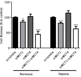
Figure 18|Effect of MCT inhibition on endothelial cell proliferation at 24 hours, under normoxia and hypoxia ... 85 Figure 19|Effect of MCT inhibition on cell cycle distribution at 24 hours, under normoxia and hypoxia.. ... 86 Figure 20|The effects of CHC on the assembling of capillary-like structures by HBMEC cells up to 24 hours, under normoxia.. ... 87 Figure 21|The effects of inhibition of MCT activity on the formation of capillary-like structures by HBMEC cells up to 24 hours, under hypoxia.. ... 88 Figure 22|Representative image of MCT expression, under normoxia and hypoxia, four days after transfection of HBMEC cells with siRNA ... 89 Figure 23|Influence of MCT knockdown on endothelial cell viability at 24 hours, under normoxia and hypoxia.. ... 90 Figure 24|Influence of MCT knockdown on endothelial cell death at 24 hours, under normoxia and hypoxia ... 90 Figure 25|Influence of MCT downregulation on endothelial cell metabolism at 24 hours, under normoxia and hypoxia.. ... 91 Figure 26|Influence of MCT downregulation on endothelial cell proliferation at 24 hours, under normoxia and hypoxia.. ... 92 Figure 27|Influence of MCT downregulation on cell cycle distribution at 24 hours, under normoxia and hypoxia.. ... 93 Figure 28|Influence of MCT downregulation on the assembling of capillary-like structures by HBMEC cells up to 24 hours, under normoxia. ... 94 Figure 29|Effects of MCT downregulation on the development of capillary-like structures by HBMEC cells up to 24 hours, under hypoxia.. ... 94 Figure 30|Representative image of MCT expression, under normoxia and hypoxia, five days after transfection of U251 cells with siRNA ... 95 Figure 31|Glucose and lactate contents in glioma cells’ conditioned media 48 hours after MCT downregulation, under normoxia and hypoxia.. ... 96 Figure 32|Qualitative evaluation of VEGF-A mRNA levels in glioma cells, upon MCT downregulation, under normoxia and hypoxia.. ... 97 Figure 33|Effects of glioma cells’ CM on endothelial cell biomass at 24 hours. ... 97
xxvii
Figure 34|Effects of glioma cells’ CM on endothelial cell metabolism, at 24 hours.. ... 99 Figure 35|Effects of glioma cells’ CM on endothelial cell proliferation at 24 hours. ... 100 Figure 36|Representative assay of cell cycle analysis for HBMEC cells, after 24 hours of growth in MCT-silenced glioma cells’ CM from normoxia and hypoxia. ... 101 Figure 37|Influence of MCT-silenced glioma cells’ CM from normoxia in the development of capillary-like structures by HBMEC cell line.. ... 102 Figure 38|Influence of MCT-silenced glioma cells’ CM from hypoxia in the development of capillary-like structures by HBMEC cell line.. ... 102 Figure 39|Effects of scramble-derived glioma cells’ conditioned media from normoxia and hypoxia on CAM vascularization, in vivo.. ... 103 Figure 40|Influence of MCT-downregulated glioma cells’ CM from normoxia on CAM vascularization, in vivo. ... 104 Figure 41|Effects of MCT-downregulated glioma cells’ CM from hypoxia on CAM vascularization, in vivo ... 104 Figure 42|Influence of hierarchical fibrous scaffolds on cell viability. ... 105 Figure 43|Influence of hierarchical fibrous scaffolds in human endothelial cells’ metabolism.. 106 Figure 44|Influence of hierarchical fibrous scaffolds in human glioma cells’ metabolism. ... 106
31
1 Introduction
1.1 Angiogenesis
1.1.1 Development of the vascular network: an overview
According to their size, humans and other vertebrate organisms require a specialized circulatory system [1], which comprises heart, major vessels (arteries and veins), as well as a subset of thin vessels known as capillaries [2]. This vascular network mainly functions to deliver an adequate nutrient and oxygen flow to whole body (arteries) and also to withdraw all the waste products from cellular metabolism (veins) [1-3]. Furthermore, it allows immune cells to travel through the body to carry out their function of protection [4].
In developing mammalian embryos, the heart and blood vessels are the first organs to be assembled and the recently formed vasculature is continuously remodeled in order to achieve a functional system [5].
During embryogenesis, there are two different ways of blood vessel formation: vasculogenesis and angiogenesis [1, 3, 5]. Vasculogenesis is thought to take place only throughout early stages of embryo development, where new blood vessels are assembled de novo. In opposition, angiogenesis promotes blood vessel assembling from the pre-existing vasculature. It occurs during wound healing and repair processes in adulthood [5, 6]. The formation of primitive vasculature during embryogenesis have been well described by Ema and Rossant, using mouse models [7]. Indeed, they showed key evidence for a common progenitor for both vascular and hematopoietic cell lineages, demonstrating that both cell subsets lack vascular endothelial growth factor receptor 2 (VEGFR-2 or Flk-1) in mutants. Accordingly, Choi and colleagues have previously described that Flk-1-positive cells, from differentiating embryonic stem cells, were able to differentiate into both endothelial and hematopoietic colonies [8]. Nevertheless, not all endothelial cells are derived from those progenitor cells, as have been demonstrated using different animal models [7, 9].
As embryo develops, the hemangioblasts in the posterior primitive streak can differentiate into both hematopoietic cells and angioblasts (Figure 1a), according to the signaling
32
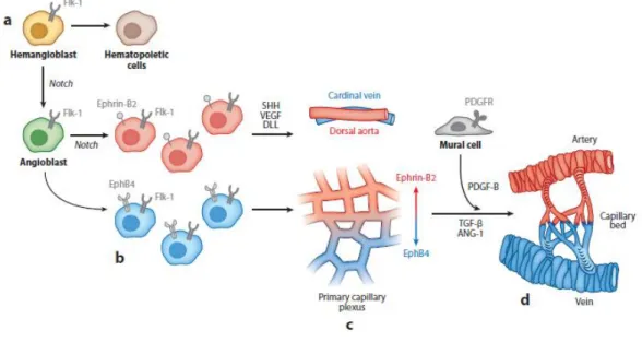
pathway involved [7, 10]. These precursors then migrate to the embryo and yolk sac, where early blood vessels develop by aggregation of angioblasts into a primitive network of simple endothelial tubes (Figure 1b) [5, 11]. In the yolk sac, the angioblasts aggregate into a vascular structure known as blood islands, including endothelial cells in the outer side, whereas the inner side is composed of hematopoietic cells. Next, blood islands fuse themselves into a primary capillary plexus (Figure 1c). Contrarily, in the embryo, angioblasts aggregate into the dorsal aorta or cardinal vein, in a process mediated by vascular endothelial growth factor-A (VEGF-A), Sonic Hedgehog (SHH) and Notch signaling (Figure 1c). The final event of vasculogenesis is known as arterial-venous specification, which is reported to be dependent on the Eph-Ephrin system. EphrinB2 is expressed in both arterial endothelial cells (ECs) and smooth muscle cells (SMCs), while its receptor, EphB4, is only expressed in veins [11].
Altogether, both intra- and extra-embryonic plexuses assemble a de novo vascular network [5, 11, 12]. Further remodeling of the primary vascular network, through recruitment of mural cells or pericytes, gives rise to a stabilized and mature vascular system. This process, known as arteriogenesis, is mediated by several molecules, including platelet-derived growth factor B (PDGF-B), angiopoietins (Agn) and transforming growth factor (TGF-) (Figure 1d) [13].
Figure 1|Development of embryonic vascular network in mammals. (a) In the developing embryo, hemangioblasts become restricted to hematopoietic or angiogenic fate. (b) The activation of Notch signaling leads to the aggregation of angioblasts either in the intra- or extra-embryonic tissues. (c) In the extra-embryonic side, the angioblast aggregation assemble a primary capillary plexus whereas, in the embryo itself, the angioblasts fuse into dorsal aorta or cardinal vein in process mediated by VEGF-A, SHH and Notch signaling. (d) Several markers are involved in the maturation of the established nascent vessels by recruitment of mural cells or pericytes. From [5].
33
After the vasculogenic process, new blood vessels are formed from the preexisting ones, in a process known as sprouting angiogenesis (Figure 2A), which includes localized extracellular matrix (ECM) degradation and subsequent proliferation, migration and tissue infiltration by endothelial cells. At the end, new capillary tubes are assembled and a new ECM is reconstituted [12,14, 15].
Under normal physiological conditions, there is a balance between angiogenic activators and inhibitors. While these pro-angiogenic factors induce angiogenesis, the anti-angiogenic ones are released to counterbalance angiogenesis induction promoting its downregulation [16]. The most well studied angiogenic regulator of vascular development is the VEGF/VEGFR system [17]. However, there are other molecules, like angiopoietin/Tie system [18], Eph/Ephrin system [19] as well as acidic and basic fibroblast growth factors (FGF) [20], playing a central role as angiogenic activators. In opposition, the most well described anti-angiogenic regulators are angiostatin [21], endostatin [22] and thrombospondin [23].
In healthy organisms, the greater part of ECs are quiescent or in a non-dividing phase, collectively assembling a monolayer of interconnected phalanx cells covered by mural cells, SMCs or pericytes, which together constitute the basement membrane. The establishment of this tightly organized cell layer inhibits endothelial cell proliferation and promotes endothelial cell survival through the release of VEGF and Ang-1 [24]. In response to an angiogenic stimulus, the quiescent vessel becomes enlarged due to mural cells detachment from the vessel wall and their subsequent release from the basement membrane in a matrix metalloproteinase (MMPs)-mediated process [24]. As a result, the endothelial cell layer becomes more permeable, enabling plasma protein extravasion that, in turn, is responsible for the assembling of a transient ECM [24]. Then, some signaling molecules induce endothelial cell adherence to the ECM, allowing its migration [24].
Importantly, to avoid the endothelial cell movement en masse onto the angiogenic signal, a tip-cell is selected to guarantee the new vessel branching. Behind the tip-cell, other endothelial cells, the stalk cells, proliferate and elongate, consequently establishing the vessel lumen. Stalk cells, through the release of Agn-1, PDGF-B and TGF-recruit mural cells, giving rise to functional and mature vessels. Finally, a new basement membrane is synthesized and the endothelial junctions are re-established to ensure optimal flow distribution [24].
34
In healthy tissues, besides sprouting angiogenesis, there is another mechanism of angiogenic events known as intussusceptive angiogenesis (Figure 2B) [24, 25]. The intussusceptive angiogenesis was firstly described in postnatal remodeling of capillaries in the lung [26]. Here, endothelial cells from opposite walls make a kissing-like contact, creating a transluminal bridge which, in turn, is further remodeled, giving rise to two daughter vessels from the progenitor one (Figure 2B) [27, 28]. The intussusception process is faster than sprouting-like angiogenesis because it does not require endothelial cell proliferation. Instead, endothelial cells increase their volume becoming thinner [27].
Figure 2|Mechanisms of angiogenesis in health tissues. During sprouting angiogenesis (A), the daughter vessels are formed through endothelial cell proliferation in two opposite progenitor vessels while, in the intussusceptive-like angiogenesis (B) a transvascular structure is assembled into the progenitor vessel after the kissing-like contact between endothelial cells in opposite walls. From [4].
1.1.2 Angiogenesis and disease
Besides developmental angiogenesis, some angiogenic events may occur at specific stages of adulthood, as wound healing and repair processes, pregnancy, as well as skeletal growth, which are strikingly regulated [5]. Nevertheless, the balance between pro- and anti-angiogenic factors may be, somehow, disrupted, giving rise to pathological conditions, the “angiogenic switch” [12, 29, 30].
In general, those pathologies arise from excessive angiogenesis, inadequate vessel growth or abnormal vessel regression, as well as abnormal vessel remodeling. For instance, heart and brain ischemia, neurodegeneration, hypertension and respiratory distress are a result
35
of insufficient angiogenesis rate. In opposition, there are other diseases like psoriasis, arthritis and blindness, which are caused by the upregulation of the angiogenic process. However, the best example of excessive angiogenesis is still cancer [5, 12].
1.1.3 Angiogenesis in cancer
The first observation of angiogenic events in tumors was made about 100 years ago [31]. Afterwards, in 1971, Judah Folkman and collaborators suggested that tumor growth and metastasis are angiogenesis-dependent processes, and that angiogenesis blockade could be a great strategy to prevent tumor progression [32]. Few years later, Gullino et. al. described that pre-cancerous lesions acquire angiogenic capacity during their evolution to malignant phenotype [33].
During the early stages of tumorigenesis (Figure 3a), there are several genetic and epigenetic modifications which, in turn, lead to activation of oncogenes or inhibition of tumor suppressors. Subsequently, the balance between cell proliferation and apoptosis rates is disrupted and tumors enter in a stage of early hyperplastic growth (Figure 3b) [34].
Figure 3| The onset of tumor angiogenesis. (a) Initially, most tumors develop in avascular sites. (b) The disruption between cell proliferation and apoptosis rates triggers early hyperplasic growth of the tumor. (c) When tumor cells, located far away from vessels, become deprived of nutrients and oxygen, the onset of tumor angiogenesis occurs (d) Tumor vasculature is further remodeled by recruitment of mural cells. (e) Tumor vasculature becomes mature and functional. From [29]
36
When the tumor reaches around 1 – 2 mm of diameter, there is an impairment in both nutrient and oxygen supply to the neoplastic cells, located far away from blood vessels, which will influence tumor progression (Figure 3c) [5, 34]. Nevertheless, through evolution, tumor cells acquired the ability to escape to growth inhibition, developing their own vasculature, commonly from the preexisting blood vessels – tumor angiogenesis (Figure 3c-e) [34]. Besides its role of supporting tumor growth, tumor angiogenesis also provides a key mechanism through which cancer cells disseminate via blood stream to distant organs to develop metastases [34].
In opposition to normal tissues, where the mechanisms of angiogenesis are limited to vasculogenesis, sprouting angiogenesis and intussusception, tumors exhibit three more modes of angiogenesis including vessel option, vascular mimicry and transdifferentiation [25]. Vessel co-option mainly occurs in well-vascularized tissues like brain and lung [35, 36], where tumor cells recruit the existing blood vessels to sustain their growth [27]. The first experiments describing the occurrence of vessel co-option in tumors were made upon the implantation of C6 glioma cells in the rat brain [37]. Few weeks after implantation, tumors became well-vascularized with blood vessels, exhibiting similar features to functional vessels of normal brain. However, these blood vessels start to retract themselves along time, without any angiogenic response as a compensatory mechanism. Consequently, in the center of the tumor, extensive areas of cell death were found due to the lack of functional vessels around glioma cells. Contrarily, in the tumor periphery, a burst of angiogenesis was further detected [37]. Indeed, these findings indicate that some tumors are able to incorporate the existing host blood vessels to support their growth, at least during early stages of tumor development [37, 38].
Later, the concept of vasculogenic mimicry was described in melanomas. Aggressive melanoma cells assembled vascular channels de novo, in an angiogenesis-independent mechanism, i.e., without the interference of endothelial cells [39]. Furthermore, tumor blood vessels lined by tumor-derived cells were discovered in mice transplanted with human glioma tumors - a process also known as vasculogenic mimicry. Altogether, these observations demonstrate that, due to their plasticity, tumor cells on the vicinity of the nascent blood vessel are recruited for the angiogenic process, acquiring some endothelial cells’ features [25].
The discovery of cancer-stem cells (CSCs) in solid tumors, mainly in glioblastoma, has contributed to the better understanding of the biology of cancer. Recently, it was shown that glioma-stem cells and endothelial progenitors share some characteristics and, therefore, CSCs
37
have the ability to differentiate into functional endothelial cells, giving rise to tumor endothelium – cancer-stem cell transdifferentiation [40-42].
Independently of the mechanism by which tumor vasculature is built up, tumor blood vessels are normally irregular, dilated and tortuous. Additionally, the standard organization into well-defined venules, arterioles and capillaries is not found in tumor-associated vessels. Also, they are sometimes hemorrhagic, partially due to the VEGF overexpression, and leaky, because mural cells become more loosely associated or less abundant [43]. Furthermore, the blood flow in tumors is irregular, moving slower comparatively with normal tissues which, in turn, lead to dysfunctional capillaries [29].
Despite their structural and functional abnormalities, tumor endothelial cells are believed to be genetically normal [25]. However, recently, cytogenetic aberrations such as aneuploidy and the existence of multiple chromosomes were found in uncultured tumor endothelial cells. The aneuploid phenotype was exacerbated in culture, indicating that the cytogenetic abnormalities might be acquired while in the tumor microenvironment [44].
Taking together, these abnormalities in tumor endothelium are likely to occur as a result from an imbalanced expression and function of pro-angiogenic factors [29].
1.1.4 Regulatory mechanisms of angiogenesis in cancer: molecular insights
As already mentioned, in health adult organisms, blood vessel growth is a well-regulated process, in which endothelial cells remain quiescent. Thus, some physiological factors, such as hypoxia, as well as numerous endogenous molecules, mainly VEGF, are likely to perform an active role on angiogenesis induction [3, 17, 24].
1.1.4.1 Hypoxia
Hypoxia – a decrease in the normal oxygen levels within a tissue – is a hallmark of several vascular diseases, including cancer. There are essentially two types of hypoxia - acute and chronic hypoxia. Acute hypoxia occurs when abnormal blood vessels are shut down, leading to a reversal blood flow, while chronic hypoxia appears as a result from uncontrolled cell proliferation, overpassing the oxygen diffusion limit [45].
38
Hypoxic regions are frequently found within tumors, namely because of the abnormal features of tumor blood vessels. These hypoxic environments confer to cancer cells the ability to survive and proliferate in hostile conditions, contributing to the malignant phenotype and tumor aggressiveness [45]. Indeed, hypoxia has emerged as a primary regulator of angiogenic switch [46], facilitating tumor progression and metastasis [47].
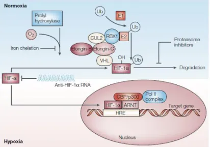
The cellular response to hypoxia is mediated by the hypoxia-inducible factor (HIF) family [48, 49]. So far, three different transcription factors belonging to this family (HIF-1, HIF-2 and HIF-3) were identified [50]. HIF-1is ubiquitous and is considered the major factor regulating responses to hypoxia. It is a heterodimeric protein composed of two subunits, an oxygen-inducible subunit, HIF-1, and a constitutively expressed one, HIF-1(Figure 4)[45, 48, 49].
In the presence of oxygen, the activity of the prolyl hydroxylase domain protein 2 (PHD2) promotes hydroxylation in two HIF-1 proline residues which, in turn, facilitates its binding to a tumor suppressor protein, Von Hippel-Lindau (VHL). This interaction results in polyubiquitination of HIF-1,targeting it for proteossomal degradation (Figure 4) [51]. In opposition, in the absence of oxygen, HIF-1 is stabilized and, consequently, dimerized with HIF-1Then, the recently formed complex is translocated from the cytoplasm to the nucleus, where it binds to the hypoxia-responsive elements (HRE) in the promoter region of several target genes (Figure 4) [48, 49].
Figure 4|HIF-1 signaling pathway. Abbreviations: Ub: Ubiquitin; E1: Ubiquitin-activating enzyme; E2: Ubiquitin-conjugating enzyme; RBX1: RING-box protein 1; CUL2: Cullin-2; VHL: Von Hippel-Lindau; CBP/p300: CREB-binding protein/p300 co-activator family; ARNT: Aryl hydrocarbon receptor nuclear translocator protein From [45].
39
In addition, two more proteins have been described to mediate the cellular response to hypoxia, HIF-2 and HIF-3. Similarly with HIF-1, HIF-2also interacts with HRE in the nucleus, enhancing gene transcription [52]. However, it has not a ubiquitous distribution, being expressed mainly in lung, carotid body and endothelium [52, 53]. There are few studies concerning HIF-3but it has been reported as an inhibitor of HIF-1 activity [52].
HIFs are essential molecules, performing a role in several aspects of cancer biology, including angiogenesis [54]. In fact, the angiogenic process is regulated by alterations in oxygen levels, and endothelial cells (ECs) have oxygen sensors, as well as HIF-related proteins, which, collectively, allow them to detect differences in oxygen levels [55].
HIF-1 directly activates the transcription of the vascular endothelial growth factor (VEGF) gene and the one of its receptor, vascular endothelial growth factor receptor 1 (VEGFR-1) [52], stimulating endothelial cell proliferation, migration and vascular sprouting [24, 55]. Indeed, HIF-1 and VEGF overexpression was correlated with more aggressive lesions in both breast cancer and gliomas [56, 57]. Additionally, HIF-1 knockdown in endothelial cells disrupted the autocrine loop required for the hypoxic induction of both VEGF receptors (VEGFR1 and VEGFR2) through VEGF signaling. Further impairments in endothelial cell proliferation and tube formation were observed in vitro. Similarly, a decrease in tumor angiogenesis and a consequent reduction of tumor growth were also observed in vivo [58].
Although further studies concerning HIF-2function should be performed, it has been associated with the maintenance of vascular integrity [24, 54, 55].
1.1.4.2 VEGF family
It is widely accepted that VEGF signaling pathway is the major cascade regulating angiogenic events. The VEGF family consists of only few homologous members, including VEGF-A, VEGF-B, VEGF-C, VEGF-D, VEGF-E, and also PlGF [59, 60]. VEGF-A is the main component of this family, regulating physiological angiogenesis during embryogenesis and at specific stages in adulthood. Furthermore, VEGF-A has also been implicated in pathological angiogenesis, being associated, for instance, with tumor growth [61].
Besides its role in angiogenesis induction, VEGF-A has other activities including vascular endothelial cell mitogenesis, endothelial cell survival and induction of hematopoiesis [62].
40
Additionally, Dvorak and colleagues described VEGF-A as a tumor-secreted molecule that increases tumors’ vascular permeability [63].
The human VEGFA gene is located on chromosome 6 and is organized into eight exons, separated by seven introns [61, 64, 65]. Since VEGF-A pre-mRNA undergoes alternative splicing, two different families of proteins are produced, one pro-angiogenic and another anti-angiogenic [66]. Concerning the pro-angiogenic family, the alternative splicing of VEGFA gene generates seven different variants (VEGF121a, VEGF145, VEGF148, VEGF165a, VEGF183, VEGF189a, VEGF206), whereas
only three anti-angiogenic variants (VEGF165b, VEGF121b and VEGF189b) were identified, so far. Those
spliced variants are termed according with their aminoacid content after signal sequence cleavage [61, 64, 66].
Importantly, VEGF is a homodimeric glycosylated protein containing heparin-binding sites. The potential binding between each VEGF-spliced variant and heparin derivatives determines its diffusion degree [54, 61, 62], as well as contributes for the mitogenic activity of VEGF [67]. In fact, the larger VEGF variants, VEGF206 and VEGF189, are highly basic, exhibiting high
affinity for heparin and, hence, are tightly bound to cell surface and extracellular matrix (ECM) [54, 61]. By contrast, the intermediate variants (VEGF165, VEGF148 and VEGF145) are moderately
diffusible, because of their weak affinity for heparin/heparin sulphate. Indeed, although these variants are secreted, a significant part of them remained attached to the ECM [61, 64]. Additionally, the shorter VEGF-spliced variant, VEGF121, has acidic properties and is freely
diffusible, due to the lack of heparin binding sites [61]. Importantly, the VEGF-A-spliced variants are differentially expressed in tissues. VEGF165a is expressed in several normal and transformed
cells. Furthermore, both VEGF121 and VEGF189 variants are found in the majority of cells and tissues
expressing the VEGF gene. In opposition, the largest variant (VEGF206) in only expressed in human
fetal liver [64]. Among all the pro-angiogenic subtypes, VEGF165a has been reported as the most
important spliced variant of the VEGF gene [68]. Indeed, some authors described that the homolog protein in mice (VEGF164), per se, was enough to trigger the angiogenic process, as
transgenic VEGF164 mice in a VEGF-A null genetic background were essentially healthy [69].
All the VEGF-A variants bind to specific protein-tyrosine kinase receptors (RTKs), the vascular endothelial growth factor receptor 1 (VEGFR1) and vascular endothelial growth factor receptor 2 (VEGFR2) (Figure 5) [17, 61]. Firstly, these RTKs were identified in vascular endothelial cells [17, 61]. However, their expression was also reported on bone marrow-derived
41
cells [61], macrophages [64, 70], some malignant cells [70] and vascular SMCs [71]. Structurally, both VEGF receptors contain seven immunoglobulin-like extracellular domains, a single transmembrane domain and a consensus tyrosine kinase sequence, which is interrupted by a kinase insert domain of 70-100 aminoacids [72, 73]. Additionally, VEGF-A binds to the specific non-enzymatic co-receptors neuropilin-1 (NRP1) and -2 (NRP2) [17, 61], which are believed to enhance VEGF-A signaling through VEGFR2, ultimately increasing angiogenesis [17].
Although VEGFR1 had been firstly identified, its exact function still remains unknown [61]. VEGFR1 has higher affinity for VEGF-A than VEGFR2. However, this interaction ligand-receptor triggers a weak tyrosine kinase phosphorylation [17]. Actually, VEGFR1-mediated signaling cascade does not induce significant mitogenic effects in endothelial cells. Nevertheless, during early embryonic development, VEGFR1 seems to sequestrate VEGF-A, preventing its binding to VEGFR-2 and consequently inhibiting further angiogenic events [61]. Contrarily to VEGFR1, VEGF-A binding to VEGFR2 induces a powerful tyrosine kinase activity, stimulating endothelial cell migration, proliferation, survival and enhancing vascular permeability [17, 61]
Figure 5|Interactions between VEGF family ligands and their receptors involved in the angiogenic process. Abbreviations: VEGF: vascular endothelial growth factor; PlGF: placental growth factor; VEGFR: vascular endothelial growth factor receptor. Adapted from [74].
VEGF-A, mainly VEGF165, is believed to be the major inducer of pathological angiogenesis
42
gastrointestinal tract, kidney, bladder, ovary and uterine cervix carcinomas, angiosarcomas and intracranial tumors like glioblastoma multiform (GBM) [64, 65]. Furthermore, increased expression of VEGF-A was also reported in some hematological malignancies and multiple myeloma [65]. Indeed, an association between increased VEGF-A mRNA levels and malignant progression has been identified [64].
Tumor cells exhibit higher mRNA levels of VEGF-A than tumor-associated endothelial cells, where its expression is negative. In opposition, both VEGFR1 and VEGFR2 are upregulated in tumor-endothelial cells [75]. These observations suggest that VEGF-A act as a paracrine molecule [64]. Interestingly, angiosarcoma cells co-expressed both VEGF-A and VEGFR1, indicating that an autocrine VEGF-A loop may also occur in some tumor types. However, angiosarcomas cannot be considered such a good model because these tumor cells are endothelial-derived ones [64]. Importantly, higher levels of VEGF-A have been correlated with decreased relapse-free survival rates of breast cancer patients [76]. Also, in gastric carcinomas, VEGF-A upregulation was associated with vessel involvement, lymph node metastasis and liver metastasis which, collectively, contribute for poor clinical outcomes [77].
Recently, the anti-angiogenic VEGF-A variants have gained focus in cancer. These molecules are highly expressed in normal tissues, but they are reported to be downregulated in certain tumor types including renal-cell, prostate and colorectal carcinomas, as well as in malignant melanoma [66]. In fact, overexpression of VEGF165b in those tumor specimens efficiently
impaired tumor growth in xenografted mouse models [66].
1.1.5 Anti-angiogenic Therapy
Judah Folkman firstly identified the involvement of angiogenesis in tumor development and metastasis through VEGF-A. Hence, blocking such powerful pro-angiogenic factor might be an excellent strategy for cancer therapy [78].
Numerous preclinical studies concerning angiogenesis inhibition have been made using VEGF-A blockers. These strategies efficiently decreased tumor progression in a range of cancer models [79]. In patients with metastatic colorectal cancer, the combined administration of a monoclonal antibody against VEGF-A (bevacizumab) and 5-fluorouracil (5-FU) demonstrated
43
benefits for the patients [79]. Consequently, in 2004, bevacizumab was approved by Food and Drug Administration (FDA) for the treatment of metastatic colorectal, metastatic non-squamous non-small lung carcinoma, metastatic breast cancer, recurrent GBM and metastatic renal cell carcinoma [24].
Additionally, multi-targeted RTKs inhibitors (e.g. sorafenib, sunitinib and pazopanib), which prevent several signaling pathways such as the one induced by VEGF-A, were further approved for the treatment of different tumor types. Sorafenib is indicated for the treatment of metastatic renal cell carcinoma and hepatocellular carcinoma whereas, both sunitinib and pazopanib, are currently used only in the treatment of metastatic renal cell carcinoma [24, 79]. Recently, sorafenib has also been proposed for the treatment of advanced pancreatic neuroendrocrine tumors [24].
The blockade of VEGF-A signaling has been demonstrated as effective in a range of tumors. However, some patients with metastatic disease have acquired resistance to VEGF-A signaling inhibitors [24, 79]. Such evidence raises the question whether cancer cells become more malignant upon treatment with these drugs. In fact, the inhibition of vascular supply might increase tumor hypoxia, increasing local invasion and distant metastasis. Furthermore, the impairment of tumor vasculature results in decreased effectiveness of conventional radio- and chemotherapy [24, 80].
In spite of all the advances targeting tumor vasculature, anti-angiogenic therapy must be improved, optimizing the dosage, as well as the treatment periods of VEGF-A blockers. In addition, the combination of these VEGF-A blockers with other pharmacological agents targeting, for instance, the escape pathways (e.g. PlGF) might be further implemented [24].
44
1.2 Monocarboxylate Transporters
1.2.1 Monocarboxylate Transporters family
Monocarboxylates such as pyruvate, ketonic bodies (acetoacetate and -hydroxybutyrate) and lactate have been described as key players in cellular metabolism (Figure 6). Some mammalian tissues, such as white muscle fibers, erythrocytes and the majority of neoplastic cells, rely on glycolysis for energy production, synthesizing lactate as end product. Thus, in order to maintain high glycolytic rates, lactate efflux becomes essential. Contrarily, other tissues like brain, heart and red skeletal muscle rapidly metabolize lactate, which constitutes the major fuel for these organs. Hence, large amounts of lactate must be canalized into these cells [81-83].
Figure 6|The role of monocarboxylate transporters in cellular homeostasis. Abbreviations: Glc-1-P – Glucose-1-phosphate; Glc-6-P
– Glucose-6-phosphate; Ac+HB – acetate + -hydroxybutyrate. From [81]
Due to their negative charge, monocarboxylates, including lactate, cannot freely diffuse through plasma membrane in large amounts, requiring a specialized transport system [81, 82, 84, 85]. Accordingly, cells express membrane transporters belonging to both proton-linked monocarboxylate transporters (MCTs) (Figure 6) and sodium-linked monocarboxylate transporters
45
(SMCTs) families, which carry those simple organic carboxylates coupled with protons (H+) or
sodium ion (Na+), in an equimolar manner through a symport mechanism, respectively [81, 86].
Regarding the MCT family, it comprises 14 members of proteins encoded by the SLC16 gene family, which is highly conserved between different species like rodents (e.g. rat and mouse) and chicken [87, 88]. So far, solely seven members of MCT-family have been functionally characterized and, from these seven only four (MCT1-MCT4) have been shown to perform the proton-coupled monocarboxylate transport [86, 87, 89]. The major differences between each of these MCT isoforms are their affinities for substrates and inhibitors, the regulation of their expression and tissue distribution, as well as their cellular localization [90].
Poole and colleagues, using hydropathy analysis, first described the amino acid sequence of rat MCT1, demonstrating a structure of twelve transmembrane (TM) helices, with both the amino and carboxyl termini located in the cytoplasm [91]. Further studies were performed, corroborating the previous results and, additionally, identifying a large hydrophilic loop between the 6 and 7 TM domains, extended from the plasma membrane into the cytoplasm [91]. Furthermore, it must be highlighted that these TM regions are highly conserved between MCT isoforms, with the greatest sequence variations located in the C-terminus and in the loop [86, 89]. Such variation is thought to be related with either the substrate affinity or the regulation of transport activity [86]
1.2.1.1 MCT1
Although MCT1 expression has been described in almost all tissues in all species studied, its major expression has been found in the heart and muscle [82, 84, 87]. Additionally, it is the only MCT isoform expressed in erythrocytes [87]. The main function of this transporter is to perform both the L-lactate entry into and efflux out of cells, depending on the intracellular and extracellular substrate concentration, as well as the pH gradient across plasma membrane [89, 90]. However, some studies in mouse tumor cell lines and Xenopus oocytes demonstrated that MCT1 isoform exhibits high affinity for a wide range of short-chain monocarboxylates (lower Km
value) [92, 93].
Besides its plasma membrane expression, MCT1 has also been reported in mitochondria of the heart, skeletal muscle and brain, enabling these tissues to oxidize lactate to pyruvate
46
though mitochondrial lactate dehydrogenase (LDH) activity [90, 94]. However, such hypothesis is still not well accepted by some researchers because oxidation of lactate within the mitochondria is both energetically unfavorable and incompatible with the nicotinamide adenine dinucleotide (NADH) redox compartmentalization in cells [94].
1.2.1.2 MCT2
Comparatively with MCT1 isoform, MCT2 exhibits higher affinity for pyruvate and lactate [89, 90]. This isoform was firstly isolated and functionally characterized from hamster liver [95]. Subsequently, a homolog protein was identified in humans [96]. However, there are several differences in expression patterns of this specific isoform among species [89, 90]. MCT2 expression in human tissues is relatively low compared with other isoforms and its expression is confined to those tissues with high lactate uptake rates (e.g. postsynaptic membrane of neurons) or gluconeogenic tissues (e.g. liver and kidney).
1.2.1.3 MCT3
Concerning all MCT isoforms, MCT3 is thought to have the most restricted distribution. Its expression has been reported in specific tissues like retinal pigment epithelium (RPE) and choroid plexus epithelia, facilitating the lactate efflux of the retina [86, 89, 90]. However, some evidence for MCT3 expression in other tissues, such as vascular smooth muscle cell lines, human aorta [97] and human kidney [98], have arisen, indicating a more widely expressed MCT3 mRNA.
1.2.1.4 MCT4
MCT4 isoform exhibits several similarities with MCT1, but the differences in both tissue distribution and substrate affinities must be highlighted [86]. MCT4 is widely expressed in glycolytic tissues, including skeletal muscle, astrocytes, white blood cells and also in some mammalian cell lines [90, 99]. Thus, MCT4 plays an important role in lactate efflux, which is clearly corroborated by the high Km value for monocarboxylates, such as pyruvate and lactate.
The low affinity, namely for pyruvate, ensures that there is no pyruvate loss from the cell, which is essential for the maintenance of the NADH pool, allowing the maintenance of the glycolytic pathway [83, 84].
47
1.2.2 Monocarboxylate transporter regulation
Despite the lack of studies concerning MCT regulation, some groups have recently been examining the regulation mechanisms of these transporters at several levels, including both transcriptional and post-transcriptional regulation, as well as regulation of transport activity.
Although putative N-glycosylation sites have been described for some members of the MCT family [85], sequence analysis demonstrate that MCTs are unlikely to be glycosylated [91, 100]. Additionally, membrane expression of MCTs become essential to perform their transport activity; thus, an ancillary protein, belonging to immunoglobulin (IgG) superfamily, is required for the correct translocation of MCTs to the plasma membrane [84, 85, 89, 90]. These molecular chaperones consist of a single transmembrane domain containing a conserved glutamate residue, a short intracellular C-terminus, and a large glycosylated extracellular domain with the immunoglobulin domains attached [101]. The correct association between MCTs and its chaperone supports MCT folding and stability in cell membranes, besides assisting MCT activity [102].
In a first study, using erythrocytes, interactions between MCT1 and an integral plasma membrane glycoprotein, gp-70 or embigin, were reported [103]. Secondly, an embigin-related protein, CD147 or basigin, was shown to coprecipitate with both MCT1 and MCT4 isoforms, emerging its potential role on MCT modulation [81, 85]. Afterwards, Kirk and collaborators, using both MCT1- and MCT4-transfected cells, demonstrated cytoplasm compartmentalization, which was clearly reverted upon CD147 overexpression, trafficking MCTs towards the plasma membrane [102]. In addition to MCT1 and MCT4, also MCT3 expression in plasma membrane is dependent on its association with the mature and glycosylated form of CD147 [104].
When available, CD147 is the favorite chaperone binding MCT1 [105] but, if CD147 is absent, an interplay between embigin and MCT1 may occur, at least in rat erythrocytes [103]. Contrarily, MCT2 expression in plasma membrane colocalize with embigin in mammalian cells, suggesting that embigin is involved in MCT2 regulation rather than CD147 [106]. Thus, it is currently accepted that both MCT1 and MCT4 isoforms have CD147/basigin as ancillary protein, while gp-70/embigin perform the molecular chaperone activity for MCT2 isoform [81].
48
Recently, another molecular chaperone, CD44, has been implicated in MCT regulation [107]. Indeed, CD44 was associated with both MCT1 and MCT4 at the plasma membrane in human cancer specimens. Accordingly, CD44 and MCT1 were co-expressed in lung carcinoma [108], whereas in prostate cancer, CD44 expression was associated with both MCT1 and MCT4 [109]. Considering recent findings, there is a reciprocal regulation between MCTs and their chaperones recognizing MCTs as indirect players in tumor growth and angiogenesis, tumor cell migration and invasion, as well as mediators in chemotherapy resistance [88].
Additionally, in recent years, some groups have reported MCT regulation through gene expression. The analysis of slc16a1 (MCT1) promoter showed the absence of the classical TATA-box region. However, it contains several binding sites for a variety of transcription factors, including those related with the regulatory effects of butyrate [110]. The transcriptional slc16a1 regulation has been well described in skeletal muscle. In this model, MCT1 appears to be upregulated after chronic stimulation or exercise in rats and humans, which seems to be associated with increased levels of calcium and adenosine-monophosphate (AMP), leading to the activation of calcineurin and AMP-activated protein kinase (AMPK), respectively. Calcineurin is believed to activate the transcription factor nuclear factor in activated T-cells (NFAT), which in turn binds to enriched-NFAT binding sites located in the promoter region of slc16a1 gene, increasing its expression. In addition to calcineurin, the activation of AMPK stimulated the activity of slc16a1 promoter, in both L6 myoblasts and hepatoma cells (HepG2). However, MCT4 expression upon AMPK activation is still controversial. In both L6 myoblasts and HepG2 cells, the promoter activity of slc16a3 (MCT4) was decreased by AMPK [90] while, in skeletal muscle MCT4 expression was revealed as increased upon AMPK stimulation [111].
Beyond the above-mentioned regulatory mechanisms, hypoxia, through hypoxia-inducible factor 1- (HIF-1) remains the major regulatory factor concerning MCT4 expression [112], which is consistent with the role of this MCT isoform in lactate efflux. Thomas C. and colleagues demonstrated upregulation of both mRNA and protein levels of MCT4 upon hypoxic stimulation in several cell types, and such effect might be mimicked using cobalt chloride (CoCl2) implying
transcriptional regulation via HIF-1. By contrast with MCT4, in this study, neither MCT1 nor MCT2 expression were found to be regulated by hypoxia [113], which might be associated with the absence of hypoxia response elements (HRE) in their 5’ untranslated regions (UTR) [87]. The promoter activities of slc16a1 (MCT1), slc16a7 (MCT2) and slc16a3 (MCT4) were measured
49
using luciferase promoter constructs, indicating that hypoxia stimulates slc16a3 promoter activity, but no effect was observed in both slc16a1 and slc16a7 promoter activity [113]. Despite this evidence, MCT1 regulation by hypoxia still remains unclear. Recently, both MCT1 and MCT4 were found to be upregulated after hypoxia induction in human adipocytes [114].
In addition to transcriptional regulation, concomitant measurements of mRNA and protein levels have suggested that MCTs might be regulated in a post-transcriptional manner. For instance, post-transcriptional MCT1 regulation might involve regulation of its translation. The 3’UTR region of MCT1, where post-translational modifications may occur, is very long and it is thought to be involved in the control of MCT1 expression [90]. Wilson and Halestrap demonstrated a post-transcriptional upregulation of MCT1 protein expression throughout cell cycle. According to these authors, the MCT1 protein levels increased in both post-mitotic and G1 phases of the cell cycle, where mRNA levels remained unchanged [90]. These results might be explained because of the existence of a putative cytosolic polyadenylation element in the 3’UTR of MCT1 which is responsible for the regulation of polyadenylation process in the post-mitotic phase [90, 115].
Finally, another regulatory mechanism must be considered, upon the discovery of a C-terminus truncated form of MCT4, which was the major form in a variety of tissues according to Western Blot analysis. Immunohistochemistry procedures revealed the existence of two cellular compartments from distinct cell types, lymphocyte cytoplasm and the capsule of muscle spindle, containing the truncated form, but not the full-length one. This evidence might indicate the existence of a specific function for this shorter MCT4 isoform, which is still unknown [116].
1.2.3 MCT inhibitors
Due to their association with specific glycosylated molecular chaperones, MCTs are correctly targeted to the plasma membrane, in order to perform their transport activity. Thus, they are attractive targets for systemic therapy [117]. Indeed, several chemical compounds have been developed as non-physiological competitive MCT inhibitors [85, 89]. The best well-studied classes of compounds are the cinnamic acid derivates (e.g. -hydroxy-4-cianocinnamate (CHC)), the stilbene dissulfonates (e.g. di-isothiocyanostilbene-2,2’-dissulfonate (DIDS) and
4,4’-50
benzamidostilbene-2,2’-dissolfunate (DBDS)) and thiol reagents (e.g. para-chloromercuribenzenesulfonic acid (pCMBS)) (Figure 7) [82].
Figure 7|Classical MCT inhibitors. Abbreviations: -CHC – -hydroxy-4-cyanocinnamate; DIDS - 4,4’-di-isothiocyanostilbene-2,2’-dissulfonate; DBDS - 4,4’-benzamidostilbene-2,2’-4,4’-di-isothiocyanostilbene-2,2’-dissulfonate; pCMBS - para-chloromercuribenzenesulfonic acid. Adapted from [118].
CHC was firstly described as an inhibitor of the mitochondrial pyruvate transport [119] and also as an inhibitor of lactate transport in Ehrlich ascite tumor cells (Ki=0.5mM) [120].
Taking into account its potential inhibition of lactate transport, CHC was further evaluated for MCT1 inhibition [118]. Actually, CHC inhibits MCT1 isoform (Ki=166M), in spite of lacking
MCT1 specificity, as revealed by Ki=24M and K0.51mM for MCT2 and MCT4, respectively
[121].
In mouse models, CHC exhibited antitumor activity, per se or in combination with radiotherapy, without relevant side effects [122]. Furthermore, CHC induced a decrease in intracellular pH of human melanoma cells, leading to impaired cancer cell survival [123]. Treatment of glioma cells with CHC disturbed their glycolytic metabolism, re-sensitizing tumor cells to radiotherapy [124].
Stilbene dissulfonate compounds (e.g. DIDS and DBDS) are competitive inhibitors for many anion channels, including the chloride/bicarbonate exchanger AE1 and MCTs (Ki=22.4M) [118]. Once again, MCT2 was reported to be more sensitive than MCT1 for both DIDS and DBDS [89]. DIDS may be considered an irreversible inhibitor upon long-lasting incubation periods, probably due to its reactive isothiocyanate groups which covalently bind to the transporters [125].
51
Also, noncompetitive inhibitors, as the organomercury-related compounds were examined as potential inhibitors for MCT activity. Indeed, p-CMBS inhibited MCT1 activity with a Ki value around 112M [126]. More recently, Wilson et al. revealed that basigin was the real
target for p-CMBS, through its thiol groups on cysteine residues, thus blocking the anion transport [106]. Furthermore, this organomercurial derivative did not demonstrate specificity for MCT1, also inhibiting MCT4 isoform (K0.5=21M) [121].
Some natural compounds, e.g. quercetin and phloretin, have also been reported as potential inhibitors of MCT activity. Actually, in quercetin-treated glioma cells (50M), there was an increase in intracellular lactate accumulation, which was corroborated by a decrease in intracellular pH of glioma cells [127]. Also, phloretin decreased MCT activity in a non-specific manner (MCT1: Ki=5M; MCT2: Ki=14M; MCT4: K0.5=41M) [121].
Recently, a new class of MCT inhibitors has been developed by AstraZeneca. These compounds were firstly synthesized for immunosuppression, thanks to the relevance of MCT1 for T-lymphocytes [128]. These inhibitors demonstrated high affinity for MCT1 with Ki values in the
nanomolar (nM) range [128, 129]. Further structure optimizations were performed, in order to overcome their solubility problems, giving rise to a new compound, AR-C155885, which efficiently binds to MCT1 in human erythrocytes [130].
1.2.4 MCTs in cancer
According to the World Health Organization (WHO), cancer has been the main cause of death worldwide, accounting with around 7.6 million deaths in 2008. Furthermore, this number is estimated to increase to over 13.1 million in 2030 [131].
Firstly, six hallmarks of cancer had been proposed as biological abilities acquired during the multi-step tumorigenic process, contributing for malignant progression, such as self-sufficiency in growth signals, insensitivity to growth suppressor signals, resistance to programmed cell death (apoptosis), limitless replicative potential, sustained angiogenesis and tissue invasion and metastasis (Figure 8), which are consequence of genomic instability underlying tumors [132]. However, more recently, the concept of tumor microenvironment has gained focus. In fact, tumors are not only insular masses of proliferating cancer cells [133]. They
52
are made up from several cell types including fibroblasts, endothelial and immune cells, which collectively give rise to tumor-associated stroma and perform an active role in tumorigenesis [133, 134]. Taking together such evidence, novel hallmarks were recently introduced, namely reprogramming of energy metabolism and evading immune destruction, which are believed to promote the development and progression of human cancers (Figure 8) [133].
Figure 8|The hallmarks of cancer. Here are depicted a range of functional abilities that cancers acquired during the multistep-tumorigenic process. From [133]
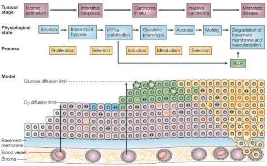
In normal tissues, cells are exposed to different oxygen levels, depending on their distance to the blood vessels, constraining cells to anaerobic glycolysis – the Pasteur effect [135, 136] . However, when the oxygen levels are re-established, non-malignant cells are able to convert glucose into pyruvate, via glycolysis in the cytoplasm, and then canalize it to the mitochondria for further oxidation into carbon dioxide (CO2) and water through oxidative
phosphorylation (OXPHOS) [133]. In contrast, tumors present an alternative metabolism to accomplish their high energetic demands and fulfill the biosynthetic requirements of proliferating cells [117, 137]. During the development of malignant lesions, which mostly occurs in avascular regions, tumor cells are both glucose- and oxygen-dependent. Hence, these substrates must diffuse from the closest blood vessel through the carcinoma basement membrane, as well as across the layers of tumor cells (Figure 9) [138].