Aleksandra Inês Mazurek Vieira de Azevedo
Bachelor Degree in Biotechnological Engineering
Molecular studies on HSV: replication rate,
infection capacity and progeny
Dissertation to obtain the Master of Science Degree in Molecular
Genetics and Biomedicine
Supervisor: Sílvia Maria Milheiro Lopo Esteves, PhD,
Instituto Nacional de Saúde Doutor Ricardo Jorge
Co-Supervisor: Alexandra Isabel Cardoso Nunes, PhD,
Instituto Nacional de Saúde Doutor Ricardo Jorge
Jury:
President: Prof. Doutora Ilda Maria Barros dos Santos Gomes Sanches
Arguer: Prof. Doutora Filomena Ribeiro Alcobia da Silva Trabucho Caeiro
Member: Doutora Sílvia Maria Milheiro Lopo Esteves
iii
NOVA University of Lisbon
Faculty of Sciences and Technology
Department of Life Sciences
Aleksandra Inês Mazurek Vieira de Azevedo
Molecular studies on HSV: replication rate, infection
capacity and progeny
Dissertation to obtain a Master of Science Degree
in Molecular Genetics and Biomedicine,
from the NOVA University of Lisbon,
Faculty of Sciences and Technology
Supervisor:
Sílvia Maria Milheiro Lopo Esteves, PhD,
Instituto Nacional de Saúde Doutor Ricardo Jorge.
Co-Supervisor:
Alexandra Isabel Cardoso Nunes, PhD,
Instituto Nacional de Saúde Doutor Ricardo Jorge.
v
Molecular studies on HSV: replication rate, infection capacity and progeny
Copyright © Aleksandra Inês Mazurek Vieira de Azevedo, FCT/UNL, UNL
The Faculty of Science and Technology (Faculdade de Ciências e Tecnologia) and the NOVA University of Lisbon
(Universidade NOVA de Lisboa) have the right, perpetual and without geographical boundaries, to archive and
publish this dissertation through printed copies reproduced on paper or digital form, or by any other means known
or hereafter invented, and to disseminate through scientific repositories and allow the copy and distribute for
vii
Notes of the author
The work described in this MSc thesis was carried out at the National Reference Laboratory
of Herpes Simplex Virus, Citomegalovirus and Parvovirus B19 of the Infectious Diseases
ix
Acknowledgments
First, I would like to thank Dr. Fernando de Almeida, president of the National Institute of Health INSA
and Dr. Jorge Machado, head of the department of infectious diseases, for allowing me to develop my
theses in the institute and for the opportunity given to work in its laboratories.
I would like to express my special appreciation and gratitude to my supervisor, Dr. Silvia Lopo for
always keeping an open door for me, for advising me, for helping with my many questions. Thank you
for the support and encouragement words, and especially for your patience. I can assure you that
without your help, I wouldn’t have able to finish this work with a sane mind!
To my co-supervisors, Dr. Alexandra Nunes and Dr. João Paulo, for always pushing me to be better
and aim higher. Thank you for the help, for stimulating interesting discussions and for teaching me the
ways of this new world.
To Dr. Maria José, Carla, Ana Rita, Rita, Marta and Vitor from INSA for the help and the patience,
for the lunches and the talks, for the jokes and the cheerful times. To Miguel, for the much needed help,
and for the perseverance when I had almost lost hope!
To Professor Jaime Mota, from FCT, for helping me with one of the most important parts of this
experimental work, and without whom it wouldn’t have been done.
I would like to specially thank my family. Words cannot express how grateful I am for having you in
my life, whether you’re far or near. First, to my parents, for always supporting me in the decisions I made, and for advising me when stress got in the way; for believing in me, for saying that everything
would eventually work out and for listening to me in my most frustrated moments. I know life hasn’t been
fair to us, but we somehow manage to overcome it!
To my sister Kika for always keeping a sane and clear mind towards me, and for annoying me in that
singular way that only she knows (!); for the late McDonalds meals and ice-creams to overcome the
worst days. Your endless source of support was vital to finish this work!
To my Portuguese grandmother, for always listening to me, and for distracting me in the most
stressful times, even though it wasn’t always easy for her.
To my polish grandparents, aunt and cousin, for the cheering, the support and the encouragement
words. Maybe one day I’ll get a job in Poland and we’ll see each other more often!!
To my friends, for always being there, never complaining about hearing me babbling about my
numerous problems, for the dinners and the lunches, for the dancing and the cheering.
xi
Resumo
Os vírus herpes simplex (HSV) são vírus ubíquos, adaptados ao hospedeiro, e responsáveis por
diversas patologias. Existem dois subtipos: HSV-1, tradicionalmente associado a infeções a nível
orofacial, e HSV-2, o qual é maioritariamente associado a úlceras genitais. Esta distinção é cada vez
menos evidente, dada a emergência de infeções genitais causadas pelo HSV-1, associada sobretudo
a fatores sociais, demográficos e migratórios, o que torna o herpes genital uma das infeções
sexualmente transmissíveis mais prevalentes mundialmente.
Uma melhor compreensão das infeções genitais causadas pelo HSV-1 e HSV-2 é mandatória para
entender a patogenicidade das doenças herpéticas. Esta tese teve como objetivo avaliar o ciclo de vida
de vários isolados clínicos genitais de HSV-1 e HSV-2 com diferentes cargas virais, em linhas celulares
distintas, dando ênfase à capacidade e eficiência da infeção viral, em termos de taxa de replicação e
progenia.
Os nossos resultados mostraram que: i) ambos os subtipos do HSV apresentam um padrão de
infeção semelhante independentemente da MOI, com DNA a ser sintetizado 6-12h pós-infeção; ii)
independentemente do subtipo, a concentração inicial do vírus aparentemente não afeta a sua
capacidade de adesão a qualquer linha celular; iii) as células Vero E6 mostraram-se as mais
apropriadas para a infeção do HSV-2; iv) as células HeLa229 mostraram ser as mais apropriadas para
a infeção do HSV-1 com cargas virais menores; e v) as células Vero apresentaram os piores resultados
de infeção viral para ambos os subtipos. Em geral, o HSV-2 mostrou sempre menores capacidades de
aderência e taxas de crescimento que o observado para o HSV-1, apesar de se terem observado
maiores progenias na linha celular Vero E6.
Em conclusão, os resultados apresentados nesta tese de mestrado irão certamente contribuir para
um melhor conhecimento da patogenicidade das infeções genitais causadas pelo herpes.
xiii
Abstract
Herpes simplex viruses (HSV) are ubiquitous host-adapted pathogens that cause a variety of
different disorders. There are two sub-types: HSV-1, which is traditionally associated with oro-facial
infections, and HSV-2 that is mostly associated with genital ulcers. This distinction, however, is
becoming less evident since HSV-1 frequency in genital infections is increasing due to social,
demographic and migratory tendencies, making genital herpes one of the most prevalent sexually
transmitted infections worldwide.
A better understanding on genital HSV-1 and HSV-2 infections is mandatory to the pathogenesis of
human herpes disease. The scope of this thesis was to evaluate the life cycle of various HSV-1 and
HSV-2 genital clinical isolates with different viral loads in three distinct host cell lines, giving special
focus on both capacity and efficiency of viral infection, in terms of replication rate and progeny.
Our results showed that: i) both HSV-1 and HSV-2 isolates exhibited similar infection patterns
regardless MOI, with DNA starting to be synthesized nearly at 6-12h post-infection; ii) regardless HSV
subtype, initial viral concentrations do not apparently affect adherence to any host cell line nor the
generated progeny; iii) Vero E6 cells seemed the most appropriated cell line for HSV-2 infection; iv)
HeLa229 cells appeared to be the most suitable for HSV-1 infection for smaller inoculums; and v) Vero
cell line had the worst viral growth results for both HSV subtypes. In general, HSV-2 displayed always
lower both attachment capacities and growth rates than HSV-1, although higher progenies were seen
in Vero E6 cell line.
Overall, the findings presented in this MSc thesis will certainly constitute a step forward for the
understanding of the pathogenesis of the human herpes genital infections.
xv
Thesis Outline
This MSc thesis is divided into 4 chapters that encompass:
i) a general introduction (Chapter 1) that intends to briefly describe the biology of Herpes Simplex
Virus (HSV), in particular the current knowledge on HSV infections, clinical outcomes and infectious
life cycle. The issues focused on this chapter aim to put into context and to emphasize the relevance
of the study described throughout the thesis. Ultimately, the detailed aims of the present MSc thesis
are presented;
ii) a section of materials and methods (Chapter 2) describing, in detail, the methodology used in
the present thesis, including preliminary assays done for optimizing the experimental conditions for
the current study;
iii) a results section (Chapter 3), where the main findings of the current study are presented, in
order to better understand whether the HSV host cell invasion process depends on the genital HSV
sub-type/strain or on the infected cell line, in terms of capacities of infections, replication rates, and
progenies.
iv) an overall discussion and final conclusions (Chapter 4) where the major findings of the
performed study are highlighted and discussed in a general context, taking into account the scope
of this thesis.
The study presented on this thesis resulted on the following scientific communication:
A. Azevedo, A. Nunes, C. Roque, I. Costa, JP Gomes, S. Lopo. 2016. Molecular studies on HSV:
replication rate, infection capacity and progeny. 19th Annual European Society for Clinical Virology
xvii
Table of contents
Acknowledgements ... ix
Resumo ... xi
Abstract... xiii
Thesis Outline ... xv
Table of contents ... xvii
Figure index ... xix
Table index ... xxi
Abbreviation List ... xxiii
1. Introduction ...1
1.1. Transmission, Clinical manifestations and Prevention/Therapy ...1
1.2. Epidemiology ...3
1.3. Virion structure and Genimoc organization ...3
1.3.1. Central core and Genome ...4
1.3.2. Capsid ...5
1.3.3. Tegument ...5
1.3.4. Envelope ...5
1.4. HSV life cycle ...5
1.4.1. Entry into the host cell...5
1.4.2. Viral gene expression ...7
1.4.3. Viral DNA replication ...8
1.4.4. Viral maturation ...9
1.4.5. Viral egress ... 10
1.4.6. Latency-Reactivation cycle ... 11
1.5. Scope of the thesis ... 12
2. Material and Methods ... 15
2.1. Biological Samples ... 15
2.2. Cell line handling and maintenance ... 15
2.3. Preliminary assays ... 16
2.3.1. Cell counting ... 16
2.3.2. Cultute conditions ... 16
2.3.3. Viral stocks ... 17
2.3.4. Inoculation conditions ... 18
2.4. Evaluation of HSV infection in distinct cell lines ... 19
2.5. DNA extraction ... 20
2.6. Cloning ... 20
2.7. Absolute Quantification assays ... 23
3. Results ... 25
xviii
3.2. Evaluation of HSV infections in distinct cell lines ... 27
3.2.1. HSV-2 infections ... 27
3.2.2. HSV-1 infections ... 30
3.3. Evaluation of HSV infection yields in distinct cell lines ... 34
4. Discussion ... 37
5. References ... 41
Appendices ...i
Appendix 1 ... i
Appendix 2 ... v
xix
Figure index
Figure 1.1: Herpesvirus induced lesions ...2
Figure 1.2: The HSV-1 virion ...4
Figure 1.3: HSV genome structure ...4
Figure 1.4: Pathways of HSV entry into the cell ...6
Figure 1.5: Multiple receptors upon HSV entry ...7
Figure 1.6: General pattern of gene expression during a HSV infection ...8
Figure 1.7: HSV origins of replication ...8
Figure 1.8: Model of HSV DNA replication ...9
Figure 1.9: HSV capsid maturation ... 10
Figure 1.10: HSV viral egress ... 11
Figure 1.11: Neural trafficking during latency-reactivation cycle ... 12
Figure 2.1: Nutrient availability assay plate scheme ... 17
Figure 2.2: CPE on Vero cells after a one-day incubation with a HSV-2 virus ... 17
Figure 2.3: Inoculation conditions plate schemes ... 19
Figure 2.4: Virus inoculation plate schemes... 20
Figure 2.5: pJET1.2/blunt vector map ... 22
Figure 3.1: Growth curves of two HSV-2 clinical isolates with different viral loads ... 26
Figure 3.2: Growth curves of two HSV-2 clinical isolates with different viral loads ... 26
Figure 3.3: Evolution of a HSV infection over time, in a monoclonal antibody staining assay ... 27
Figure 3.4: HSV-2 isolate C infection in vitro, at a MOI of 1, 30 hours p.i. ... 28
Figure 3.5: HSV-2 isolate C growth curves in HeLa229, Vero and Vero E6 cells at four MOIs ... 29
Figure 3.6: HSV-2 isolate C growth curves in distinct cell lines, at four different MOIs ... 30
Figure 3.7: HSV-1 infection in vitro ... 31
Figure 3.8: Clinical isolates A and B growth curves in the three cell lines, at a MOI of 0.1 ... 31
Figure 3.9: HSV-1 isolates A and B growth curves the three cell lines, at MOIs of 1, 10 and 100 ... 32
Figure 3.10HSV-1 isolates A and B growth curves the three cell lines, at four different MOIs ... 33
Figure 3.11: Viral yield at four MOIs ... 35
Figure A.1: UL31 amplification curves ... i
Figure A.2: UL31 standard curve ... i
Figure A.3: UL53 amplification curves ... ii
Figure A.4: UL53 standard curve ... ii
Figure A.5: UL27 amplification curves ... iii
Figure A.6: UL27 standard curve ... iii
Figure A.7: US2 amplification curves ... iv
xxi
Table index
Table 2.1: Epidemiological, laboratory and clinical data ... 15
Table 2.2: Final viral charges, after viral stock production ... 18
Table 2.3: Sets of primers tested ... 21
Table 2.4: Pipetting instructions (per reaction) ... 21
Table 2.5: PCR cycling conditions ... 21
Table 2.6: Location and sequence of the primers used to amplify the desired DNA fragments ... 22
Table 2.7: Pipetting instructions (per reaction). ... 22
Table 2.8: Real-Time PCR cycling conditions ... 22
Table 2.9: Absorbance reading and plasmid quantification ... 23
Table 2.10: Determination of plasmid molecular weight ... 23
Table 3.1: Viral yield values for each sample, in three distinct cell lines and four MOIs ... 34
Table A.1: Preliminary assays qPCR results for each time-point and both inoculation processes tested ... v
Table A.2: Sample A qPCR results for each time-point, in three cell lines and four different MOIs ... vi
Table A.3: Sample B qPCR results for each time-point, in three cell lines and four different MOIs ... vii
xxiii
Abbreviation List
3-OS-HS - 3-O sulfated heparan sulfate
ATCC - American Type Culture Collection
bp – Base pair
CNS - Central nervous system
CPE – Cytopathic effect
DPBS –Dulbecco’s phosphate buffered saline
dsDNA – Double-stranded DNA
E.coli–Escherichia coli FBS - Fetal bovine serum
g – Glycoprotein
H/P - Helicase/primase
HIV - Human immunodeficiency virus
HS - Heparan sulfate
HSV – Herpes simplex virus
HSV-1 - Herpes simplex virus type 1
HSV-2 - Herpes simplex virus type 2
HVEM - Herpesvirus entry mediator
ICP - Infected cell protein
INM - Inner nuclear membrane
kPCR – Quantitative polymerase chain
reaction
LAT - Latency-associated transcripts
MEM– Minimum essential medium
MOI - Multiplicity of infection
ONM - Outer nuclear membrane
p.i.- Post-infection
PCR - Polymerase chain reaction
pPCR - Amplified PCR product
Primer-F– Forward primer
Primer-R– Reverse primer
RNase – Ribonuclease
rpm – Rotations (revolutions) per minute
RT-PCR– Real-time polymerase chain reaction
STI - Sexually transmitted infection
TG - Trigeminal ganglia
TGN- Trans-Golgi network
UL–Unique long
US– Unique short
1
1. Introduction
Herpes simplex viruses (HSV) are important human pathogens that cause disease on a variety of
different tissues. As members of the Herpesviridae family, they comprise four major components: (i) a
core containing a linear double-stranded DNA (dsDNA) molecule (Pellet et al., 2003); (ii) an icosahedral
capsid surrounding the core with 162 capsomers (Kukhanova et al., 2014); (iii) an amorphous structure
asymmetrically distributed around the capsid designated tegument; and (iv) a host-derived lipid
envelope with embedded viral glycoproteins on its surface (Kukhanova et al., 2014). Based on their
biological properties, HSV can be further placed in the alphaherpesviridae subfamily due to their variable
host range, short reproductive cycle, rapid spread in culture and ability to destroy infected cells (Wagner
et al., 1997).
Like other herpesviruses, HSV have the ability to remain latent in the host after a primary infection.
During latency, the genome acquires a “dormant” state as a circular episome within cells and persists
for the lifetime of the infected individual, evading detection by the immune system. Reactivation from
latency is often induced by internal/external stimuli leading to productive infection and recurrence of
disease (Boehmer et al., 2003, Nicoll et al., 2012).
HSV can be divided into two sub-types, herpes simplex virus type 1 (HSV-1) mostly associated with
epithelial cells of the skin and mucosa near the mouth; and herpes simplex virus type 2 (HSV-2), more
related to genitalia (Stevens et al., 1971, Singh et al., 2005, Palmer, 2010).
1.1. Transmission, Clinical Manifestations and Prevention/Therapy
HSV transmission occurs by intimate contact with an infected individual and depends on the direct
exposure of infectious mucous membranes or abraded skin with the virus, as infected individuals shed
it mainly through saliva, tears and genital secretions (Chilukuri et al., 2003, Beauman, 2005, Lee, 2008).
HSV-1 and HSV-2 are usually transmitted through different routes of infection, affecting different areas
of the body, although the signs and symptoms can overlap (Whitley et al., 2001, Pellet et al., 2003,
Roizman et al., 2003). Primary infection by both HSV has, in general, a more severe symptomatology
than recurrent infections, leading to the establishment of long-term latency after the infection of sensory
nerve endings (Chilukuri et al., 2003, Lee, 2008). Depending on whether the immune system has been
compromised, HSV infections can have more severe consequences.
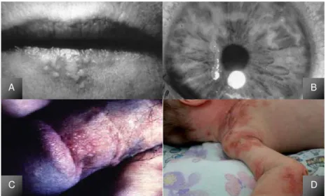
Regarding HSV-1, the most common sites of infection are the skin and mucosal membranes.
Oro-facial lesions are characterized by the development of vesicles at the border of the lip (Figure 1.1A) that
progress to a pustular or ulcerative state within 3 days. The infection is completely healed in 8 to 10
days; however, viral shedding continues for 3 to 5 days after the lesions have healed (Esmann, 2001,
Sciubba, 2003). Ocular lesions (Figure 1.1B) caused by HSV-1 are linked to a variety of ocular
complications, being one of the leading causes of corneal blindness and keratoconjunctivitis (Whitley et
al., 1998, Rudnick et al., 2002).
On the other hand, HSV-2 infections are more associated with genital herpetic lesions and are
currently one of the most prevalent sexually transmitted infections (STIs) worldwide (Smith et al., 2002).
2
within a few days (Figure 1.1C), for a period that can last up to 6 weeks. Consequences in men are rare,
but women can develop serious complications including aseptic meningitis or urinary retention. Primary
infections can be associated with fever, dysuria, localized inguinal adenopathy, malaise and headaches
(Whitley et al., 2001, Beauman, 2005). Over the past few years, there has been an increased
consciousness on genital herpes due to the discovery of its association with the risk of human
immunodeficiency virus (HIV) acquisition. The interactions between HSV-2 and HIV may result in a more
efficient HIV transmission and in an increase in its replication rate and shedding during a HSV
reactivation. The relationship between HSV-2 and HIV seems to be reciprocal, since HIV infections also
increase HSV-2 reactivation and probably its acquirement (Beauman, 2005, Mayaud et al., 2008).
Educational efforts must be developed for those at greater risk to prevent virus transmission: the use of
condoms, for instance, should be promoted, as its use significantly decreases the probability of acquiring
STIs.
Neonatal HSV infections are also a very important matter, as they can result in serious morbidity and
mortality (Rudnick et al., 2002). Neonate infections can occur in utero (5%), intrapartum (85-90%) or
postnatally (5-10%)(Straface et al., 2012). Infections are usually symptomatic, with sores appearing in
the skin, eyes and mouth (Figure 1.1D), and the possibility to affect multiple organs, leading to the baby’s
death. Most of these infections are due to HSV-2, but 15 to 30% are found to be caused by HSV-1
genital infections (Nahmias, 1970, Rudnick et al., 2002, Lee, 2008).
Additionally, both HSV can infect the central nervous system (CNS) and cause diseases such as
encephalitis and meningitis, being one of the most common causes of fatal sporadic encephalitis in
humans. Patients may experience fever, malaise, headaches and personality fluctuations. The mortality
among untreated patients exceeds 70%, decreasing in treated patients to 19%. However, more than
50% of the patients who survive are left with moderate or severe neuropsychiatric sequels, as only 2.5%
of the patients who survive regain normal neurological function(Whitley et al., 1998, Whitley et al., 2001,
Meyding-Lamadé et al., 2012).
Figure 1.1: Herpesvirus induced lesions.
[A] Oral herpes; [B] Ocular herpes; [C] Genital Herpes; [D] Neonatal herpes.
[A][B] (Pellet et al., 2003, Roizman et al., 2003); [C] (Beauman, 2005); [D] (Bittencourt et al., 2016)
A
C D
3
Despite an increasing awareness regarding both HSV-1 and HSV-2 infections, many cases remain
undiagnosed, as they are usually asymptomatic (silent transmission to partners or to the neonate) or
unrecognized, when symptomatic infections are present in an unusual or atypical way, making the
diagnosis harder (Roizman et al., 2003).
Currently, there are several systemic antiviral agents against HSV. The most effective treatment is
acyclovir or its prodrugs valacyclovir and famciclovir (Chilukuri et al., 2003, Pellet et al., 2003, Lee, 2008,
Martinez et al., 2008). Even though these drugs can treat herpes disease, they cannot prevent
reactivations. In addition, prolonged prophylaxis and treatment can result in the development of drug
resistance, particularly in immunocompromised patients (Bacon et al., 2003).
1.2. Epidemiology
HSV infections occur worldwide, without seasonal variation. Despite the fact that infections caused
by HSV are a serious health problem worldwide, few updated epidemiological data exist, with the most
recent ones dating from 2012. Nevertheless, it is known that the prevalence of both HSV infections
varies according to the subject’s age, country, regions and population subgroups (Smith et al., 2002). Regarding HSV-1, it has been estimated that 3.7 billion people under the age of 49 are globally
infected with this subtype (Looker et al., 2015a). This prevalence was clearly higher in Africa,
South-East Asia and Western Pacific than for Europe, Americas and Western Mediterranean. However, a
different scenario was seen for the HSV-1 genital infections, which seem to be globally increasing. Of
the 3.7 billion prevalent cases, ~140 million correspond to prevalent genital HSV-1 infections, primarily
in the Americas, Europe and Western Pacific (Looker et al., 2015a).
Concerning HSV-2, the estimated worldwide prevalence among people between the ages of 15 and
49 was 417 million (Looker et al., 2015b). Prevalence was higher in Africa (31.5%) and generally higher
in females compared to males (14.8% vs. 8%). This difference is due to the fact that women have greater
biological susceptibility to HSV-2, although different patterns of sexual behavior between the sexes can
expose women to a higher risk of infection. Estimates also indicate that ~ 23 million new cases are
diagnosed every year (Looker et al., 2008)
Neonatal HSV has an estimated incidence of around one in 2000 to 5000 births per year, although
in some areas there can be as many as one neonatal infection in 1500 deliveries. This incidence is
directly related to the seroprevalence of HSV-2 (Looker et al., 2015a).
1.3. Virion structure and Genomic organization
The mature infectious HSV virion has four distinct structural components: the dsDNA genome is
enclosed within an icosahedral capsid, surrounded by a proteinaceous tegument and a host-derived
4
Figure 1.2: The HSV-1 virion.
Electron micrograph of a negatively stained HSV-1 virion and a cartoon representation showing the
components comprising the virion. Adapted from Roizman et al., 2003.
1.3.1. Central core and Genome
The HSV core contains a dsDNA molecule, with approximately 152 to 155 kilo base-pairs (kbp) and
a 68.3%-70.4% G+C content (Szpara et al., 2014, Kolb et al., 2015). The genome comprises two
covalently linked components, L (long) and S (short), with unique sequences UL (107.09 kbp) and US
(12 kbp), respectively, flanked by large inverted repeats (Figure 1.3). The UL and US units of the genome
can invert relative to one another, producing four different types of DNA molecules (Kukhanova et al.,
2014). The genes of the long and short unique sequences are designated UL1 to UL56, and US1 to
US12, respectively. UL and US encode about 70 proteins, designated virion proteins (VP) and infected
cell proteins (ICP) (Dolan et al., 1998, Palmer, 2010).
Figure 1.3: HSV genome structure.
The long component of the genome (UL) is flanked by inverted repeats designated as a (black) and b (dark
grey), and b’a’; the short component (US) is flanked by a’c’ and ca sequences. Adapted from Kukhanova et
al., 2014.
The genetic maps of both HSV genomes confirms the co-linearity and shows an overall identity of
83% (Roizman et al., 2003). Nevertheless, the two genomes differ in the location of endonuclease
cleavage sites in their genomic DNAs, in the apparent sizes of the encoded proteins, as well as in some
5 1.3.2. Capsid
During an HSV infection, the capsid plays a central role in delivering the genome of the virus into the
host cell nucleus. The capsid, where DNA is stored, consists mainly of four proteins: VP5 (coded by the
UL19 gene), VP19C (UL38), VP23 (UL18) and VP26 (UL35). VP5 is the major capsid protein and is
present in both pentons, forming the vertices, and hexons, forming the faces (Baines, 2011). VP26 is
present as a ring on top of the VP5 subunits of each hexon (Zhou et al., 1995), whereas triplexes made
of one VP19C and two VP23 molecules link adjacent capsomers. One of the vertices is unique,
consisting of 12 copies of the portal protein UL6 through which viral DNA is sent into the nucleus
(Bowman et al., 2003, Roizman et al., 2003, Brown et al., 2011).
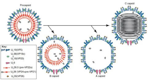
Four types of capsids can be isolated from infected cells. First, spherical procapsids are formed,
containing the internal scaffold. During infection, the maturation of procapsids gives rise to A-, B- and
C-capsids. A-capsids lack both scaffold proteins and viral DNA, and are incapable of maturation;
B-capsids have scaffold proteins and are thought to be in an early stage of viral assembly; C-B-capsids
contain the viral genome and can become infectious virions (Gibson et al., 1972, Sheaffer et al., 2001,
Palmer, 2010, Tandon et al., 2015).
1.3.3. Tegument
The tegument occupies the space between the capsid and the envelope, and can be divided in inner
tegument, which is tightly bound to the capsid, and outer tegument, connected to the viral envelope
(Scrima et al., 2015). It occupies about two thirds of the volume enclosed within the virion (Grunewald
et al., 2003) and is made of, at least, 20 different proteins. The tegument functions as a delivery compartment for proteins during the early course of infection and participates in virion assembly (Lee,
2008).
1.3.4. Envelope
The outer layer of the HSV virion is a host-derived envelope, consisting in a lipid bilayer with around
13 virally encoded glycoproteins (g) embedded in it. HSV glycoproteins have roles in attachment and
entry into the host cell, even though the function of each glycoprotein varies according to the overall
pathogenesis and immune evasion strategy of the virus (Kukhanova et al., 2014). Nevertheless, it is
known that gB, gC, gD, gH and gL are essential for the infection process (Reske et al., 2007).
1.4. HSV life cycle
HSV is thought to have a relatively short life cycle of about 15 to 24 hours (Jenkins et al., 1996,
Lehman et al., 1999, Roizman et al., 2003, Schiffer et al., 2013) that can be divided in six major steps:
(i) entry into the host cell, (ii) viral gene expression, (iii) viral DNA replication, (iv) viral maturation, (v)
viral egress and (vi) latency.
1.4.1. Entry into the host cell
HSV has two ways of getting into the host cell: endocytosis, not yet fully understood; and membrane
6
Figure 1.4: Pathways of HSV entry into the cell.
Two major entry pathways are known for HSV entry: (I) entry by viral envelope fusion at the plasma
membrane and (II) endocytosis. Both processes lead to the delivery of the HSV genome into the host cell
(Salameh et al., 2012).
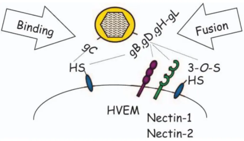
Initial attachment/entry is mediated via gC (or gB, in case gC is unavailable), that binds to the cell
surface’s glycosaminoglycans, in particular heparan sulfate (HS) (Campadelli-Fiume et al., 2012). Afterwards, gD interacts with one of the three entry receptors: (i) the herpesvirus entry mediator
(HVEM), a component of T and B-lymphocytes, epithelial cells and fibroblasts; (ii) both Nectin-1 and
Nectin-2, members of the immunoglobulin family, expressed in human tissues including epithelial cells,
fibroblasts and neurons, or (iii) the 3-O sulfated heparan sulfate (3-OS-HS), a polysaccharide present
in endothelial cells (Figure 1.5) (Spear, 2004, Salameh et al., 2012, Kukhanova et al., 2014). Despite
structural disparities and different interactions with gD, the binding to one of the three receptors
described above leads to the same endpoint: membrane fusion (Heldwein et al., 2008, Gianni et al.,
2009, Kukhanova et al., 2014).
The next step requires gB and the heterodimer gH/gL (Figure 1.5), resulting in the release of the
capsid and of approximately 20 tegument proteins into the cell’s cytoplasm. The capsids and several
tegument proteins are transported through the cell to the nucleus via microtubules, mediated by the
retrograde motor dynein (Döhner et al., 2002, Palmer, 2010, Kukhanova et al., 2014). Once the capsids
have reached the proximity of the nucleus, they dock at the nuclear pore, releasing the viral genome
through the UL6 portal (Roizman et al., 2003, Kukhanova et al., 2014, Fay et al., 2015). Upon entry into
7
Figure 1.5: Multiple receptors upon HSV entry.
gC mediates the initial attachment of virus particles to HS. The next step involves the interactions between
gD with HVEM, Nectin-1/Nectin-2 or 3-OS-HS, resulting in membrane fusion, later mediated by the gH-gL
heterodimer, resulting in the release of the viral nucleocapsid into the host cell cytoplasm (Spear, 2004).
1.4.2. Viral gene expression
Once the nucleocapsid reaches the nucleus and the viral DNA ejects, transcription begins
(Ramachandran, 2003). HSV, as all herpesvirus, has a controlled temporal cascade of gene expression,
with three classes of genes, designated α (or IE –immediate early), β (or E –early) and γ (or L – late).
All genes are transcribed using the host’s RNA polymerase II (Honess et al., 1974, Lee, 2008, Palmer,
2010). The α genes begin to be expressed very soon after viral entry and α proteins reach their peak 2
to 4 hours p.i., encoding proteins involved in the transcription of other viral genes. Transcripts are
transported to the cytoplasm and translated into five proteins, being the most important one ICP0.
Immediate early proteins accomplish multiple functions and perform dramatic reorganization of cellular
processes in the interest of the virus (Boehmer et al., 2003, Kukhanova et al., 2014). β genes are
expressed following the accumulation of α proteins, and their expression peaks around 5 to 7 hours p.i. encoding proteins responsible for viral DNA replication, and signaling the onset of viral DNA synthesis,
which results in the expression of γ genes. The expression of the later genes typically peaks between 8
and 12 hours p.i., and can be further divided in two sub-classes, γ1 and γ2, depending on their
8
Figure 1.6: General pattern of gene expression during a HSV infection.
The first genes to be expressed are αgenes (purple) followed by β genes (blue). Meanwhile, DNA replication begins (dotted line). At this point, γ genes start to be expressed: γ1 (orange) can be expressed prior to DNA
replication, and γ2 (green) requires DNA synthesis to be expressed. Once the proteins have accumulated,
progeny viruses (red) assemble (Ramachandran, 2003).
1.4.3. Viral DNA Replication
DNA replication takes place after the expression of α and β genes, using the circularized DNA as
template. The HSV genome contains three origins of replication (Figure 1.7): OriS, present twice in the
c inverted repeats flanking the US region, and OriL located in the middle of the ULregion (Weller et al.,
2012).
Figure 1.7: HSV origins of replication.
The figure shows the organization of the HSV genome, with the origins of replication shown as OriL and
OriS. Both OriS and OriL are located in the promoter-regulatory regions of divergently transcribed genes:
OriL between genes encoding replication proteins and OriS between genes encoding α proteins. Adapted
from de Silva et al., 2009.
The DNA is replicated using a virally-encoded replication complex comprising an origin-binding
protein UL9; a single stranded DNA-binding protein ICP8 (UL29); the DNA polymerase and DNA
polymerase processivity factor, UL30 and UL42, respectively; and the DNA helicase/primase (H/P)
complex UL5, UL52 and UL8 (Lehman et al., 1999, Weller et al., 2012).
The first stage of DNA replication involves an origin-UL9-dependent step. Upon DNA circularization,
UL9 binds to specific recognition sites in either OriL or OriS and begins to unwind the viral DNA. UL9
then recruits ICP8, resulting in the recruitment of the helicase-primase heterodimer, followed by the
polymerase complex (Figure 1.8). The second stage of replication is UL9-independent, and leads to the
formation of longer-than-unit length concatemers, later packaged into procapsids in the form of
9
Figure 1.8: Model of HSV DNA replication.
Replication fork model. The H/P complex, made of proteins UL5, UL8 and UL52, unwinds the DNA; the
binding protein ICP8 binds to single-stranded template DNA; the polymerase (UL30) and its accessory
protein UL42 promote leading and lagging-strand DNA synthesis. Adapted from Crumpacker et al., 2002.
1.4.4. Viral maturation
Following repeated rounds of DNA synthesis, concatemeric genomes accumulate within replication
compartments. The synthesis of γ genes, encoding late proteins that include structural proteins,
envelope proteins incorporated in nuclear membrane and several other structural and packaging
proteins, lead to the assembly of new progeny virions (Lehman et al., 1999). Capsids are initially formed
as procapsids with an internal protein scaffold made of UL26 and UL26.5 gene products, which is lost
upon DNA packaging. The protease UL26 cleaves itself, generating capsid proteins VP24 and VP21,
and UL26.5, generating VP22a (Newcomb et al., 2000, Mettenleiter et al., 2006). Capsid formation also
requires the capsid proteins VP5, VP23 and VP19C (Mettenleiter et al., 2009). The procapsids differ
from mature capsids in several aspects: while the procapsids are spherical, have a more porous
structure with holes between capsomers and have oval hexons, mature capsids (A-, B- or C-) are
icosahedral, have a less porous composition and have hexagonal hexons (Newcomb et al., 2000,
Mettenleiter et al., 2006). Procapsids transform into mature capsids in a process in which the shell
undergoes structural changes (Figure 1.9). Capsid maturation overlaps with viral DNA packaging,
resulting in a mature C-capsid containing the viral genome. C-capsids are the only ones able to undergo
10
Figure 1.9: HSV capsid maturation.
The spherical procapsid is the precursor of all other capsid types. It is made primarily of VP5 pentons (dark
blue) and hexons (light blue) linked by triplexes (green), with 12 copies of UL6 that will eventually form the
portal. The inner scaffold shell is lost or degraded in A capsids, retained in B capsids, and replaced with
DNA in C capsids. Adapted from Baines, 2011.
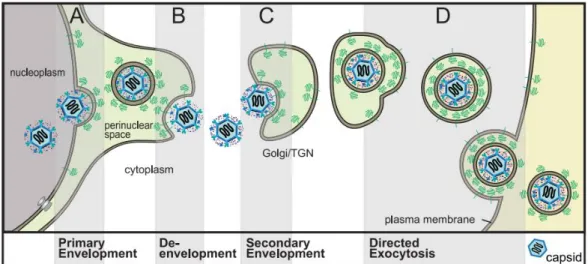
1.4.5. Viral egress
Successful encapsidation of the viral genome is followed by budding through the modified inner
nuclear membrane (INM) into the perinuclear space to acquire the primary envelop and continue the
maturation process (Kukhanova et al., 2014). Several models have been proposed over the years to
explain the exact egress pathways; however, the most exact one is the envelopment-deenvelopment
model. According to this, capsids dispersed in the nucleus approach and attach the INM, through
intranuclear movement via nuclear actin filaments, induced by HSV infection (Mettenleiter et al., 2009).
Proteins UL31 and UL34 are crucial for this process, since the UL31/UL34 nuclear envelopment
complex induces conformational changes in the nuclear lamina. This disruption of the lamina allows the
capsids to bud into the perinuclear space gaining a primary tegument and envelope (Figure 1.10A)
(Newcomb et al., 2006, Kukhanova et al., 2014).
Next, nucleocapsids undergo a de-envelopment process, by fusing with the outer nuclear membrane
(ONM), resulting in the loss of the primary envelope and nucleocapsid release into the cytoplasm, for
continuing maturation (Figure 1.10B) (Roizman et al., 2003, Mettenleiter et al., 2006, Mettenleiter et al.,
2009).
After nuclear egress, the nucleocapsids have to acquire tegument proteins and the final (secondary)
envelope in order to exit the cell (Mettenleiter et al., 2009). This process occurs predominantly in the
cytoplasm, and is controlled by a complex network of protein-protein interactions (Owen et al., 2015).
HSV nucleocapsids make their way toward cytoplasmic vesicles derived from the trans-Golgi Network
(TGN) to reacquire their envelop (Figure 1.10C).
Currently, little is known about the mechanism by which mature virions are released from cells, but
it is thought that these viruses use proteins from the host cell’s secretory pathway to facilitate egress at
11
they fuse and release mature virions, by exocytosis, resulting in the death of the host cell (Figure 1.10D)
(Mingo et al., 2012).
Figure 1.10: HSV viral egress.
[A] Primary Envelopment - After capsids are formed in the nucleus, they bud into the INM to form an
enveloped particle in the perinuclear space;
[B] De-envelopment - HSV particles reside in the perinuclear space. HSV glycoproteins gB and gH/gL
mediate fusion of the viral envelope with the ONM;
[C] Secondary Envelopment - In the cytosol, capsids coated with numerous tegument proteins bind onto
the surface of the TGN that contains HSV glycoproteins. Interactions between all of these proteins promote
envelopment;
[D] Egress - Virions are transported to cell surface in exocytic vesicles, leading to the death of the host cell.
Adapted from Johnson et al., 2011).
1.4.6. Latency-Reactivation Cycle
Herpesvirus have the ability to establish latency, one of the most intellectually challenging aspects
of HSV biology (Stevens et al., 1971). Despite a vigorous immune response during an infection, HSV
establishes latency in sensory neurons, typically trigeminal ganglia for HSV-1 and sacral ganglia for
HSV-2. Remarkably, up to 40% of sensory neurons can be latently infected (Kramer et al., 1998, Perng
et al., 2010, Nozawa et al., 2014).
The latency-reactivation cycle can be divided into three major steps: (i) establishment, (ii)
maintenance, and (iii) reactivation. Establishment of latency includes the entry of the viral genome into
a sensory neuron, through retrograde axonal transport (Figure 1.11). Viral gene expression is silenced
with the exception of one set of transcripts that continue to be expressed known as latency-associated
transcripts (LAT) (Nicoll et al., 2012, Ma et al., 2014). Maintenance of latency is a phase that lasts as
long as the host lives, and is defined as a period when infectious genes are not detected, but LATs
continue to be expressed. The full length 8.3 kbp LAT is transcribed from the latency associated
promoter and is sliced, giving rise to two stable 2.0 kb and 1.5 kb introns (Nicoll et al., 2012, Kukhanova
12
process that results in the decrease of α genes expression (Jackson et al., 2003). During latency, the viral genome circularizes and is maintained as an episome within the nucleus (Ramachandran, 2003).
In Humans, latency is maintained throughout the host’s life, indicating that a well-conceived strategy exists, allowing periodic reactivation, while maintaining the viral genome in sensory neurons.
Reactivation from latency is initiated by an external stimulus, such as stress or immunosuppression,
culminating in viral gene expression. Clinically, depending on the host immune status, reactivation can
be symptomatic, where the virus can be detected in peripheral tissues and disease is recognized,
resulting in lesions, or asymptomatic, where the virus can be laboratory detected but signs of disease
are unrecognized. In both cases, the virus is carried by anterograde axonal transport (Figure 1.11) to
peripheral tissues through the motor protein kinesin, usually to cells at, or near the site of initial infection.
Reactivation leads to viral expression and generation of progeny virions (Perng et al., 2010).
Figure 1.11: Neural trafficking during latency-reactivation cycle.
HSV establishes latent infection in the nuclei of peripheral ganglia, following retrograde transport along
microtubules, through the dynein transport protein. Reactivation results in the production of new virions that
undergo anterograde transport back to peripheral tissues with the transport protein kinesin. Adapted from
Owen et al., 2015.
1.5. Scope of the Thesis
HSV infects a variety of host cells, including lymphocytes, epithelial cells, fibroblasts, and neurons
(Connolly et al., 2011). Although there are multiple studies that try to evaluate the infection patterns of
HSV, including among sub-types, none of them is conclusive and reproducible, being even sometimes
contradictory (Docherty et al., 1971, Tada et al., 1977, Nguyen et al., 2005, Aguilar et al., 2006). For
instance, even though HSV is considered to have a relatively short life cycle when compared with other
viruses, there doesn’t seem to be a consensus regarding the duration of the infectious process, as
cycles ranging from 15 to 24 hours can be currently found throughout the literature (Jenkins et al., 1996,
Lehman et al., 1999, Roizman et al., 2003, Schiffer et al., 2013). Moreover, it is also not known whether
13
infected tissue (oro-facial tissue versus genitalia), even though it is known that HSV-1 genital
reactivations appear to be less common and timely shorter than those of HSV-2. Indeed, as seen for
other human pathogens (Nunes et al., 2013, Martines et al., 2015), the observed cellular tropism doesn’t
seem to be straight to a particular HSV type. Differences in viral evolutionary dynamics, such as
evolution rates, can explain why certain viruses have the capacity to adapt to new host species, increase
in virulence, or develop resistance to antivirals (Hicks et al., 2014).
In order to contribute for this knowledge, the scope of this thesis was to evaluate the life cycle of
various genital HSV-1 and HSV-2 clinical isolates with different viral loads in distinct host cell lines,
giving special focus to the invasion process. Considering that there has been an increase in the number
of cases of genital herpes caused by HSV-1, especially among the younger population, it is of utmost
importance to understand whether HSV-1 in the genitalia replicates the same way as it does in mouth
sores (not evaluated in the present study), or if it has an alternative route of infection that may be similar
to that of HSV-2. In particular, understanding whether capacities of infections, replication rates, and
progenies depend on the genital HSV sub-type/strain or on the infected cell line is a mandatory step for
the understanding of the pathogenesis of human herpes infections and may be important for the
development of a more effective antiviral chemotherapy.
To achieve robust and reliable conclusions, four genital HSV samples isolated from genital swabs,
two HSV-1 and two HSV-2, were used in the present study, as well as three host cell lines with different
phenotypic and molecular characteristics. Hence, specific objectives of these assays were:
1) To assess whether genital infections caused by HSV-1 and HSV-2 have the same molecular
characteristics, in a particular cell line;
2) To test if both HSV display the same infection capacity in all three cell lines tested;
3) To measure whether there is a viral saturation in a HSV infection in the three cell lines;
4) To determine the duration of the HSV-1 and HSV-2 life cycle;
5) To determine when does DNA replication begin;
15
2. Materials and Methods
2.1. Biological samples
The viruses used in the present study were obtained from ulcer and genital/urethral swabs of four
patients suspected of having a HSV infection, from a Sexually Transmitted Diseases Outpatient Clinic.
The viral swabs (with viral transport medium) were sent for confirmation of diagnosis to the National
Reference Laboratory of Herpes simplex virus, Cytomegalovirus and Parvovirus B19 of the Portuguese
National Institute of Health (INSA), in Lisbon, and treated according to laboratory protocol. After swab
agitation and content filtration with a 0.45µm filter, DNA was extracted using the automatic nucleic acid
extractor NucliSENS® EasyMag® (BioMérieux). HSV-1 and HSV-2 DNA was quantified with a
commercial real-time PCR (RT-PCR) kit (HSV1 HSV2 VZV R-gene®, ARGENE®, BioMérieux), that
targets a 142 bp fragment of the US7 gene from HSV-1 and a 177 bp fragment of the US2 gene from
HSV-2. PCR reactions were carried out with the amplification platform ABI PRISM® 7500 (Applied
Biosystems®). Positive results were reported as copies/mL, taking into consideration sample extracted
volume, final elution volume and DNA volume used in the amplification reaction.
The epidemiological, laboratory and clinical data of the selected clinical isolates are described in
Table 2.1:
Table 2.1: Epidemiological, laboratory and clinical data.
Clinical
isolates Virus
Patients information Viral Load
copies/mL
Gender Age Episode Location
A HSV-1 Female 22 Primary infection Vulva,
perineum 46 058 248
B HSV-1 Female 22 Symptomatic reactivation Vulva 87 474 493
C HSV-2 Male 32 First symptomatic episode,
but not primary infection Penis 526 335 906
D HSV-2 Female 24 --- Not available --- Vulva 302
2.2. Cell line handling and maintenance
The present experimental work was performed using three different adherent epithelial cell lines that,
due to their susceptibility to a wide range of hosts, are usually used in virology studies, in particular for
HSV: Vero (ATCC® CCL-81™) and Vero E6 (ATCC® CRL-1586™) from the kidney of the African Green
monkey, and HeLa229 (ATCC® CCL2.1™) from a cervix adenocarcinoma of a 31-year old black
woman. Vero E6 cells differ from regular Vero cells since they show some degree of contact inhibition,
being suitable for supporting the growth of slowly replicating viruses. All cell lines were purchased from
16
Each cell line was grown in 175 cm3 tissue culture flasks (T175; Sarstedt) with Dulbecco’s Minimum
Essential Medium (MEM 1x + GlutaMAX™; Gibco, USA) supplemented with 10% fetal bovine serum
(FBS; Gibco, USA). For both Vero and Vero E6 cells, the medium was further complemented with 1%
HEPES buffer solution 1M (Gibco, USA), 1% Non-Essential Amino Acids (NEAA 100x; Gibco, USA) and
0,2% Antibiotic Mixture (PSN 100x; Gibco, USA), while for HeLa229 cells, Gentamicin (50 mg/mL;
Gibco, USA) and Fungizone (250 µg/mL; Gibco, USA) were added. Cell cultures were incubated in a
CO2 incubator (Binder) at 37ºC with a 5% CO2 atmosphere, and cellular growth was followed by
observing cultures under an inverted optic microscope (Leica DMIL LED).
In order to produce sufficient cellular stocks for all assays, each cell culture was subjected to
continuous passages whenever a 90% confluent cell monolayer was reached. Briefly, after the medium
was removed and the cells washed twice with 10 mL of Phosphate Buffered Saline (DPBS 1x; Gibco,
USA), 5 mL of trypsin (0.5% Trypsin-EDTA 10x; Gibco, USA) were added to the flask, following an
incubation of 5 minutes in the CO2 incubator, in order to detach the cells. After that time, the cells were
ressuspended and added to a new flask with fresh medium.
2.3. Preliminary assays
For each cell line and HSV isolate, preliminary assays were performed to determine the optimal
cellular concentration, culture conditions and viral loads to be used for the subsequent quantification
assays. The inoculation process was also evaluated in order to better mimic an in vivo HSV infection.
2.3.1. Cell counting
For each cell line, in order to ensure that HSV inoculation occurred on a 90% confluent cell
monolayer, cell density was verified 24 hours before the assays, using the trypan blue method on a
hemocytometer (Spencer-Buffalo, USA). Briefly, confluent T175 culture flasks were treated as stated in
step 2.2 and resuspended; then, 50 µL of the cell suspension were added to 200 µL of Hanks’ Balanced
Salt Solution (HBSS 1x; Gibco, USA). Following vortex agitation (PV-1 Vortex Mixer, Grant), 50 µL of
this solution were added to 50 µL of trypan blue (Sigma), which stains dead cells. Finally, 10 μL of this
solution were placed in each of the two hemocytometer chambers, and the number of viable cells (not
stained by the trypan blue) were counted under the microscope (Leica DM IL LED). Cell density was
determined through equation (1):
(1)
[
𝑁𝑢𝑚𝑏𝑒𝑟 𝑜𝑓 𝑐𝑒𝑙𝑙𝑠𝑚𝐿
] = (
∑ 𝐶𝑒𝑙𝑙𝑠 𝑝𝑒𝑟 𝑞𝑢𝑎𝑑𝑟𝑎𝑛𝑡
4
) x 10
4 (Chamber volume x dilution factor)2.3.2. Culture conditions
Considering that the success of any microbial infection depends, among other factors, on the
availability of nutrients in the medium, the optimal FBS quantity to be added to each cell culture was
determined, in order to guarantee, not only that a confluent monolayer was achieved at the time for the
experiments, but also that this monolayer was stable enough to support the entire period of a viral
infection. Basically, suspensions of each cellular culture were seeded into two 24-well plates, with half
17
placed in a CO2 incubator (Binder) at 37ºC with a 5% CO2 atmosphere, one for 48h and the other for
72h. Cellular growth was evaluated by both visual assessment on an inverted optical microscope and
cell counting.
Figure 2.1: Nutrient availability assay plate scheme.
2.3.3. Viral stocks
For each HSV under evaluation, viral stocks were produced by inoculating 100% confluent Vero cells
monolayers. Infected cells were incubated at 37ºC with a 5% CO2 atmosphere for 1-4 days, until they
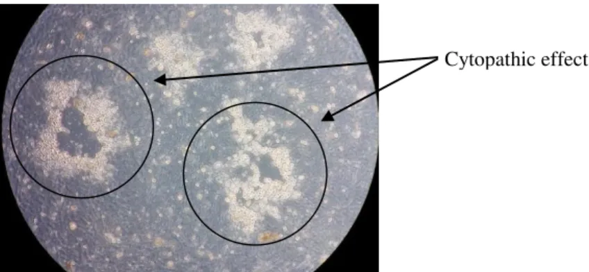
showed cytopathic effect (CPE), i.e., severe morphological alterations in infected cells and lack of
adherence (Figure 2.2).
Culture flasks were frozen and defrozen in order to detach all cells from the walls of the flask. Virus
suspensions were well homogenized, aliquoted into 2 mL tubes and stored at -80ºC, until use. In
average, a total of 100 mL of stock were produced for each virus. For all four viral stocks, two aliquots
were randomly selected and subjected to DNA extraction followed by subsequent absolute quantification
by RT-PCR, as described in 2.1.
Figure 2.2: CPE on Vero cells after a one-day incubation with a HSV-2 virus.
The final viral loads, after viral stock production, and based on which further calculations were made
(for step 2.4) are presented in table 2.2.
4% FBS 10% FBS
1 2 3 4 5 6
18 Table 2.2: Final viral charges, after viral stock production.
Clinical
isolate Virus
Final viral charge copies/μL
A HSV-1 6.05 x 10⁶
B HSV-1 2.76 x 10⁶
C HSV-2 2.16 x 10⁷
D HSV-2 3.68 x 10¹²
2.3.4. Inoculation conditions
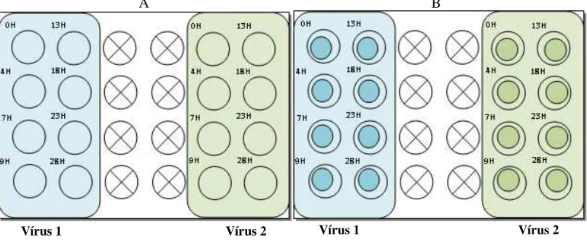
In order to better mimic the HSV infection in vivo, two different in vitro inoculation conditions were
tested in parallel: one involved centrifugation and the other agitation. To achieve robust conclusions,
two HSV-2 clinical isolates, with distinct viral loads (3-fold difference) were used. Briefly, quadruplicate
24-well plates with confluent Vero cells monolayers were prepared, after adding coverslips to two sets
of plates. Inoculations were performed by adding 200 μL of each HSV-2 virus per well to 1/3 of the
wells/plate (Figure 2.3).
After inoculation, two sets of plates (one with and another without coverslips) were centrifuged at
700g for 45 min, at 4ºC (Centrifuge 5810 R, Eppendorf), in order to mechanically promote only viral
attachment to the host cells. At 4ºC, no cellular metabolism occurs, preventing any viral entry. The entry
process is then synchronized, ensuring that attached viruses will later enter (at a higher temperature) at
the same time. The other two plates (with and without coverslips) were subjected to agitation at 20 rpm
for 1 hour (Rocking Platform VWR®), at 37ºC (Memmert), allowing the viruses to attach and possibly
enter host cells in a less artificial way, at different rates. At the end of both processes, inoculum and
dead cells were removed, and 1 mL of new media complemented with 4% FBS was added, thus
accounting time-point “0”.
Plates were then incubated for ~ 30 hours at 37ºC with a 5% CO2 atmosphere. Infection was followed
at different time-points (4h, 7h, 9h, 13h, 18h, 23h and 29h p.i.) in order to evaluate the putative HSV
growth curve. The plates without coverslips were subjected to RT-PCR absolute quantification (Figure
2.3A) for determining viral load at each time-point, while the others were used for monoclonal antibody
staining to monitor eventual changes at a cellular level (Figure 2.3B).
Regarding RTPCR, the wells were scratched at the indicated timepoints of infection and kept at
-20ºC until use. Extractions were later performed using the QIAamp® DNA Blood Mini Kit (Qiagen),
according to the manufacturer’s instructions. The HSV-2 DNA was amplified and quantified using the
HSV1/HSV2/VZV R-gene®” ARGENE® kit, as instructed, using the amplification platform ABIPRISM
7500 (Applied Biosystem). For immunofluorescence, the medium was removed at each time-point,
washed twice with 1 mL of DPBS and fixed with methanol. After a 10 min air-dry, DPX (Fluka,
BioChemika) was added for the coverslip to adhere to a slide. 50 μL of the monoclonal antibody specific
for HSV-2 (Pathfinder™ HSV 1/2, Biorad) were added to the coverslips, following a 30 min incubation
19
the slides with DPBS, 50 μL of Mounting Medium were added. All slides were later observed in the fluorescence microscope (Axioscop 2 plus, Zeiss).
Figure 2.3: Inoculation conditions plate schemes.
[A] – kPCR plate; [B] – Monocolonal antibody staining plate.
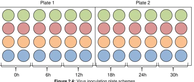
2.4. Evaluation of HSV infection in distinct cell lines
Once optimization processes regarding inoculation conditions, time-points of infection, cell counting,
and cell conditions, were established, assays were performed in order to determine the capacity of
infection of the four viruses under evaluation (Table 2.1), in three distinct cell lines, in terms of progeny
and infection efficiency. To do this, different multiplicities of infections (MOI) were tested (0.1, 1, 10 and
100) for each virus, to evaluate if different viral loads had any influence on the success of the infection
process. To calculate the MOIs, we counted the DNA particles and not the viable viral particles per se,
as we assumed that by the end of the viral stock preparation all viral replication and assembly had
already stopped, thus giving rise to infectious viruses, once the monolayer had been completely
destroyed.
For each cell line, two 24-well plates with confluent cell monolayers were prepared (as described in
step 2.3.1; for further information, see Results) and inoculated with each virus at 4 different MOIs (Figure
2.4). After agitation (step 2.3.4), the inoculum was removed, the wells were washed twice with DPBS
and 1 mL of fresh media was added, complemented with 4% FBS for Vero and Vero E6 cells, and 10%
for the HeLa229 cell line. These steps were made in order to guarantee that all the viruses that were
not able to attach/enter host cells would not affect the infection process. Infected cultures were then
incubated at 37ºC with a 5% CO2 atmosphere for 30 hours. The infectious cycle was interrupted at
selected timepoints (0h, 6h, 12h, 18h, 24h and 30h p.i.), where cells were harvested and stored at
-20ºC until subsequent extraction and quantification. Time-point “0” refers to the time-point after agitation,
following the addition of fresh medium.
A B
20
Figure 2.4: Virus inoculation plate schemes.
Two sets of plates were prepared for each virus per cell line. Time-points were prepared in duplicate.
Each line corresponds to a different MOI: 0.1 (green), 1 (red), 10 (orange) and 100 (blue).
2.5. DNA extraction
Total DNA was extracted using the QIAamp® DNA Blood Mini Kit (Qiagen), with minor changes
regarding the given protocol. Briefly, after thawing the samples harvested in step 2.4, the tubes were
centrifuged at low speed, at 2500 rpm for 5 minutes, in order to isolate non-infected cells and cellular
debris from viral particles. The supernatant was then collected and from this, 500 µL were added to 50
µL of Protease or Proteinase K, followed by 500 µL of buffer AL, or ATL, and agitated (Maxi Mixer Vortex
714). The samples were incubated at 56ºC for 30 minutes in a thermoblock (Thermomixer® Comfort,
Eppendorf) when using AL, and 15 minutes for ATL. 500 µL of ethanol were added and the whole
mixture was transferred to a mini-spin column and centrifuged (Sigma 1-14). The columns were washed
with 500 µL Buffer AW1 and Buffer AW2, with spins between each treatment. Finally, the samples were
eluted in 100 µL of buffer AE and stored at -20ºC until use.
2.6. Cloning
In order to develop an in-house HSV-1/HSV-2 RT-PCR system and to design a standard curve for
RT-PCR experiments, a cloning assay was performed using a single copy gene from each HSV. Two
genes for each HSV were chosen based of whether they were single copy genes. For HSV-1, a literature
search (Szpara et al., 2014) was performed to identify highly conserved genes; regarding HSV-2, both
the US2 and UL27 genes were selected, as the first is the target of the standard HSV commercial kit,
while the other is often used in the diagnosis and quantification of HSV DNA in the CNS (Tang et al.,
1999). Primers were designed using the Primer Express® Software (Applied Biosystems) and further
alignments were made using Blast (available in: http://blast.ncbi.nlm.nih.gov/Blast.cgi) to guarantee that
they were HSV specific and fell in conserved gene regions. The two primers tested for both HSV are
displayed in Table 2.3.
Plate 1 Plate 2
21 Table 2.3: Sets of primers tested.
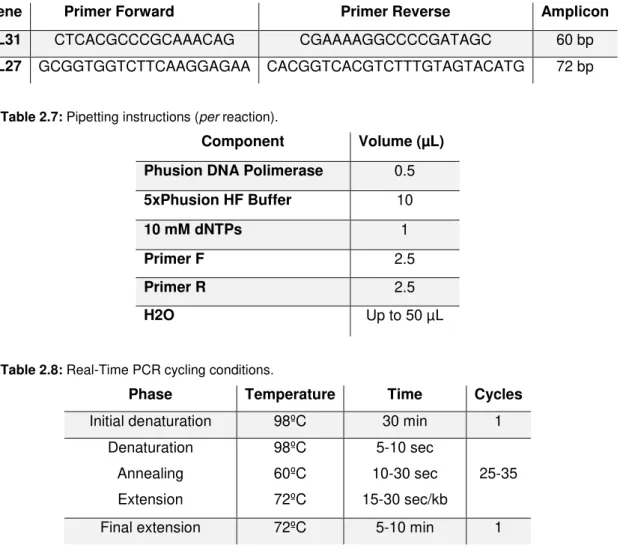
Virus Gene Protein Reference
HSV-1 UL53 Envelope glycoprotein gK (Dolter et al., 1994)
HSV-1 UL31 Nuclear protein (Szpara et al., 2014)
HSV-2 UL27 Envelope glycoprotein gB (Tang et al., 1999)
HSV-2 US2 Tegument protein (Kang et al., 2013)
Primer stock solutions were diluted to a concentration of 25 pmol/μL concentration. DNAs had
already been extracted and quantified from two HSV clinical isolates (one HSV-1 and one HSV-2) from
the viral stocks previously prepared (step 2.3.3). DNA serial dilutions were performed based on viral
load, from a concentration of 1x10⁸ cop/μL up to 1x101 cop/μL. Components for RT-PCR reactions
(Table 2.4) and PCR conditions (Table 2.5) are described below:
Table 2.4: Pipetting instructions (per reaction).
Component Volume (µL)
SYBR Green I 12.5
Primer F 2
Primer R 2
H2O 3.5
Sample 5
Table 2.5: PCR cycling conditions.
Phase Temperature Time Cycles
Pre-incubation 95ºC 10 min 1
Amplification
95ºC 15 sec
40
60ºC 1 min
Melting Curve
95ºC 15 sec
1
60ºC 20 sec
95ºC -
Cooling 40ºC 30 sec 1
For each HSV, primer selection was based on both efficiency and slope of the obtained HSV standard
curves. Primers where the respective calibration curve presented a slope of approximately - 3.3 and an
efficiency of nearly 100% were chosen (Svec et al., 2015).
Once chosen the fittest primers, new RT-PCR reactions were performed to amplify the two conserved
gene fragments, one for each HSV, using the Thermo Scientific Phusion High-Fidelity DNA Polymerase
kit (Thermo Fisher): UL31 for HSV-1 and UL27 for HSV-2. The set of primers used for this reaction